
Abstract
Due to both the avascularity of the cornea and the relatively immune-privileged status of the eye, corneal transplantation is one of the most successful clinical transplant procedures. However, in high-risk patients, which account for >20% of the 180,000 transplants carried out worldwide each year, the rejection rate is high due to vascularization of the recipient cornea. The main reason for graft failure is irreversible immunological rejection, and it is therefore unsurprising that neovascularization (NV; both pre and post grafting) is a significant risk factor for subsequent graft failure. NV is thus an attractive target to prevent corneal graft rejection. OXB-202 (previously known as EncorStat®) is a donor cornea modified prior to transplant by ex vivo genetic modification with genes encoding secretable forms of the angiostatic human proteins, endostatin and angiostatin. This is achieved using a lentiviral vector derived from the equine infectious anemia virus called pONYK1EiA, which subsequently prevents rejection by suppressing NV. Previously, it has been shown that rabbit donor corneas treated with pONYK1EiA substantially suppress corneal NV, opacity, and subsequent rejection in an aggressive rabbit model of cornea graft rejection. Here, efficacy data are presented in a second rabbit model, which more closely mirrors the clinical setting for high-risk corneal transplant patients, and safety data from a 3-month good laboratory practice toxicology and biodistribution study of pONYK1EiA-modified rabbit corneas in a rabbit corneal transplant model. It is shown that pONYK1EiA-modified rabbit corneas (OXB-202) significantly reduce corneal NV and the rate of corneal rejection in a dose-dependent fashion, and are tolerated with no adverse toxicological findings or significant biodistribution up to 13 weeks post surgery in these rabbit studies. In conclusion, angiogenesis is a valid target to prevent corneal graft rejection in a high-risk setting, and transplanted genetically modified corneas are safe and well-tolerated in an animal model. These data support the evaluation of OXB-202 in a first-in-human trial.
Introduction
S
Corneal rejection is associated with vascularization of the corneal bed. So once a graft has failed, prior graft failure then becomes a risk factor for rejection of subsequent grafts, 2 with a much higher likelihood of rejection and a poorer prognosis for these patients. Each subsequent ipsilateral graft is associated with an increasing risk of rejection. 3 A repeat transplantation is associated with only a 20–45% graft survival rate, 4,5 and regrafting a failed transplant is one of the top two indications for corneal transplantation in many centers. In some of these patients, the prognosis is extremely poor, with grafts failing at an accelerating rate to the point where patients are no longer considered suitable for further transplants, despite an otherwise normally functioning visual system, and are subsequently left blind. Corneal neovascularization (NV) within the transplant is one of the most important risk factors associated with corneal graft rejection. 6 Thus, prevention of NV would be a pivotal step toward addressing the unmet need in preventing graft failure and rejection in these high-risk patients, but no specific anti-angiogenic treatment has yet been licensed to treat corneal NV.
An ex vivo gene therapy has been developed for the treatment of patients at high risk of corneal graft rejection due to NV. OXB-202 is a donor cornea that has been genetically modified ex vivo to be inherently angiostatic due to the secretion of two naturally occurring anti-angiogenic proteins: endostatin and angiostatin. These two proteins function to prevent aberrant angiogenesis and are specifically involved in maintaining corneal avascularity following injury. Secretion of these anti-angiogenic proteins will lead to suppression of blood and lymph-vessel invasion into the corneal graft after transplant to maintain the immune privilege of the eye and reduce the possibility of graft rejection due to NV.
Previously, efficient gene transfer has been demonstrated to human, primate, and rabbit corneal-scleral tissue using equine infectious anemia virus (EIAV)-based vectors expressing either green fluorescent protein (GFP) reporter or endostatin/angiostatin (pONYK1EiA), resulting in detectable and sustained transgene expression (either GFP fluorescence or release of both endostatin and angiostatin; Parker et al. 7 and data not shown). Corneas genetically modified to secrete endostatin and angiostatin have been shown to inhibit NV significantly (by up to 80%), maintain corneal clarity, and subsequently reduce the rejection rate in an acute rabbit model of corneal rejection. 7,8 Biopotency in models of corneal angiogenesis has also been demonstrated for both endostatin 9,10 and angiostatin (kringle 1–3). 11,12 Together, these data support the scientific rationale for the use of OXB-202 in the treatment of patients at high risk of corneal graft rejection due to NV.
This study reports that OXB-202 is able to reduce corneal NV significantly, as well as reduce corneal opacity and thickness, thereby significantly reducing the rate of corneal graft rejection in a high-risk rabbit corneal rejection model. This model closely mirrors the clinical setting where patients have substantial vascularization of the corneal bed prior to PK. The toxicity, biodistribution, and shedding characteristics of OXB-202 were examined >13 weeks following PK in a rabbit model of corneal transplant, and the data show that the genetically modified cornea is safe and well-tolerated and that residual vector is minimal and localized to the cornea.
Materials and Methods
Plasmids
The pONYK1EiA EIAV vector genome plasmid contains cDNAs for secreted human angiostatin and endostatin linked by an internal ribosomal entry site element (described previously in Binley et al.
14
). It contains a cytomegalovirus (CMV) constitutive promoter driving transgene expression, a woodchuck posttranscriptional regulatory element, and self-inactivating long-terminal repeats (LTRs; Supplementary Fig. S1; Supplementary Data are available online at
Viral vector production and characterization
Vector manufacture was as described previously. 13 Briefly, vector was prepared by transient transfection of vector genome plasmid, gag-pol packaging plasmid, and vesicular stomatitis virus envelope glycoprotein (VSV-G) plasmid into HEK293T cells using Lipofectamine 2000 (Invitrogen, Carlsbad, CA) in 10-layer cell factories (Nunc). For the safety study, vector was purified and concentrated by anion exchange chromatography and hollow-fiber technology, and manufacture was carried out in a manner analogous to Good Manufacturing Practice and characterized as described previously. 14 For the efficacy study, vector was purified and concentrated by double centrifugation (6,000 g for 18–24 h, and then 50,000 g for 1.5 h).
Animals
All animals were treated in accordance with the Animal Welfare Act, as promulgated in U.S. Code Sections 2131–2159, in its current form, and the PHS Policy on Humane Care and Use of Laboratory Animals. All animal procedures were performed under Institutional Animal care and Use Committee (IACUC)-approval, which also conforms to the ARVO Statement for the Use of Animals in Ophthalmic and Vision Research standards for humane animal care.
The efficacy study was performed with naïve 3- to 4-month-old female New Zealand white rabbits (n = 36) supplied from Western Oregon Rabbit Company (Philomath, OR), and the safety and biodistribution study was conducted with naïve 17- to 28-week-old Hra:(NZW)SPF rabbits (36 male and 36 female) supplied by Covance Research Products, Inc. (Denver, PA).
Rabbit model of high-risk rejection: efficacy study
Donor cornea modification and transplantation
Donor rabbits were euthanized by intracardial injection of Euthasol Euthanasia Solution (Virbac Animal Health, Bury St. Edmonds, United Kingdom) 1 mg/kg of body weight following a ketamine/xylazine/acepromazine cocktail (dose: ketamine [100 mg/mL] 25 mg/kg + xylazine [20 mg/mL] 5 mg/kg + Acepromizine [10 mg/mL] 2.5 mg/kg subcutaneously). Cornea scleral buttons (7.5 mm) were removed and genetically modified by overnight exposure to pONYK1EiA vector (2 × 10⁶ TU/mL for high dose or 4 × 10⁵ TU/mL for low dose) in 1 mL of an animal component–free version of the corneal storage media MegaCell DME (Sigma–Aldrich, St. Louis, MO) supplemented with 4 mM L-Gln (Sigma–Aldrich) and 1 × antibiotic-antimycotic solution of penicillin, streptomycin, amphotericin (Sigma–Aldrich) to generate OXB-202 corneas, which were washed briefly the next day in storage media to remove residual vector. Control corneal scleral buttons were incubated with storage media alone. Prior to transplantation, the corneal-scleral button was washed carefully in 1 mL sterile balanced salt solution (BSS; Alcon, Fort Worth, TX).
Recipient rabbit corneal beds were pre-vascularized by the addition of two 6-0 silk sutures placed in the middle of the cornea to a depth of approximately two-thirds the thickness of the cornea 2 weeks prior to surgery. This corneal pre-vascularization (PV) results in the rejection of a high percentage of allografts placed in the vascularized graft sites. 15
On the day of surgery, recipient rabbits were anesthetized, the 6-0 silk pre-vascularizing sutures were removed, and a 7 mm corneal button was removed from the host eye using a trephine (Katena, Denville, NJ). The modified donor cornea (OXB-202) was grafted into the host eye with a single 10-0 nylon (Sharpoint™; Surgical Specialties Corp., Wyomissing, PA) running suture (burying the knot within the wound). Cardinal sutures were placed at each quadrant to secure the corneal button in place until the running suture was placed. Immediately following the corneal transplantation, the host eyes were administered with 0.1 mL Gentamicin sulfate (80 mg/2 mL; Hospira, Inc., Lake Forest, IL) and 0.1 mL dexamethasone (10 mg/mL sodium phosphate injection, USP; West-Ward, Eatontown, NJ). The animals were also administered with topical atropine (1%) and a single injection of carprofen (2.5 mg/kg Rimadyl®; Pfizer Animal Health, New York, NY). After surgery, each recipient rabbit was treated twice daily for 5 days with one drop of tobramycin (0.3%, Bausch & Lomb, Rochester, NY). Sutures were removed 14 days after surgery.
Measuring corneal NV, opacity, thickness, and graft rejection
Corneal NV was measured by angiography performed on weeks 1, 3, and 6 post transplantation. Rabbits were sedated with a ketamine injection (35 mg/kg administered intramuscularly), and fluorescein solution (10%, 100 mg/mL stock) was injected via an ear vein with a 27G butterfly needle. One drop of proparacaine (proparacaine hydrochloride ophthalmic solution 0.5%; Akorn, Lake Forest, IL) was placed on the cornea to minimize any discomfort. Corneal blood vessels were visualized and recorded with a fundus camera (FF 450; Carl Zeiss AG, Oberkochen, Germany). Images were taken every 2–10 s for approximately 2 min, at which time the fluorescein solution was cleared from the blood stream. The area of NV was measured using Image Pro Plus and Image J software. As there was a range of preoperative PV, animals were assigned to cohorts to ensure a similar range in each group based on total NV.
Corneal opacity was assessed postoperatively using portable slit-lamp microscopy weekly for the first month then fortnightly thereafter. Ocular photographs were taken at each examination. Masked, trained observers were employed to grade corneal opacification on a 0–4+ scale.
Corneal thickness was measured postoperatively using optical coherence tomography (OCT) with a Zeiss Visante OCT weekly for the first month then fortnightly thereafter.
Corneal graft rejection following surgery was assessed by slit-lamp microscopy weekly for the first month then fortnightly thereafter by estimation of presence of persistent corneal graft edema with opacification encompassing 100% of the graft. Serial corneal photographs were taken. When a cornea was rejected, the animal was euthanized and the cornea removed and placed in formalin for histopathological examination. If there was no evidence for corneal rejection, the remaining animals were either terminated at postoperative day 90 and the corneas excised for endothelial cell quantitation and histopathological examination or kept on study for an extended period to ascertain the long-term outcome.
Immune cell counting was performed by immunohistochemistry on corneas after hematoxylin and eosin (H&E) staining, sectioning, and microscopic examination to determine levels of immune cell infiltration to the cornea.
A Mann–Whitney U-test was used for the statistical analysis of NV and opacity measurements. Student's t-test was used for the statistical analysis of corneal thickness (unpaired, two-tailed). To test for statistical significance of rejection, the data were used to plot survival curves using GraphPad Prism v5 (GraphPad Software, La Jolla, CA), which were compared using the log-rank test.
Rabbit safety and biodistribution study
The safety and biodistribution studies were performed on adult male and female (3.0–3.5 months old) New Zealand rabbits. All animal studies were in accordance with the U.S. Food and Drug Administration (FDA) good laboratory practice (GLP) regulations. Groups of equal numbers of male and female rabbits (n = 36) were assigned to two groups (control and OXB-202 transduced corneas; Table 1). They were euthanized at three time points on days 8, 29, and 92 to collect tissues, and gross pathology and histologic examination, together with urine and blood chemistry analysis, of a wide variety of tissues was performed to determine signs of toxicity.
Study design for 3-month toxicology studies in New Zealand white rabbit
M/F, male/female; TS1, terminal sacrifice 1; TS2, terminal sacrifice 2; TS3, terminal sacrifice 3.
Donor cornea transduction
Freshly harvested donor rabbit corneal scleral buttons were genetically modified by incubation with vector (125 μL pONYK1EiA and 375 μL CorneaMax® corneal storage media; Eurobio, Les Ulis, France) overnight in a humidified incubator (37°C and 5% CO2), adding 2.0 mL fresh storage media after 4 h. Prior to transplantation, the corneal scleral button was washed three times in 1 mL sterile BSS (Alcon).
Corneal transplant surgery
Prior to surgery, animals were anaesthetized appropriately. A topical anesthetic (0.5% proparacaine) and a mydriatic agent (1% tropicamide) were instilled in each eye before the surgery. Transplant surgery was conducted by an ophthalmologist supported by a board-certified veterinary ophthalmologist. A 5 mm corneal button was removed from the recipient eye using a trephine (Katena), and a 5.5 mm donor corneal button removed from the appropriately treated donor cornea was grafted into the recipient eye with a single 9-0 nylon running suture (burying the single knot within the wound). Cardinal sutures were placed at the quadrants of the button to hold the corneal button in place until the running suture was placed. The treatment regimen post surgery was to provide palliative treatment of inflammation related to the corneal transplant procedures and was applied and adapted accordingly on an individual animal basis.
To confirm successful transduction of the OXB-202 corneas, remaining corneal/scleral tissue from treated donor corneas was maintained for 21 days in Cornea Max® media in an humidified incubator (37°C, 5% CO2), and the media was replaced regularly (every 24–48 h). Endostatin protein was measured by enzyme-linked immunosorbent assay. As angiostatin protein is translated from the same mRNA as endostatin, it has been shown in previous studies to mirror endostatin levels so these measurements are not shown. 16
Toxicological assessment
The assessment of toxicity was based on mortality, clinical signs, body weight, and qualitative food consumption. In addition, regular in-life assessments of toleration were performed by slit-lamp biomicroscopy, indirect ophthalmoscopy, recording of intraocular pressure (IOP) by Tono-Pen Vet application tonometry on days 1 (following surgery), 4 or 5, 8, 15, 22, 29, 43, and 92 of the dosing phase (subject to scheduled euthanasia). In addition, noncontact specular microscopy (NCSM) by a masked board-certified veterinary ophthalmologist was conducted once during the pre-dose phase, once between days 26 and 29, and once between days 89 and 92 of the dosing phase. A grading scheme adapted used to assess the transparency of the graft (0, clear graft; 1, slight opacity with iris/lens details easily visible; 2, mild opacity, iris/lens details still visible; 3, moderate opacity with no iris/lens details; 4, opaque cornea).
Necropsy and macroscopic observations
Histologic endpoints on ocular and nonocular tissues were performed on days 8, 29 or 32, and 92 of the dosing phase. All tissues were fixed and preserved in neutral-buffered 10% formalin. Tissues from each animal were embedded in paraffin and sectioned, and slides were prepared and stained with H&E.
Absolute body weight, body weight change, continuous clinical pathology values, immunophenotyping data, terminal body weight, absolute organ weight, organ:body weight percentage, and organ:brain weight data for each sex were analyzed separately. Means and standard deviations of data for groups were compared by one-way analysis of variance to determine significant differences. A p-value of <0.05 was considered statistically significant.
Immunogenicity assay
Blood was collected from all rabbits at baseline and on days 8, 29, and 92 for the detection of potential immune responses against OXB-202 corneas. Immunogenicity was tested for the presence of antibodies against components of OXB-202 inclusive of the envelope protein (VSV-G2), transgene product angiostatin and endostatin and EIAV-p26 capsid protein, neomycin components, phosphotransferase (Neo PT), and HEK293T-packaging cell constituents by Western blot analysis, as described previously. 14
Vector persistence, shedding, and biodistribution
Vector persistence and shedding were assessed by analyzing RNA extracted from plasma, urine, cerebrospinal fluid (CSF), vitreous and aqueous fluid, and swabs of saliva and tears (right eye and left eye) by quantitative reverse transcription polymerase chain reaction (RT-qPCR) to determine the presence of OXB-202-derived EIAV RNA sequences in these samples. The validated RT-qPCR assay used has an approximate lower limit of quantification (LLOQ) of 100 RNA copies per reaction.
Biodistribution was assessed by DNA extraction from target and non-target tissues and buffy coat samples at day 2 (buffy coat) and day 8 (tissue samples). The qPCR assay used has an approximate LLOQ of 10 DNA copies per reaction containing up to 1 μg of DNA.
Results
Reduction in corneal NV and corneal graft rejection in OXB-202-treated rabbits
OXB-202 efficacy was measured in a high-risk rabbit model of corneal graft rejection in which eyes were pre-vascularized prior to receiving an OXB-202 or control cornea. This mirrors the clinical setting in high-risk patients where rejection is driven by presurgical NV, often as a result of previous failed grafts. 17 Following PV of the recipient cornea, rabbits received either OXB-202 (incubated overnight with pONYK1EiA vector) or a control donor cornea (incubated overnight with formulation buffer only). One animal from the control group was excluded due to poor PV (suture lost), and two animals were excluded from the high-dose group (one animal had a narrow anterior chamber, which caused a postoperative synechia, and the other developed a postoperative infection). Rabbits receiving OXB-202 showed a dose-dependent reduction in NV, opacity, and to a lesser extent corneal thickness. These mean reductions were statistically significant (relative to the control group) in the high-dose group for corneal NV only (p = 0.05 at week 6 and p = 0.07 at week 6 in the low-dose group), although they approached significance for opacity (p = 0.07 at week 6; see Fig. 1 and Supplementary Fig. S2 for representative images). However, it should be noted that although effort was made to distribute the level of PV in recipient animals between the groups (based on total NV area), the extent of PV of the recipient corneas was slightly higher in the high-dose group in terms of total NV area (61.3% in the high vs. 51.2/49.3% in untreated/low-dose groups), which may have diminished the comparative efficacy (reduction in NV, corneal opacity, and thickness) in this treatment group compared to the control group (see Supplementary Fig. S3).
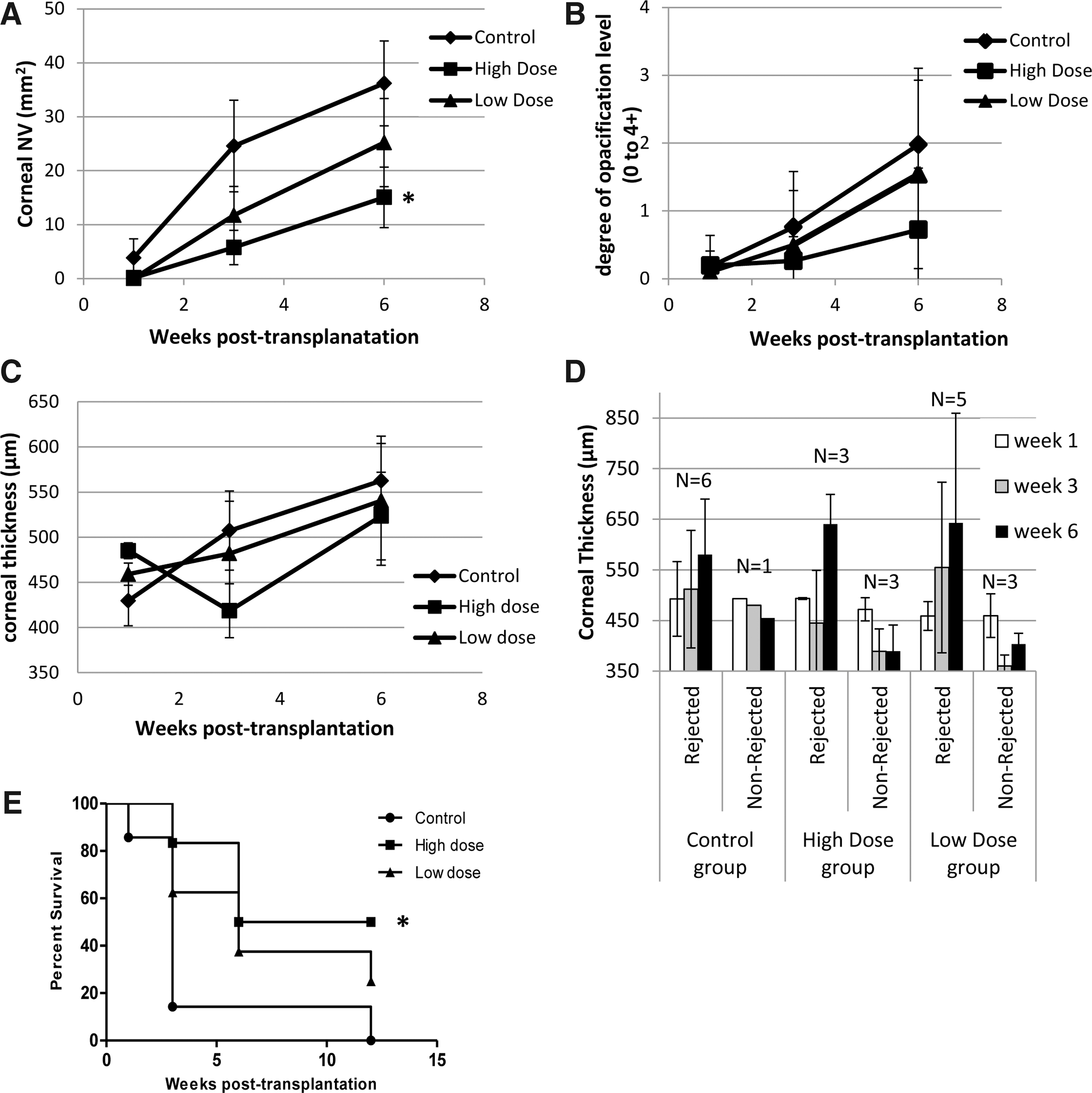
Corneal neovascularization (NV), opacity, thickness, and rejection following transplant into a rabbit model of high-risk penetrating keratoplasty (PK).
Mean corneal thickness was lower at later time points in the treatment groups but appeared less affected (see Fig. 1C). However, this may be due to the impact of the rejected corneas in this analysis. A clear trend for a reduction in corneal thickness over time was seen in corneas, with no signs of rejection, whereas increasing corneal thickness was observed in corneas experiencing rejection in all groups (see Fig. 1D).
Corneas were assessed for rejection at weeks 1, 3, and 6 and months 3 and 5 based on a rejection index, as previously published. 18 In the control group, signs of rejection were first detected 1 week post transplantation, with most corneas experiencing rejection by week 3 and all rejected by week 12, as expected in this model (Fig. 1E). The rate of rejection of high-dose OXB-202 was rescued (at 3 months) from 100% to 50% (p = 0.02), which is the expected rate of rejection for non-pre-vascularized (i.e., low risk) rabbit recipient eyes. 19 Rejection was also reduced in the OXB-202 low-dose group. However, this did not reach significance (p = 0.07).
At termination, corneas were removed, sectioned, H&E stained, and examined microscopically for immune infiltrates. These corneas were harvested once fully rejected at either 3 or 5 months after transplantation. Two rabbits still had clear, unrejected corneas at 5 months within the OXB-202 high-dose group and were therefore not included in the histological analysis. One of these rabbits died due to complications associated with the cornea angiography at this time point. The donor cornea of the other remained clear and unrejected out to 13 months post surgery at which point the rabbit was sacrificed. Importantly, there was no difference in endothelial cell density 13 months after surgery compared to the contralateral (nonsurgery) eye in this animal (see Supplementary Fig. S4), confirming no long-term detrimental effect of the genetic modification of this layer.
There was no difference in the number of immune cells quantified in corneal sections from different groups (see Supplementary Fig. S5). This is likely to be because all of the corneas analyzed at this stage (3 or 5 months) were fully rejected. The degree of immune cell infiltration was much lower than observed previously (see supplementary figure 3 in Parker et al. 7 ). This may be due to the later time points assessed in this study (3–5 months compared to 1 month) or may reflect the lower level of inflammation associated with this model.
This efficacy study shows that OXB-202 reduced the three hallmarks of rejection, namely, vascularization, opacification, and corneal thickening of the transplanted graft tissue, which resulted in a significant reduction in the rate of rejection in this model of high-risk corneal graft rejection.
Three-month GLP toxicology, immunogenicity, biodistribution, and shedding study to evaluate OXB-202
The GLP study was designed to examine ocular toleration to donor rabbit cornea genetically modified by overnight exposure to 3.25 × 10⁶ TU/mL pONYK1EiA vector.
Clinical assessments and ophthalmic observations
There were no unscheduled deaths or any statistically significant differences in body weights and relative organ weights at the scheduled necropsy at 3 months in either group. Furthermore, there were treatment-related changes in blood chemistry at any of the sampling time points, and there were no significant treatment-related microscopic observations in non-ocular tissues in the rabbits.
There were no clinical ophthalmic findings related to OXB-202, with most expected post-corneal transplant. Suture pulling through the host cornea correlated with anterior displacement of the graft in most cases and occurred in both groups (OXB-202 and control corneas). Corneal edema was present in the host cornea and in the graft cornea around the incision for both OXB-202 and control corneas. Three animals in both control and OXB-202 groups were noted with epithelial haze by the end of study. Out of 19 animals treated with OXB-202, four had fluorescein stain retention over 1–25% of the graft. Fluorescein stain retention and the development of epithelial haze likely represented a mildly unstable epithelium over the graft. Anterior cortical lens opacities were likely due to touching the lens during surgery and occurred at a similar frequency in both treatment and control groups, suggesting no OXB-202-related effect. Two animals with OXB-202 developed fibrin in the anterior chamber at different times between days 29 and 43, which resolved by the next examination interval. Mean IOP was slightly lower in both OXB-202 and control groups compared to untreated contralateral eyes but remained within the normal range (measurement taken from untreated eye, 10–15 mmHg) for the duration of the study in both groups (Fig. 2A). Therefore, the IOP decrease was considered a surgery-related effect.
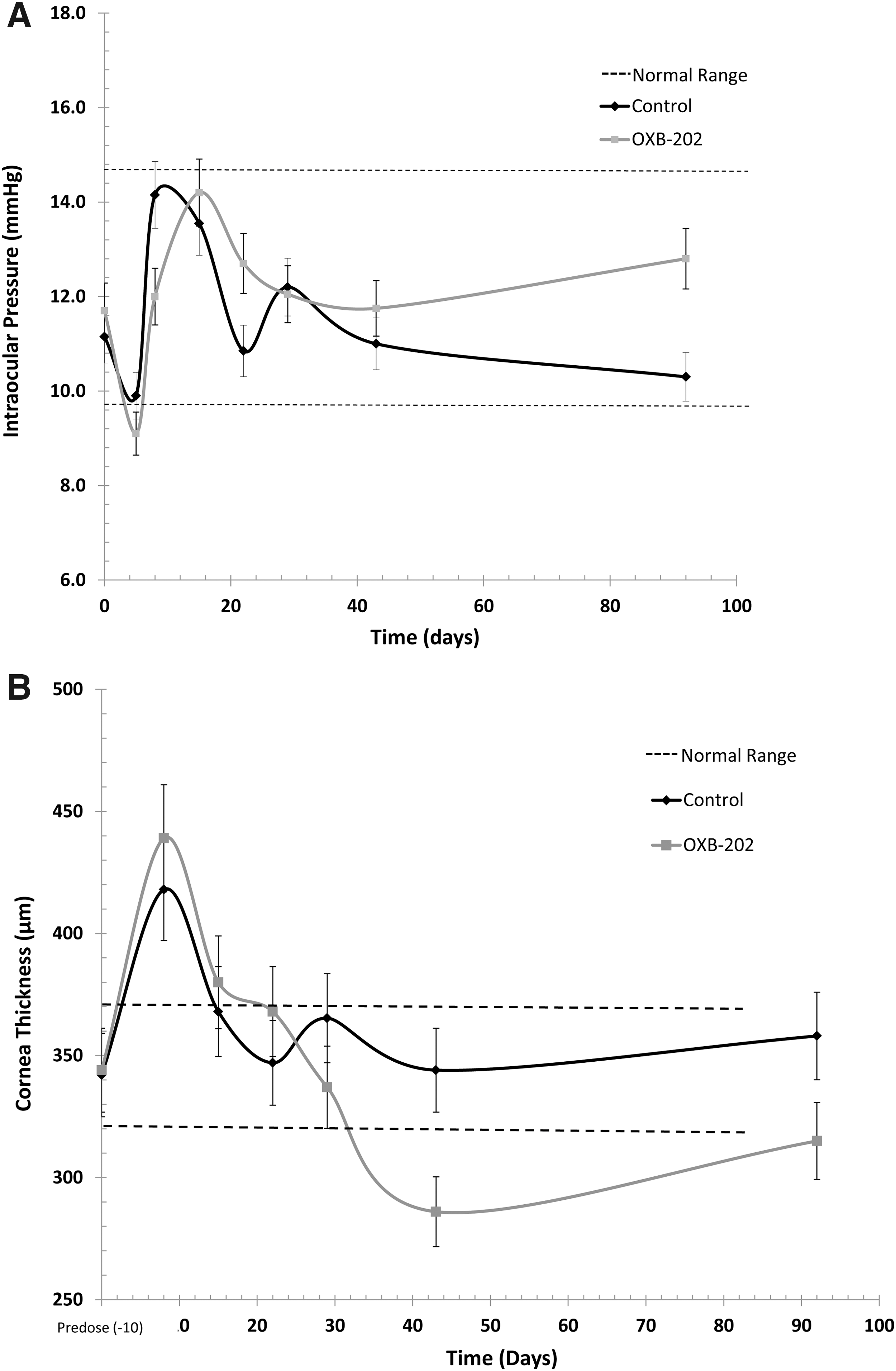
Measurement of mean intraocular pressure (IOP) and corneal thickness following transplant of OXB-202 or control corneas in a rabbit model of PK.
Mean corneal thickness of the donor corneas at 1 week post surgery was higher in both OXB-202 and control groups, probably as a result of the edema that occurs during overnight incubation of the cornea prior to transplant. This fell below the normal range (compared to the contralateral eyes) at later time points in the OXB-202 group only. This effect was similar between groups, indicating no OXB-202-related effect. A similar decrease in central corneal thickness is appreciated clinically after PK: cornea thickness can decrease over the first 6 months after transplant, which is thought to be due to recovery of the donor endothelium after the initial insult of surgery. 20 By the end of the study, both OXB-202 and control cornea thickness returned to normal range (measurement taken from untreated eye, 325–360 μm; Fig. 2B).
No difference was noted between the endothelium of OXB-202- and control grafts, as evaluated by NCSM. Qualitatively, the average cell size increased, and cell density and percent of hexagonal cells decreased in the operated eyes compared to non-operated eyes (2,800 vs. 3,800 cell/mm2; 26% reduction; see Fig. 3). A similar reduction is also observed clinically following PK and is thought to be a result of the surgical procedure. 21 Decreases were similar between control and OXB-202-treated eyes, indicating this was not an OXB-202-related effect.

Mean endothelial cell density (measured by noncontact specular microscopy) following transplant of OXB-202 or control corneas in a rabbit model of PK. Dotted line shows the normal range of corneal thickness measurement taken from untreated eye.
Ocular histology
Microscopic findings related to the surgical procedure and noted similarly in all animals control and OXB-202-treated eyes at all time points (29 and 92 days) included minimal to moderate ulceration of the cornea, minimal to slight attenuation/hyperplasia of the corneal epithelium, minimal to moderate fibrosis of the corneal stroma and the presence of disrupted corneal stroma, disruption of Descemet's membrane, and suture material in the corneal stroma. These findings were generally localized in the donor cornea and adjacent portions of the host cornea.
These findings were noted clinically at the time of surgery and were considered an effect of the surgical procedure and not OXB-202 related.
RT-PCR analysis to measure residual levels of pONYK1EiA vector in washing media
To determine efficient pONYK1EiA vector removal from the modified corneas, residual levels of vector in rabbit cornea media samples post transduction were quantified. Levels of vector-associated EIAV RNA sequence were within the quantifiable range of the assay. Residual vector detected after the third wash post transduction was <0.02% starting dose (see Supplementary Fig. S6). Consistent with previous data, 7 this indicates the washing protocol removes the vast majority of residual vector sequences prior to grafting OXB-202 into the rabbit eye. No OXB-202 vector-associated EIAV RNA sequence was detected in any media samples from corneas incubated with TSSM (control group).
Biodistribution, persistence, and shedding from OXB-202
Vector persistence and shedding were assessed by analyzing RNA extracted from plasma, urine, CSF, vitreous and aqueous fluid, and swabs of saliva and tears (right eye and left eye) by RT-PCR to determine the presence of OXB-202-derived EIAV RNA sequences in these samples. Vector-associated RNA sequences were only detected at day 2 of right-eye swabs of tears taken from OXB-202-treated animals. These levels were below the assay LLOQ, and no vector-associated RNA sequences were detected at the next time point, day 8.
Biodistribution was assessed by qPCR analysis of DNA extracted from target and non-target tissues and buffy coat samples at day 2 (buffy coat) and day 8 (tissue samples; see Fig. 4). No vector DNA was detected in the buffy coat or in any of non-target (ovaries, testes, liver, heart, lung, spleen, kidney right medulla, esophagus, stomach, duodenum, brain occipital cortex, and brain optic chiasm) or target (eyelid lower and upper, retina/choroid, sclera, ciliary body, optic nerve, Harderian gland, lacrimal gland, mandibular lymph node, nictitating membrane, and superior recti and inferior recti extraocular muscles) right-eye tissues samples from OXB-202 corneas. Vector DNA was detected in only two out of six iris samples at day 8 from OXB-202 recipient animals, but was non-quantifiable (<10 copies/reaction). The donor cornea graft had quantifiable EIAV vector-associated sequences detected in two of the six animals (an average of 288.17 copies detected in 1 μg DNA analyzed). The remaining four animals had DNA levels that were non-quantifiable. No vector DNA was detected in any of the contralateral and control corneas mock-transduced with TSSM.
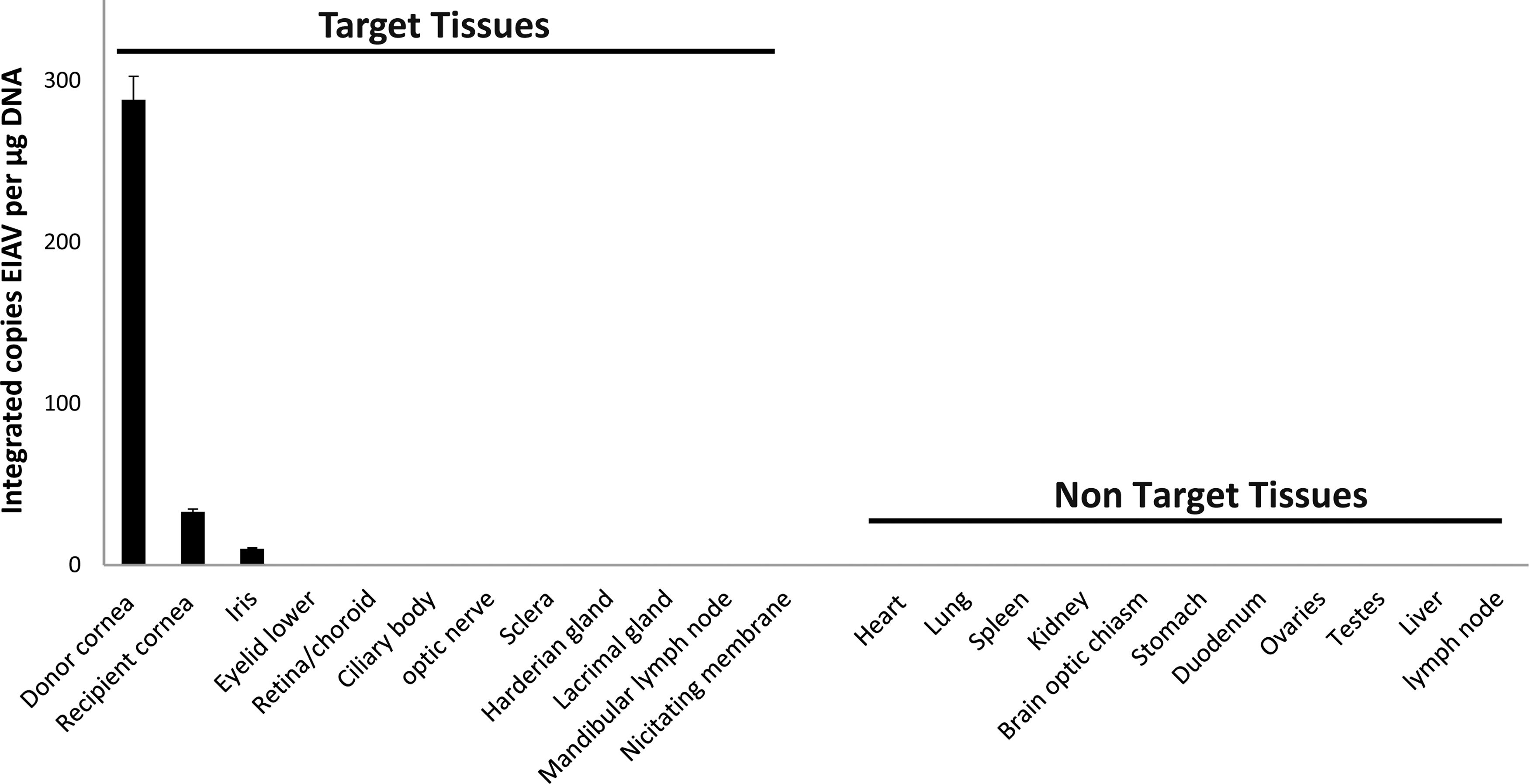
Biodistribution following the surgical implantation of OXB-202. Donor rabbit corneas treated overnight with either pONYK1EiA vector or the control media formulation buffer (TSSM) were transplanted into recipient rabbits and observed for up to day 8 post treatment prior to necropsy. Integrated copies of equine infectious anemia virus in DNA extracted from a comprehensive list of tissues were measured.
These results indicate that vector was confined to a minority of target (eye) tissues, and there was no indication of a consistent or robust presence of vector-associated RNA or DNA sequences in non-target samples.
Immunogenicity
Immunological assessment of serum samples did not show any humoral antibody responses toward any component of OXB-202, including VSV-G, EIAV p26, endostatin, angiostatin, and neomycin phosphotransferase (Neo PT) and HEK293T-packaging cell constituents, following transplantation of donor OXB-202 corneas in rabbits (Fig. 5). These data contribute to the safety and toxicity assessment of OXB-202, and indicate that immunogenicity toward OXB-202 was undetectable in peripheral serum samples from rabbits collected up to the 13 weeks following transplantation of OXB-202 donor corneal grafts.
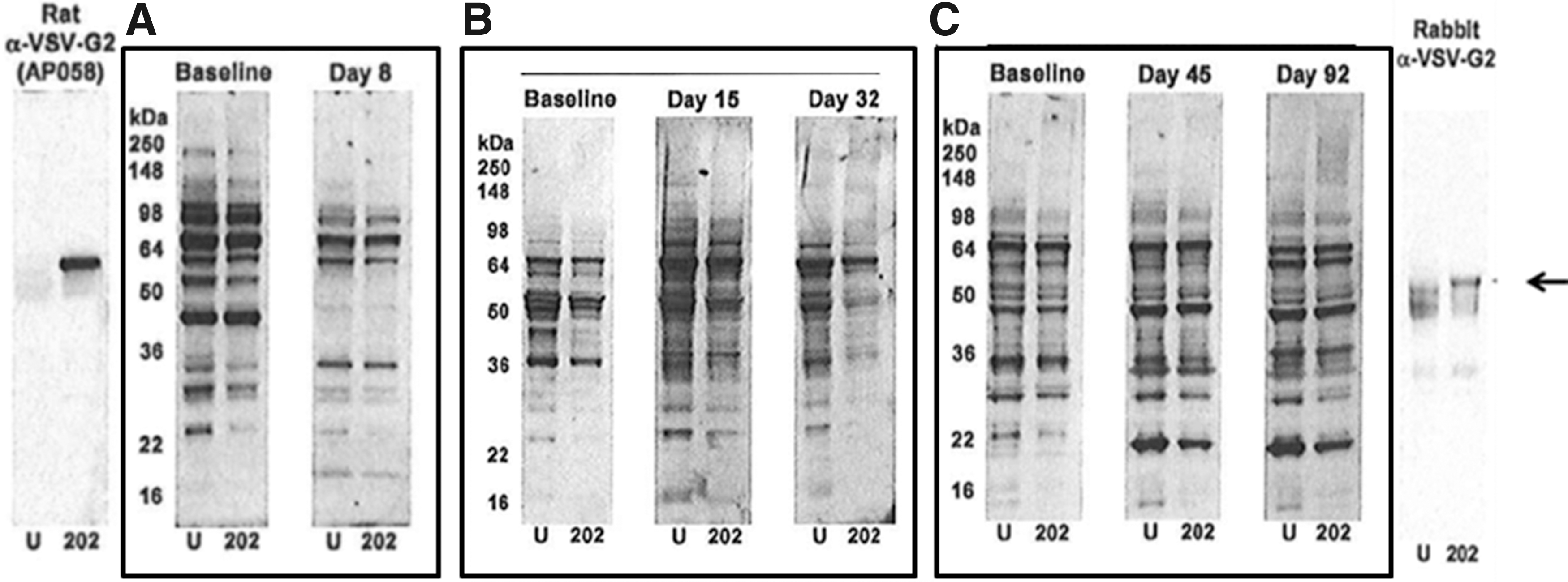
Humoral responses were not generated against any component of OXB-202 following transplantation in the toxicology study. Representative Western blot using rabbit serum samples from animals that received OXB-202. Example of serum samples from individual animals collected at baseline before transplantation and at time points for up to day 92/13 weeks following transplantation: baseline, day 8
Detection of endostatin and angiostatin in residual tissues media from OXB-202
To determine that rabbit corneas had been efficiently transduced to produce OXB-202 donor corneas, residual donor tissue was collected after trephination of the cornea scleral tissue and cultured for 3 weeks. Endostatin was then measured in a number of media samples collected throughout the culture period as a direct marker for transduction efficiency. In this study, all media samples from residual OXB-202 donor corneas showed an increase in secreted endostatin concentration over the time course studied, where levels peaked between days 15 and 22. The increase in endostatin was statistically significant (p < 0.01) as early as days 8–9 and remained significant at the last time point (days 21–23 inclusive), indicating the OXB-202 corneas were successfully modified (Fig. 6).
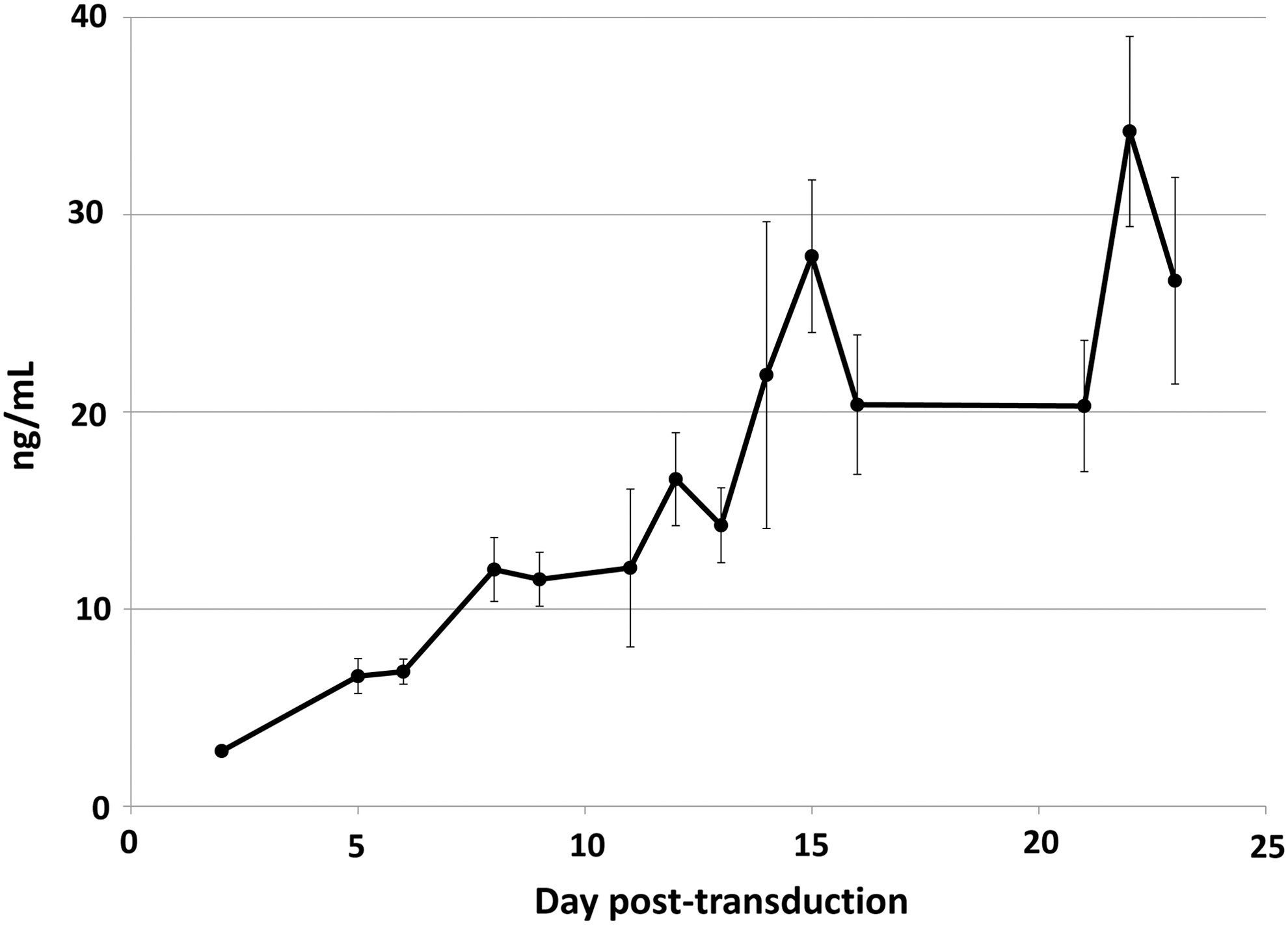
The mean levels of endostatin (ng/mL) in residual tissue media collected between days 2 and 23 for various donor residual tissue media collected from all residual donor corneas. Standard errors are indicated. The first sampling time point is day 2 (n = 11) or day 5 (n = 8) for various residual donor corneas.
Discussion
Corneal transplantation remains the most common solid-tissue transplant and is expected to increase substantially as developing countries establish their own eye banking infrastructure to meet the global demand for this type of procedure, estimated at >10 million patients worldwide. The success rate is very good in low-risk patients (94% in first year 5 ), and this has been further improved by the recent introduction of new surgical techniques that involve replacing only specific diseased layers of the cornea with donor tissue (lamellar keratoplasty [LK]). This has led to a rapid decrease in the number of PKs performed, from 95% of U.S. transplants in 2005 to 40.3% in 2015, driven by a corresponding increase in LKs from 4.6% to 59.6% over the same period. 22 However, while these LKs effectively reduce the risk of rejection by reducing the amount of allogeneic tissue transplanted, in the high-risk setting they are either unsuitable or unsuccessful. 23 It is likely therefore that PK will continue to be used in a significant proportion of corneal transplant patients in the future, such as those patients at high risk of rejection.
It has previously been shown that rabbit corneas genetically modified to secrete endostatin and angiostatin were able to reduce corneal NV significantly (and subsequently reduce opacity and immune infiltration) in an aggressive model of high-risk corneal transplantation. 7,8 In this model, rejection is rapidly mediated by the used of 16 interrupted, thick individual sutures that are retained throughout the study and cause excessive corneal NV and inflammation. Based on these encouraging data, a follow-on dose-ranging efficacy study was conducted in a more clinically relevant high-risk rabbit model of corneal rejection in which the recipient eye is pre-vascularized prior to transplantation and a single, thinner suture is used to secure the graft (burying the knot to minimize inflammation) that is subsequently removed. This model mimics corneal transplant in high-risk patients, with a high level of NV prior to surgery driving a high rate of rejection (86% of control corneas either rejected or showing clear signs of rejection by week 6), and allows much more accurate measurement of rejection markers. The treatment of rabbits with OXB-202 corneas in this second model once again reduced corneal NV, opacity, and thickness, and in a dose-dependent manner, although this only reached significance for NV. Although corneal opacity and thickness have been shown to act as a predictor of corneal graft survival, 20 the lack of significant reduction in corneal thickness may be due to the relatively modest increases generally observed during corneal rejection in the rabbit in this and previous studies. This is not the case clinically, where rejection generally leads to a substantially thickened cornea, as the pumping function of the endothelium is compromised. This difference may be due to the limited regenerative potential observed for the rabbit endothelium, which has not been observed in human corneas. The reduction in NV was significant in the high-dose group (p = 0.05) but was less than shown in the previous study 7 (∼60% compared to ∼80% previously). This may be due to the lower rate of NV development in this less aggressive model (approximately twofold lower than Parker et al. 7 ) and/or the higher level of variability in NV development in this model, possibly driven by the variation in recipient cornea PV. Alternatively, it is possible that it was due to differences in presurgical PV in this study. Although animals were distributed among groups to ensure a similar mean level of total PV, there were differences between groups in the radial pattern and the degree of PV within the surgery area. However, while total NV has been shown to correlate with the risk of graft rejection, the contribution (if any) of these more qualitative measures has yet to be determined.
The suppression of these three hallmarks of corneal rejection led to a significant reduction in the rate of rejection for the high-dose group. OXB-202 rescued the rate of rejection (at 3 months) from 100% to 50%, the rate of rejection published for non-pre-vascularized (i.e., low risk) rabbit recipient eyes. 15 OXB-202 therefore not only significantly reduced the rate of rejection, but appeared to restore the low-risk rate of rejection in this high-risk model of corneal transplantation. It is interesting to speculate that if this translates in humans, the use of OXB-202 may have the potential to lower the rejection rate from >50% in high-risk patients to <10% seen in low-risk patients.
In addition to the significant reduction in the rate of NV and rejection shown in the efficacy study, the 3-month GLP toxicology, biodistribution, shedding, and immunogenicity study in New Zealand rabbits showed that OXB-202 was safe and well-tolerated, elicited minimal immune responses, and the residual vector (shown to be marginal) was confined to the ocular compartment following transplantation. This is consistent with the well-characterized safety profile demonstrated both non-clinically 14 and clinically 16 for the same lentiviral pONYK1EiA vector product used to generate OXB-202 that is currently in development as a gene therapy treatment for an alternative ocular indication: RetinoStat® for wet age-related macular degeneration (AMD). This vector product has been administered directly into the back of the eye in a Phase I FIM study 16 at high doses (up to 1,000 times more than that expected to be shed from OXB-202) with no adverse toxicological or immunological findings or significant biodistribution outside of the eye. Importantly, this clinical trial demonstrated that the pONYK1EiA vector leads to robust expression of endostatin and angiostatin that is persistent in patients, out to >4 years in the earlier patients so far. As it may be the sustained presence of neovessels (particularly lymphatic) in high-risk host beds that is responsible for the higher risk of rejection in these patients compared to low-risk patients, it may be critical for effective anti-angiogenic therapies to act long term in this way. 24
Together, the successful dose-ranging efficacy and safety studies build on the existing safety data for the LentiVector® platform and pave the way for evaluation of OXB-202 in a Phase I/II clinical trial. This study has shown that corneas genetically modified to secrete antiangiogenic factors (OXB-202) reduce the hallmarks of rejection and subsequently significantly lower the rate of rejection in a clinically relevant acute rabbit model of high-risk corneal rejection. Formal toxicology and biodistribution studies have demonstrated that OXB-202 is safe and well-tolerated, with no associated immunogenicity, and biodistribution analysis indicated that pONY1KEiA vector was confined to the cornea after transplantation. These data support the initiation of a first-in-human clinical trial in high-risk patients indicated for corneal transplantation.
Footnotes
Acknowledgments
The study was sponsored by Oxford BioMedica (UK) Ltd., with the support of an Innovate UK grant (101622). An abstract was previously presented at the 19th annual meeting of the American Society for Cell and Gene Therapy (2016).
Author Disclosure
N.F., V.K., K.B., L.M., J.L., M.K. K.A.M., and S.E. are employed by Oxford BioMedica (UK) Ltd. No competing financial interests exist for M.P. J.T.S. is the inventor of technology that is used in this research and that has been licensed to Oxford BioMedica (UK) Ltd. This potential conflict of interest has been reviewed and managed by Oregon Health & Sciences University.
References
Supplementary Material
Please find the following supplemental material available below.
For Open Access articles published under a Creative Commons License, all supplemental material carries the same license as the article it is associated with.
For non-Open Access articles published, all supplemental material carries a non-exclusive license, and permission requests for re-use of supplemental material or any part of supplemental material shall be sent directly to the copyright owner as specified in the copyright notice associated with the article.