
Abstract
Antigen-specific tolerizing DNA vaccines are one of the most promising strategies for rheumatoid arthritis (RA) treatment. They act by inducing potent immune tolerance instead of generalized immunosuppression. Recently, we developed a novel antigen-specific tolerizing DNA vaccine pcDNA-CCOL2A1 coding for chicken type II collagen (CCII) and confirmed its potent therapeutic efficacy in an established rat model of collagen-induced arthritis (CIA). Here we report the prophylactic vaccination efficacy of a single 300 μg/kg dose of pcDNA-CCOL2A1 against CIA incidence, severity, and onset. CCOL2A1 transcripts were detected in the blood of CIA rats 14–42 days after intramuscular injection by 300 μg/kg pcDNA-CCOL2A1. The expression of CCOL2A1 transcripts increased quickly on day 21, peaked at day 28, and then gradually decreased thereafter. Importantly, a single prophylactic vaccination of pcDNA-CCOL2A1 14 days before CIA establishment significantly reduced CIA incidence and severity, deferred its onset, and was as efficacious as the current gold standard drug, methotrexate. The marked effects on CIA incidence and severity closely corresponded to the expression of CCOL2A1. Furthermore, prophylactic vaccination with pcDNA-CCOL2A1 markedly decreased serum content of anti-type II collagen (CII) immunoglobulin G (IgG) antibodies, induced Th1-to-Th2 and Tc1-to-Tc2 shifts, and decreased the percentages of CD4+CD29+ and Th17 T cells. Prophylactic vaccination with pcDNA-CCOL2A1 also downregulated various Th1 cytokines, while upregulating both the Th2-type cytokine interleukin-10 and the Th3-type cytokine transforming growth factor β. Our results indicate that the pcDNA-CCOL2A1 DNA vaccine acts as a highly efficient inducer of specific immunotolerance that could be a promising option for RA treatment in the near future.
Introduction
T
These therapeutic or prophylactic DNA vaccines target one or several RA autoantigens. Of special note is the novel DNA vaccine pcDNA-CCOL2A1, with potent therapeutic effect comparable to the current “gold standard” drug, methotrexate (MTX), in an established CIA rat model. 8 The pcDNA-CCOL2A1 DNA vaccine is safe and well-tolerated and does not cause any abnormal clinical signs and symptoms or side effects on normal physiological function. 12,13 In addition, pcDNA-CCOL2A1 was cleared quickly in most tissues approximately 4–6 weeks after a single intramuscular injection at the therapeutic dose, and CCOL2A1 cDNA was not integrated into the genomic DNA of any tissues from vaccinated normal rats. Taken together, these results strongly suggest the high druggability of pcDNA-CCOL2A1. In this study, we first systemically evaluated the prophylactic vaccination efficacy of a single dose of DNA vaccine pcDNA-CCOL2A1 against CIA incidence, severity, and deferred onset. Furthermore, we explored the immunoregulatory mechanisms responsible for the prophylactic effects of DNA vaccine pcDNA-CCOL2A1.
Materials and Methods
Animals and induction of CIA rat model
Inbred Wistar rats (female, 4–6 weeks old) were purchased and raised under specific pathogen-free conditions from the Animal Breeding Center of the Academy of Military Medical Sciences (Beijing, China) and cared for in accordance with approved guidelines by the Academy of Military Medical Sciences Animal Welfare Committee. Natural chicken type II collagen (CCII; Sigma, St. Louise, MO) was dissolved in 0.1 mol/L glacial acetic acid and then emulsified to a 1:1 mixture using complete Freund's adjuvant (Sigma) by whirl mixer, resulting in a final concentration of 1 mg/mL. Rats were primed with 0.2 mL/200 g CCII in complete Freund's adjuvant by subcutaneous injection into the plantar skin of the right rear foot and multipoint injection intracutaneously at the tail root. Seven days later, the rats were boosted intraperitoneally with 0.2 mL/200 g CCII emulsified using incomplete Freund's adjuvant (Sigma) as described above. The operating procedure induces the CIA rat model with satisfactory incidence of arthritis. 8
Prophylactic vaccination with pcDNA-CCOL2A1
The pcDNA-CCOL2A1 vaccine carrying chicken type II procollagen gene was previously constructed as a recombinant plasmid in our laboratory, including a 4,837-bp full-length cDNA (GenBank: AY046949 and AF452711) with deleted N-propeptide, signal peptide, and Kozak consensus sequences. 8,14 The plasmid for prophylactic vaccination was produced in Escherichia coli and purified using the Endo-Free Plasmid Mega kit (Qiagen, Valencia, CA). The purified plasmid pcDNA-CCOL2A1, in naked formulation not in polymer-encapsulated or nanoparticle formulation, was diluted with normal saline (NS) with a concentration of 180 μg/mL. 15,16 Biochemicals and reagents used in this study were purchased from Sigma, Takara (Dalian, China), Chondrex (Redmond, WA), BD (Franklin Lakes, NJ), eBioscience (San Diego, CA), and Affymetrix (Santa Clara, CA) unless indicated otherwise.
Wistar rats were randomized to three groups (30 animals per group) and vaccinated 14 days before the CIA model induction. The experimental timeline for CIA model induction and vaccination is shown in Fig. 1A. The pcDNA3.1-CCOL2A1 vaccine was administered intramuscularly (i.m.) via a single-dose injection of 300 μg/kg (the confirmed optimal therapeutic dose) in the left hind limb. Rats received MTX at a dosage of 0.75 mg/kg i.m. once a week for 4 weeks as the positive control and a single injection of NS i.m. as the negative control. 17
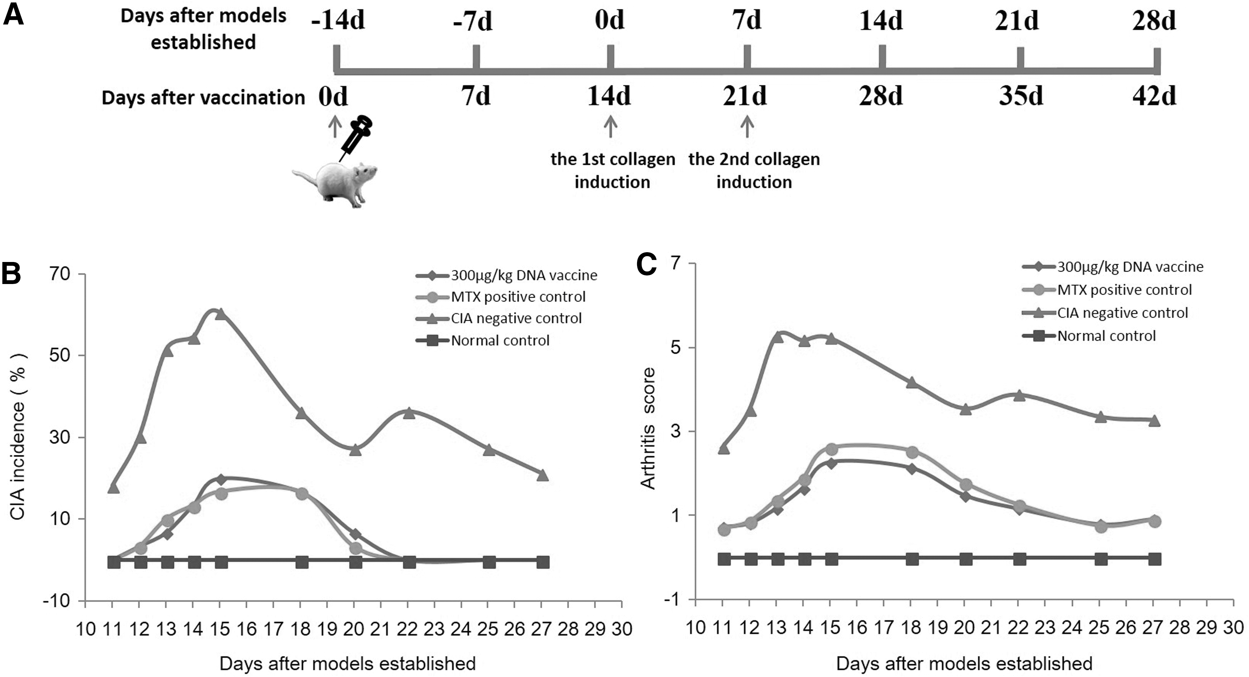
The assessment for the prophylactic vaccination efficacy of DNA vaccine pcDNA-CCOL2A1 with a single dose of 300 μg/kg intramuscularly (i.m.) in collagen-induced arthritis (CIA) rats.
Evaluation of the prophylactic vaccination efficacy of pcDNA-CCOL2A1
CIA incidence, severity, and onset were assessed on days 11–27 according to a macroscopic scoring system. 8,18 Arthritis for each paw was graded 0 if not present. If present, it was graded as follows: 1 = one to two interphalangeal (IP) joints are red and/or swollen; 2 = three to four IP joints and/or one larger joint are red and/or swollen; 3 = more than four IP joints and/or podarthrum are red and/or swollen; and 4 = entire paw is severely red and/or swollen. The score was maximized to 4 per paw and yielded 0–16 for each animal. An overall score greater than 5 points was identified as the onset of arthritis.
Expression analysis of pcDNA-CCOL2A1 in CIA rats
On days 0, 3, 7, 10, 14, 21, 28, and 35 after i.m. injection of a single dose of 300 μg/kg pcDNA-CCOL2A1 into CIA rats, blood was collected from the orbital venous plexus. Total RNA extraction was performed immediately with TRIzol reagent (Life Technologies, Frederick, MD). Subsequently, 5 μL of RNA was reversely transcribed to cDNA using a RT reagent kit (Takara). A 2-μL sample of cDNA was amplified using a Real Time-quantitative polymerase chain reaction (RT-qPCR) kit (Takara) in an Applied Biosystems 7500 Real-Time PCR System (ABI, Foster City, CA). The qPCR cycling conditions were as follows: an initial denaturation at 95°C for 30 s, followed by 40 cycles of denaturation at 95°C for 5 s and annealing at 60°C for 30 s. The forward primer (5′-GCTGGTGAGGAAGGCAAGAG-3′) and reverse primer (5′-CTTTGGGACCGGCGAGAC-3′) resulted in a 175-bp (base pairs) amplicon from CCOL2A1 gene. TaqMan Probe (5′-FAM-CTCGTGGTGAACCTGGTGCCGCC-TAMARA-3′) was used in the RT-qPCR system. Rat β-actin was used as an internal control and was amplified with up-stream primer (5′-GTGCCCATCTATGAGGGTTACG-3′), down-stream primer (5′-CACGCTCGGTCAGGATCTTC-3′) and probe (5′-HEX-CCTGCGTCTGGACCTGGCTGGC-TAMARA-3′) producing a 103-bp fragment simultaneously from the resultant cDNA.
RNA samples isolated from the blood of CIA rats after a single-dose vaccination were analyzed in parallel with negative control samples (from CIA rats injected with NS). In addition, a reaction sentinel control (using ddH2O instead of the template) and a positive control (using NS-treated rat blood cDNA spiked with pcDNA-CCOL2A1 as the template) were included as quality controls in each PCR amplification. A relative quantification assay was used to compare the RNA expression of CCOL2A1 at different time points after vaccination.
Measurement of serum antibody against type II collagen (CII) in CIA rats
Serum samples were collected from the retro-orbital venous plexus 7, 14, and 21 days after establishment of the CIA model; that is, 21, 28, and 35 days after vaccination with 300 μg/kg pcDNA-CCOL2A1, to measure anti-CII IgG antibody titers. The levels of anti-rat and anti-chicken serum antibodies against CII were determined by enzyme-linked immunosorbent assay (ELISA) with commercially available test kits (Chondrex). 8
Flow cytometric analysis of T cell subsets in CIA rats
On days 21, 28, and 35 after vaccination with 300 μg/kg pcDNA-CCOL2A1, peripheral blood was harvested and stained immediately using various specific fluorescently labeled extracellular and intracellular monoclonal antibodies successively. The percentages of CD3+CD8+CD28+ cytotoxic T (Tc), CD3+CD8+C28− suppressor T (Ts), CD4+CD25+FoxP3+ regulatory T (Tr), and CD4+CD29+ T cells were identified and assayed respectively by four-channel flow cytometry (BD FACSVerse). Th1, Th2, and Th17 cells were identified by cell stimulations, extracelluar antibodies, and intracellular cytokine staining using six-channel flow cytometry, and data were shown in the form of scatter diagrams according to interferon (IFN)-γ-APC, interleukin (IL)-4-PE, and IL-17A-PE-Cy7 levels. All isotype-specific fluorescence-conjugated antibodies were used throughout as controls for nonspecific cell surface binding. 12
Measurement of Th1, Th2, and Th3 cytokines in CIA rats
Serum was collected on days 21, 28, and 35 after receiving a single intramuscular dose of 300 μg/kg DNA vaccine pcDNA-CCOL2A1. In order to better control interbatch differences, all samples were stored at −20°C until use and tested together. The cytokine signals were batch analyzed using commercial kits (Affymetrix, Santa Clara, CA), according to the manufacturer's instructions, as described earlier. 12 The high-throughput cytokine detection technology named multifunctional flow lattice (Luminex 200TM, Austin, TX) was used to quantify inflammatory cytokines such as IL-1, IL-2, IL-6, IL-12(IL-23p40), IL-17, IFN-γ, tumor necrosis factor (TNF)-α, monocyte chemoattractant protein (MCP)-1, receptor activator for nuclear factor-κB ligand (RANKL), and regulated on activation in normal T cell expressed and secreted (RANTES), and anti-inflammatory cytokines such as IL-4, IL-10, and transforming growth factor (TGF)-β. Standard curve and quality control samples were included in each measuring process to accurately calculate and calibrate values from different time points after vaccination.
Statistical analysis
Student's t-test was used to statistical significance of differences between treatment and control groups in relation to IgG antibody levels, cytokine concentrations, and the proportions of T-cell subsets by SPSS 13.0 software (Chicago, IL). Data obtained were expressed as means ± standard deviation (SD) if they followed Gaussian distribution. The level of significance was taken as p < 0.05.
Results
Prophylactic vaccination with pcDNA-CCOL2A1 decreased CIA incidence and severity and deferred its onset
The prophylactic vaccination efficacy of a single 300 μg/kg dose of pcDNA-CCOL2A1 against CIA incidence, severity, and onset was monitored starting 11 days after establishment of the CIA rat model, daily during the first week, and once every 2 or 3 days afterwards, using a macroscopic scoring system. 8,18 As shown in Figs. 1B and 1C, the joints of CIA rats began to swell 11 days after the first collagen induction. Swelling peaked on days 14–15, then slowly recovered after day 18. Notably, compared to the CIA rat negative control group, prophylactic vaccination with pcDNA-CCOL2A1 significantly reduced CIA incidence and severity and also deferred the onset and peak of the disease. Importantly, prophylactic vaccination with a single 300 μg/kg dose of pcDNA-CCOL2A1 was as effective as MTX, currently the most extensively used therapy for RA. 19
Prophylactic vaccination with pcDNA-CCOL2A1 induces high CCOL2A1 expression
To examine dynamic pcDNA-CCOL2A1 expression in the peripheral blood of vaccinated CIA rats, the mRNA expression of CCOL2A1 was examined by RT-qPCR 0, 3, 7, 10, 14, 21, 28, and 35 days after vaccination. As shown in Fig. 2, CCOL2A1 expression was initially detected on day 14, increased markedly on day 21, and peaked on day 28, consistent with observations of CIA development and severity. In particular, the marked effects on CIA incidence and severity closely corresponded to CCOL2A1 expression. No signals were detected in the CIA negative and reaction sentinel controls, demonstrating no background interference or cross-contamination during PCR analysis.

The dynamic expression of CCOL2A1 gene in peripheral blood from CIA rats received the prophylactic vaccination with DNA vaccine pcDNA-CCOL2A1. DNA vaccine pcDNA-CCOL2A1 was injected with a single intramuscular dose of 300 μg/kg (n = 30) into the pre-established CIA rats on day −14 before the establishment of CIA rat models. Peripheral blood samples were harvested on days 0, 3, 7, 10, 14, 21, 28, and 35 after vaccination and processed for RT-qPCR determination. Data are expressed as 2−ΔΔCt by the relative quantification assay. The negative control group was from CIA rats injected with normal saline (NS).
Prophylactic vaccination with pcDNA-CCOL2A1 significantly decreased serum anti-CII antibody levels
Anti-CII IgG antibody levels are considered the most reliable marker for arthritic severity in both RA patients and the CIA model. 20 Thus, we used an ELISA to investigate the serum levels of anti-CII antibodies 21, 28, and 35 days after a single dose of either pcDNA3.1-CCOL2A1 or NS.
Two types of anti-CII antibodies were tested at three time points after the first collagen induction (i.e., 7, 14, and 21 days after model establishment), and the results are shown in Table 1. Consistent with clinical evaluation, anti-rat CII IgG antibodies did not appear before the onset of arthritic swelling, and anti-chicken CII IgG antibodies were detected at a weaker level at the same time point, 7 days after CIA rat model establishment. Moreover, vaccination with pcDNA-CCOL2A1 markedly reduced the levels of both antibody types 14 and 21 days after the establishment of the CIA rat model compared to the negative control group (p < 0.05), although there was no statistically significant difference between anti-chicken CII IgG levels on day 14. Taken together, these results provided the first direct evidence that vaccination with a single 300 μg/kg dose of pcDNA-CCOL2A1 can effectively block anti-CII IgG antibody production in CIA rats.
The serum levels of anti-type II collagen (CII) antibody from CIA rats receiving a single intramuscular dose of 300 μg/kg DNA vaccine pcDNA-CCOL2A1 at different time points (U/mL,
NC, normal control.
p < 0.05 compared to CIA negative control using the t-test. These data are representative of three experiments. Three separate experiments yielded similar results.
Prophylactic vaccination with pcDNA-CCOL2A1 significantly changed the percentages of most T-lymphocyte subsets
As stated above, pcDNA-CCOL2A1 showed remarkable preventive efficacy against CIA incidence and severity and deferred the onset and peak of the disease. On this basis, we investigated whether there were changes in peripheral blood Tc, Ts, Treg, Th1/Th2, Tc1/Tc2, Th17, and CD4+CD29+ T-cell subsets after vaccination with pcDNA-CCOL2A1. To exclude spurious fluorescence, labeled IgG-matched isotype control antibodies were used as controls for nonspecific cell surface binding in all fluorescence activated cell sorter (FACS)-based measurements. Compared with the CIA NS group, the percentage of CD4+CD29+ T cells was significantly decreased 7, 14, and 21 days after the establishment of the CIA rat model; that is, 21, 28, and 35 days after vaccination (p < 0.05). Strikingly, the level of CD4+CD29+ T cells after pcDNA-CCOL2A1 vaccination was almost the same as in the normal control group. There were no obvious differences in the percentages of Th1/Th2, Tc1/Tc2, and Th17 cells between the two groups (p > 0.05) on day 21, but their levels were significantly lower 28 and 35 days after vaccination. Vaccination with pcDNA-CCOL2A1 did not change the percentages of Tc, Ts, and Treg cells. These results and representative diagrams are shown in Figs. 3A and 3B.

The dynamic changes of T-lymphocyte subsets from CIA rats receiving a single intramuscular dose of 300 µg/kg DNA vaccine cDNA-CCOL2A1 at different time-points.

Prophylactic vaccination with pcDNA-CCOL2A1 significantly changed the levels of several cytokines
Pro-inflammatory and anti-inflammatory cytokines are highly important for regulating the balance between cellular and humoral immune responses. To study the vaccination effects with pcDNA-CCOL2A1 on these molecules, we systemically investigated the levels of all types of cytokines in vaccinated CIA rats 21, 28, and 35 days after a single dose of 300 μg/kg pcDNA3.1-CCOL2A1 (Table 2). Pro-inflammatory cytokines, such as IL-1, IL-2, IL-12, IFN-γ, MCP-1, RANKL, and RANTES, were significantly decreased 21, 28, and 35 days after vaccination compared to the CIA negative control group. In contrast, the levels of anti-inflammatory cytokines including IL-10 and TGF-β were markedly increased at all three time points. Several other key pro-inflammatory cytokines (e.g., IL-6, IL-17, and TNF-α) and anti-inflammatory cytokines (e.g., IL-4) were below the limit of detection.
The serum concentrations of inflammatory and anti-inflammatory cytokines from CIA rats receiving a single intramuscular dose of 300 μg/kg DNA vaccine pcDNA-CCOL2A1 at different time points (pg/mL,
IFN, interferon; IL, interleukin; MCP, monocyte chemoattractant protein; NC, normal control; RANKLE, receptor activator for nuclear factor-κB ligand; RANTES, regulated on activation in normal T cell expressed and secreted; TGF, transforming growth factor; UD, undetermined.
p < 0.05 compared to normal control.
p < 0.05 compared to CIA negative control using the t-test. These data are representative of three experiments. Three separate experiments yielded similar results.
Discussion
A large number of basic and clinical research studies have indicated that RA onset and progression are caused by the failure of CD8+CD28− Ts and CD4+CD25+ Treg cells to govern autoreactive CD4+CD28+ Th1 cells and autoantibody-producing B cells. 9,21 –23 Thus, the ideal strategy for treating RA should selectively restore the impaired Ts and Tr suppressor/regulatory mechanisms and interfere with detrimental autoreactive immune responses mediated by both Th1 and B cells, while not changing normal immune responses necessary to combat pathogenic microorganisms or inducing generalized immunosuppression. Theoretically, the antigen-specific tolerizing DNA vaccine pcDNA-CCOL2A1 achieves these goals through specific regulation of the immune system but not generalized immunosuppression. Practically, we have confirmed the potent therapeutic effects of DNA vaccine pcDNA-CCOL2A1 in an established CIA rat model through the induction of potent immune tolerance. 3,4,7,8 Here, we demonstrate that prophylactic vaccination with DNA vaccine pcDNA-CCOL2A1 can significantly reduce CIA incidence and severity and defer its onset and peak. Moreover, the marked prophylactic vaccination efficacy closely corresponded with CCOL2A1 expression. The present study has several main differences from our previous studies: (1) different study purposes (i.e., prophylactic versus therapeutic vaccination efficacy); (2) different models (i.e., pre-established versus established CIA rat models); (3) different vaccine delivery (i.e., intramuscular versus intravenous injection, and even intra-articular injection 24 ); and (4) different mechanisms of action (i.e., against normal naive immune cells versus activated autoimmune cells). 8
CIA is universally accepted as a unique experimental animal model for human RA, and it has been widely used to study the immunogenetics, pathophysiology, immunohistopathology, and immunopathogenesis of human RA, particularly for the development of important new strategies for its treatment. 25,26 Based on the inherent characteristics of CIA models, such as acute arthritic attack, rapid monophase course, short disease course, gradual spontaneous resolution, and seldom-relapsing course, the models have been commonly or mostly used in prophylactic studies, with the exception of a few therapeutic studies in the development of new RA drugs. The present results demonstrate that a single intramuscular dose of prophylactic vaccination with 300 μg/kg pcDNA-CCOL2A1 14 days before CIA establishment can significantly reduce CIA incidence and severity and defer its onset. Strikingly, the prophylactic vaccination efficacy of DNA vaccine pcDNA-CCOL2A1 alone appears to be as effective as MTX, currently the most extensively prescribed disease-modifying antirheumatic drug (DMARD) treatment for RA. Recently, low-dose MTX was confirmed to have significant therapeutic efficacy against the established CIA rat model both in vitro and in vivo through specific immunotolerance, not by nonspecific immunosuppression. 19,27 This finding is consistent with our previous series of studies on therapeutic vaccination in an established CIA rat model using DNA vaccine pcDNA-CCOL2A1 or DNA vaccine B7-2-PE40KDEL. 8,9 The significant prophylactic vaccination efficacy of DNA vaccine pcDNA-CCOL2A1 described in this report following only a single vaccination suggests that DNA vaccine pcDNA-CCOL2A1 could be efficacious over a long period of time, with no need for additional vaccinations when used in RA patients recovering from a high disease activity phase requiring combination therapies of conventional DMARD + biological agents or ± kinase inhibitors for prolonged periods of time. Compared with the DNA vaccine pcDNA-CCOL2A1 dosing described in this report, current therapies for RA must be administered at higher doses for a long period of time, with the added hazard of significant side effects. 28,29 The significant prophylactic vaccination efficacy of DNA vaccine pcDNA-CCOL2A1 was confirmed not only by a clinical-visual scoring system, but also by markedly decreased serum anti-CII IgG levels. This point is very important because anti-CII IgG levels are generally accepted as the most reliable objective index to reflect inflammatory activity and arthritic severity both in RA patients and in the established CIA model. 30,31 In this study, we measured both anti-chicken and anti-rat CII antibodies. Without exception, the overall anti-chicken CII antibody levels were higher than the anti-rat CII antibody levels. This outcome may be because the chicken CII, used as an inducer for the establishment of the CIA rat model, led directly to the antigen–antibody reaction, causing the production of anti-chicken CII antibodies and triggering inflammation and articular cartilage damage. 18,30 Afterwards, rat CII is released from damaged cartilaginous tissues and subsequently plays a critical role as an RA autoantigen, inducing the production of anti-rat CII antibodies.
In our previous results demonstrating the therapeutic vaccination efficacy of DNA vaccine pcDNA-CCOL2A1 against established CIA rats, the vaccine theoretically acts against activated autoimmune T and B cells. In contrast, the prophylactic vaccination efficacy shown in the present report would require the vaccine to act on normal naive T and B cells. Indeed, our previous therapeutic vaccination study indicated that DNA vaccine pcDNA-CCOL2A1 exerts its therapeutic efficacy against the established CIA by increasing the levels of CD4+CD25+ Treg cells, decreasing the specific proliferative responses of T cells to CII and causing a shift from Th1 toward Th2 cells, accompanied by the downregulation of the Th1-cytokine TNF-α and the upregulation of both the Th2-cytokine IL-10 and the Th3-cytokine TGF-β. 8 Although the exact mechanism by which DNA vaccine pcDNA-CCOL2A1 exerts its prophylactic vaccination efficacy before establishment of CIA remains to be investigated, our current findings suggest that it may take effect by partially decreasing the percentages of Th17 and CD4+CD29+ T cells and effectively inducing Th1-to-Th2 and Tc1-to-Tc2 shifts, accompanied by the downregulation of various Th1 cytokines and upregulation of Th2 and Th3 cytokines. However, the results were somewhat different from our previous therapeutic vaccination results 8 in that CD4+CD25+FoxP3+ Treg cells did not increase and the percentages of CD3+CD8+CD28− Ts and CD3+CD8+C28+ Tc cells did not significantly change compared to the CIA negative control. One possible explanation for this outcome is that considerable differences exist in the pathophysiology, immunohistopathology, and immunopathogenesis of pre-established and established CIA rat models. After prophylactic vaccination with DNA vaccine pcDNA-CCOL2A1, the evolutionary processes of pathophysiology, immunohistopathology, and immunopathogenesis from the pre-established CIA to the established CIA were markedly reduced or slowed, especially for effectors of immunologic homeostasis such as CD4+CD25+FoxP3+ Treg and CD3+CD8+C28− Ts cells. Nevertheless, regardless of the exact mechanism by which DNA vaccine pcDNA-CCOL2A1 exerts its prophylactic vaccination efficacy, fortunately and critically, our results clearly indicate that the prophylactic vaccination efficacy of DNA vaccine pcDNA-CCOL2A1 alone was comparable to the current gold standard drug, MTX.
In conclusion, this report is the first to demonstrate that prophylactic vaccination with DNA vaccine pcDNA-CCOL2A1 can significantly reduce CIA incidence and severity and defer its onset and peak. With these unique biological functions, antigen-specific tolerizing DNA vaccine pcDNA-CCOL2A1 could offer a new class of promising specific immunotherapy for patients with RA in the near future.
Footnotes
Acknowledgments
This project was supported by grants from the National Major Scientific and Technological Special Project for “Significant New Drug Development” (Nos. 2009ZX09103-624 and 2015GKS-072/139, to YZX). The funding agency took no role in the study design, experimental data collection and analysis, preparation of the manuscript or decision to publish. The authors declare no conflict of interest.
Author Disclosure
All of the authors read and approved the final manuscript. The authors declare no conflict of interest.