
Abstract
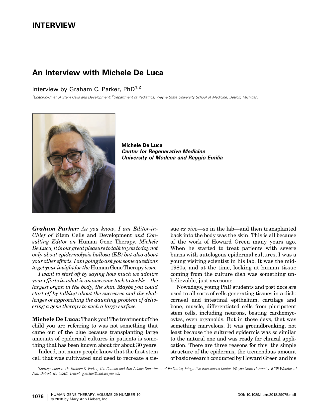
Michele De Luca
Center for Regenerative Medicine
University of Modena and Reggio Emilia
I want to start off by saying how much we admire your efforts in what is an awesome task to tackle—the largest organ in the body, the skin. Maybe you could start off by talking about the successes and the challenges of approaching the daunting problem of delivering a gene therapy to such a large surface.
Indeed, not many people know that the first stem cell that was cultivated and used to recreate a tissue ex vivo—so in the lab—and then transplanted back into the body was the skin. This is all because of the work of Howard Green many years ago. When he started to treat patients with severe burns with autologous epidermal cultures, I was a young visiting scientist in his lab. It was the mid-1980s, and at the time, looking at human tissue coming from the culture dish was something unbelievable, just awesome.
Nowadays, young PhD students and post docs are used to all sorts of cells generating tissues in a dish: corneal and intestinal epithelium, cartilage and bone, muscle, differentiated cells from pluripotent stem cells, including neurons, beating cardiomyocytes, even organoids. But in those days, that was something marvelous. It was groundbreaking, not least because the cultured epidermis was so similar to the natural one and was ready for clinical application. There are three reasons for this: the simple structure of the epidermis, the tremendous amount of basic research conducted by Howard Green and his numerous collaborators over the year, and the feasibility of a clinical translational protocol.
When I came back to Italy, again in the mid-1980s, we started to use those cultures to treat large burns. Then, during the years that followed, we developed our own line of research. You might be aware of the regeneration of the corneal epithelium.
Therefore, it is thanks to this background that many years ago we tried to address the issue of a genetic skin disease by harvesting epidermal cells and trying to correct them genetically.
EB was one of the key targets because of the severity of the disease. EB is a really devastating disease, with patients experiencing a very poor quality of life right from birth. You might know from my publications that we did the first proof of principle in 2006. As described in the 2006 Nature Medicine paper, we substituted the blistering epidermis in both legs of a patient with junctional EB by transplanting sheets of autologous, genetically corrected, cultured epidermis.
That patient is now in the 13th year of follow-up, and the epidermis is still there, renewing and not blistering, functionally equivalent to healthy skin. You might wonder why we then stopped gene therapy for many years after this promising proof of principle. The answer is very simple.
In 2007, the entire regulations in Europe changed when the EC Regulation 1394 came into force. This law stated that all stem cell–based therapies and gene therapies should be considered and managed as drugs and must comply with Good Manufacturing Practice (GMP) and GxP rules. They call it “advanced therapy medicinal products.”
At the time, we did not have a GMP facility, and so we could not treat patients. We had to stop all work on gene therapy and even on the cornea, despite many patients waiting to be treated. It took about 2 years to build a new institute—the Center for Regenerative Medicine of the University of Modena and Reggio Emilia that I am heading. Then, it took another 2 years to obtain the GMP certification. Hence, 4 years were invested in doing this.
However, GMP is not merely a law, it is a way of thinking. It is a way of working, and people in the university are not used to that. Therefore, we had to create a biotech company together with the pharmaceutical industry because if you have to follow pharmaceutical rules, you need to have experts in that field.
For about 4–5 years, the biotech concentrated—all of us concentrated—on obtaining approval for the most advanced therapy we had: the cornea. As a matter of fact, in February 2015, we received the registration for Holoclar, which is the first formal stem cell–based advanced therapy that has been approved in Europe.
Therefore, it was only after all this effort that in 2016 we were able to start again with our clinical trial on EB. This is the reason why there was so much time between the first proof of principle and the following work.
Thus, all together, the treatment of Hassan—of this Syrian kid who is in the Nature paper—is the final step of 25–30 years of research. As a matter of fact, if you look carefully at that paper, you will see that the clinical part was vitally important. The kid was in a severe state, and his prognosis was poor. To change all the skin with transgenic stem cells was an enterprise. Absolutely true. But the work of the plastic surgeons and pediatricians and nursing staff in Bochum has been spectacular.
There is also a very important biological part to this paper because by aiding the transgenic epidermis, it is like having a transgenic human skin that then we can analyze in a way that was never possible before. Therefore, by conducting clonal tracing types of experiments, we were able to prove a crucial point formally: the human epidermis is sustained by a small population of long-lived stem cells—the holoclones—that are responsible for the long-term regeneration and renewal of the epidermis. Well, we knew that from indirect evidence, but there was no formal proof of this. The transgenic skin gave us that formal proof.
I believe that the criteria emerging from this work, as well as from the great work done on gene therapy with transgenic hematopoietic cells, pioneered by the research group at the San Raffaele Hospital in Milan, hold true for all stem cells aimed at regenerative medicine. It is not sufficient to put cells into a dish, grow them, and dump them into a patient. In order to have reproducible, long-term results with stem cells, you need to have a meaningful rationale—lots of basic biology to start with and well defined targets and clinical protocols.
So, it was the combination of these basic biology studies that we, and other scientists, many of whom were from Howard Green's lab, developed in the last 30 years, together with the clinical part, that allowed us to create the large amount of transgenic, functional epidermis that we were able to transplant. The stability of the skin also comes from all of those years that were invested in developing gene therapy. The patient now has 3 years of follow-up and does not even have a single blister. The skin is fine. All the parameters of the epidermis are basically normal. So, he is allowing us to jump ahead again by about 3, 4, or 5 years, partially recovering some of the time that we have lost because of the regulations.
Is that clear?
Thus, keeping those stem cells in culture and growing them without losing their abilities was a big challenge. The other big challenge was to keep the patient alive in the meantime. This is the reason why the patient was actually in the burns unit—because in the burns unit, they know how to do that.
But it is also true that from a surgical point of view, EB is completely different from a burn—they just cannot be compared. With EB, we have a dermis, which is the ideal receiving wound bed for an epidermal culture. With a full-thickness burn, the entire skin—the epidermis, the dermis, even the fat—is destroyed. Often, the muscle is the only vital tissue left. Therefore, patients with severe burns, having lost the dermis, do not have a receiving wound bed on which the epidermis can be applied. As a matter of fact, one of the techniques used for burns is to transplant these patients first with cadaver skin, removing the epidermis of the donor by keeping the cadaver dermis in order to create a sort of receiving bed for the cultures.
All of this is not necessary in EB, since in the junctional form of the EB, the genetic defect is on the level of the junction between the epidermis and the dermis. Therefore, if we completely remove the epidermis, which is damaged by the genetic disease, we have a natural dermis that has not been destroyed. It is an ideal receiving bed for the grafts. This is the reason why the quality of the skin is so impressive—because we had a good dermis to start with, something that we do not have with a burn. Is that clear?
But if I perform a bone-marrow transplant, I know that I need to have some true hematopoietic stem cells in there for the transplant to be effective. It really does not matter how many other cells are in that transplant. I'm sure you can guess where I am going with this, Michele.
How do you achieve either the homogeneity or at least the consistent pattern of heterogeneity required to produce such a visible part of a human being?
The same principle is also applicable to the epidermis. As a matter of fact, the epidermis is a simple tissue because it is basically made by keratinocytes. There are other cell types—melanocytes, Langerhans cells—but the structure of the epidermis is the keratinocyte.
However, keratinocytes are not all alike. We have a population of epidermal long-lived stem cells that represent around 3–5% of the clonogenic keratinocytes. We have a population of short-lived transit-amplifying progenitors, which are important for epidermal renewal and mostly during the wound-healing process. Then we have all the suprabasal terminally differentiated cells, which have lost their clonogenic and proliferative capacity. Altogether, they make up the epidermis that makes the external barrier.
All these features are intrinsically present in the biology of the stem cell that we are growing. Therefore, if we put clonogenic keratinocytes, including the stem cells, in culture, we have to know what we are doing—we need to know the biology of the whole system. I often say to my students and post docs to treat those cells as if they were their children. You have to respect them. You have to make them happy and let them do what they are able to do in order to preserve those ratios of populations of stem cells, progenitors, and differentiated cells.
Once you have done that, what comes out from the dish is a real bona fide epidermis. Once these criteria are met, the transplanted epidermal culture is going to engraft, and then all the regeneration procedure starts.
If you do not treat those cells appropriately, if you do not respect them and if you want to push them in directions that are not physiological, often you will lose the stem cells. And if you lose them, if you lose those populations of holoclone-forming cells, you have an epidermis sheet that looks normal but is not normal. It does not work once you put it back on the patient.
This is one of the reasons why there have been many failures in the long-term generation of the epidermis in the burns field because those cells were cultured inappropriately—because the population of holoclones was not carefully monitored.
You know, it is tedious work. It is a lot of work because you have to analyze them at a clonal level. It is not just isolating keratinocytes, dumping them in a culture dish, growing them, and putting them back in the patient. It is not like that. You have to analyze them at the clonal level, and only after you are sure that you are maintaining the physiology of the epidermis can you use your cultures in the clinic. Is that clear?
It is true that because of the penetrance of the disease, some patients are more affected than others. It depends on the genetics in terms of the correlation between the genotype and the phenotype and also the penetrance of the disease.
Therefore, some patients have serious problems in the internal mucosa, but other patients do not. Hassan, for instance, does not have significant problems with his internal epithelia.
At the moment, there is nothing we can do for those patients with problems in the internal mucosa. We know how to grow stem cells for other squamous epithelia, but we do not know how to transplant them into tubular internal organs. We do not know how to resurface an esophagus or an upper airway.
To transplant the corneal surface or the epidermis is quite easy. They are external epithelia lining basically a flat surface. We see the cornea or the skin. We can remove a damaged corneal epithelium or a diseased epidermis and replace them with healthy epithelial cultures. But how can we transfer a graft in the esophagus? How can we transfer a graft in the upper airways? That is different. It is a difficult task, and it is a surgical problem that has not yet been solved.
How to deliver a cell culture in the body successfully is the main challenge of many cell and gene therapy approaches.
When we started the application of transgenic epidermal cultures, we hoped that the transgenic epidermis would enlarge and slowly substitute the surrounding diseased skin. That does not happen, probably because when the transgenic epidermis touches the diseased epidermis, the cells detect that there is another piece of skin there and so stop growing.
So, even within the skin itself, those cells will not migrate to cover other lesions. For internal lesions, it is a specific problem that we still have to address, and it is not solved yet, especially for those patients where those lesions are severe.
Rather, I think that the ideal approach for ex vivo gene therapy of EB, like probably other skin disease, is to try to treat the patient as soon as possible. There are a number of reasons for this.
First, if you treat a child—a small child rather than an adult—you prevent the chronic lesions and deformity that these patients develop day by day. Rather than trying to cure them, you prevent them. Second, the amount of skin you have to grow is much smaller because you are treating a small child. Third, and probably the most important, especially for junctional EB, you prevent the stem-cell loss that eventually occurs during the continuous wound healing that these patients experience during their life.
Indeed, at a certain point, the junctional EB clonogenic cells become exhausted, and sometimes it is difficult to grow the cells out from these patients because of the depletion of the stem cells.
In conclusion, for all these reasons, the best target in my opinion would be children—infants to start with. That would really change their life if the therapy works.
For these reasons, the epidermis contains many stem cells as well as progenitor cells. The Hayflick limit of epidermal stem cells can vary, depending on the intrinsic biological variability among individuals, but it is always very, very high.
Under the appropriate culture conditions, we can grow primary epidermal keratinocytes for months, and then these cells undergo up to 150, 160, 180, sometimes 200 cell doublings. It is a tremendous amount of cell doublings before undergoing replicative senescence. But eventually and invariably, they undergo replicative senescence.
In 30 years, we have never observed spontaneously immortalized clones arising from these cultures. So, the Hayflick limit stands between a minimum of 100–120 and a maximum of 180–200, depending upon the donor and upon the situation of the patient.
I am sure you can guess where I am going with this. If you had a time machine and you could start this project again, would you start with a different engine, or would you stick with your tried-and-tested vector that you are using?
Obviously, when we started 13 years ago, this was the best vector we could use. If I could start now, I would start with a new one. But we started 13 years ago. The clinical data we have are for these old vectors. We are quite happy with these old vectors because they work fine, especially for junctional EB.
It does not mean that we are not developing new vectors now. We are running two or three clinical trials now because Hassan is not an isolated case. Meanwhile, however, we are developing new classes of integrating vectors in collaboration with top molecular biologists.
However, the new vectors have to maintain the same performance in order to substitute the currently used vectors. Otherwise, we would have a powerful but useless Ferrari engine. These new vectors will be more advanced—more modern, if you will—and, at least in principle, safer. Actually, since gene insertion, gene addition technology can tackle only the recessive types of EB, we are also working on precise gene editing using the CRISPR/Cas9 technology, which will also be able to tackle dominant mutations, especially keratin 5 or keratin 15 mutations affecting simplex EB, or other monogenic genetic diseases affecting squamous epithelia.
So, in summary, in collaboration with colleagues in Germany, we are developing a new class of retroviral vectors, and we ourselves are developing gene editing technologies for dominant genetic skin diseases. Remember though that the engine is the holoclone.
This is damaging the entire field of stem cell–mediated regenerative medicine. Stem-cell biology and regenerative medicine is one of the most interesting fields of science to come up in the last couple of decades. It can be the future or at least part of the future of medicine because the concept is exciting. It is not like surgery, not like a drug controlling something. It is the regeneration of an entire tissue.
In order to achieve this, a tremendous amount of basic research is needed to start with, and not everybody does that. It is not a coincidence that the most important clinical results up to now have been achieved with blood and the epithelium because the stem cells of these tissues are very well defined.
On top of this, these tissues are simple. They are not complicated organs like the brain or the heart or the kidney or liver. Blood is a fluid that is made by specific cells—very simple. The epidermis is also quite simple—a three-dimensional structure but not even vascularized. It is just made by virtually a single cell type. The translation part is feasible, with blood and skin or with the cornea.
So, there are a number of reasons. The most important issue in order to develop appropriate regenerative medicine is to start with a good understanding of the biology of the system. The other important thing—and this comes back to your notes on the collaborative effort—is that regenerative medicine is one of those fields where people should stick together and collaborate with all sorts of expertise. It is one of the best examples where all the expertise of many people should come together.
Often, surgeons make the mistake of thinking that you can employ a young cell biologist, give him a large enough flow hood and say, “Okay, grow the skin,” and you have a therapy ready for application in humans. That is not the way it works. And vice versa—a stem-cell biologist cannot take care of the clinical part.
There is a sort of collaborative effort between the molecular biologists, the cell biologists, the microbiologists, the clinicians, the surgeons, the anesthesiologists, and today the regulatory bodies—all the people who know the disease—and they should work together to do something new. For instance, the cultivation and clinical application of epidermal cultures is totally—conceptually and practically—different from transplantation of a classical skin graft. Therefore, this reciprocal knowledge between the different experts is what is going to be successful in this field.
I'll give you a couple of examples that are emerging these days. There was a great paper that came out in Nature Biotechnology some months ago about this group in the United Kingdom that was generating retinal epithelium out from embryonic stem cells.
Now, think about it. In order to develop this advanced therapy, you need an expert in embryonic stem cells, which is a different world. You need an expert in retinal epithelial cells because then you have to check whether the epithelium that you are making from the embryonic stem cells has the same characteristics of the natural native retinal epithelium.
Then you need an expert in ophthalmology who knows the disease, age-related macular degeneration, and knows how to transplant this into the patients. Then, you have to know how to perform a proper follow-up.
This is a very good example. Something that is going to be appearing soon, I hope, in this field and which I'm really looking forward to is the work that Malin Parmar in Sweden and Lorenz Studer in the United States are doing to generate dopaminergic neurons from embryonic stem cells. If those dopaminergic neurons are well defined, they are probably going to prove to be useful for Parkinson's disease. So, you see again how much basic biology and expertise you have to put together to do this kind of regenerative medicine, which needs time—time and effort.
It has been achieved with blood. It has been achieved with the cornea. It has been achieved with the skin. It is going to be achieved, I think, on the application of pluripotent stem cells, starting with embryonic and probably induced pluripotent stem cells.
But what is important besides the basic biology is that you also have to know the target, that there is a specific target, a specific disease that you can tackle, you can address by using very specific cells. The magic bullet does not exist. These days, many people grow poorly characterized mesenchymal stromal cells, often for unpredictable and wide-ranging diseases. That is not the way to perform regenerative medicine.
So, my aim is to develop these clinical trials as fast as possible and to attempt to leave something that can be developed by other young scientists to try to bring this as a regular therapy for the skin of these patients, as we did for the cornea.
I start to be worried about the lack of time because the procedures need time, but I also worry because of the regulations. However, I can tell you that we are growing here at the Center for Regenerative Medicine. I have a few collaborators who are spectacular from this point of view. Our collaborators came from the basic research field. They were PhD students or post docs in my lab who started with basic research projects, but then they got in touch with the disease and with the clinical trials that we are doing. Most of them are changing their minds. They are putting a lot of effort into EB and developing their own career on the gene therapy of EB.
Whether we will see success in this, I do not know because it is not written in stone that we are going to have the same results with other forms of EB as we had with junctional EB—for a number of reasons that are too technical and too specific to get into now.
Dystrophic EB, for instance, despite belonging to the same group of diseases called “epidermolysis bullosa,” is in some ways another disease. We might encounter different problems in the treatment of dystrophic EB from those we faced with junctional EB. So, we have to adjust the experiments in the clinical trials and see what happens.
But I have no other aim right now apart from concentrating on this and making the Hassan story not only a major paper, which is, as nice as it is, just a proof of principle. I want to make this something that is done for all the new kids who will be affected by this disease in the future.