
Abstract
Coronary artery disease is a major cause of death and disability worldwide. New therapies are needed for patients who do not benefit or are not suitable for current treatments. Angiogenic gene therapy using vascular endothelial growth factors (VEGFs) has shown potential in preclinical trials. However, undesired side effects, such as increased permeability, limit their therapeutic potential. The aim of this study was to investigate if adenoviral gene transfer of a VEGF receptor 2 (VEGFR-2) ligand Gremlin, given simultaneously with VEGF-A, could modulate VEGFR-2-mediated increase in permeability without impairing the angiogenic effect of VEGF-A gene therapy. Gene transfers were done in pigs (n = 22) using endocardial injections with an endovascular injection catheter. Animals were divided in three groups receiving adenoviral (Ad) VEGF-A (n = 10), Gremlin (n = 6), or VEGF-A+Gremlin (n = 6) gene therapy. Animals were sacrificed and samples collected 6 days later for histological, safety, and permeability analyses. The mean capillary area was significantly increased in both treatment groups with AdVEGF-A when compared with the AdGremlin group. Also, the capillary area was significantly larger in AdVEGF-A group without AdGremlin. No significant differences in tissue permeability were observed using modified Miles assay between AdVEGF-A and AdVEGF-A+AdGremlin groups. However, cardiac tamponade and sudden cardiac deaths were observed only in the AdVEGF-A group. AdVEGF-A induces strong angiogenesis in porcine myocardium. Our results suggest that AdGremlin can limit the side effects of AdVEGF-A therapy, even though no direct effect on tissue permeability could be demonstrated. This could enable the use of larger AdVEGF-A doses to increase the treatment area and angiogenic effects without adverse side effects.
Introduction
Ischemic heart disease is a significant cause of mortality and morbidity in the western world. 1 Coronary artery disease (CAD) is a chronic condition causing hypoxia and insufficient nutrition in the heart. It typically manifests as a condition where patients experience chest pain, shortness of breath, and fatigue during physical activity. 2
Treatments for CAD have developed rapidly during the recent years. CAD treatments include lifestyle changes, pharmacological therapy, and revascularization. Lifestyle changes mainly control CAD risk factors, whereas pharmacological therapy aims to reduce symptoms, limit the disease progression, prevent the development of ischemic heart failure, and prevent acute cardiovascular events. 3 Revascularization procedures consist of surgical bypass grafting and endovascular operations. Coronary bypass is a major operation and as such not suitable for all patients with severe comorbidities. Endovascular operations are not suitable for patients with diffuse CAD, as no clear culprit lesion can be identified.
Thus, there remains many patients who are not suitable for current therapies or do not sufficiently benefit from them and thus, their quality of life is severely impaired. Novel therapies for these patients are urgently needed as the number of CAD patients is steadily increasing. Therapeutic angiogenesis using adenoviral gene therapy with vascular endothelial growth factors (VEGFs) has shown promising results in preclinical trials 4 –8 and has been demonstrated to be safe in clinical trials. 9,10 However, clinical trials have so far been disappointing 4,11 and further studies are clearly warranted. The mechanism of action for therapeutic angiogenesis is the stimulation of blood vessel enlargement and growth in the ischemic heart to increase myocardial perfusion, and as a result to improve tissue function and regeneration. 11 Angiogenesis is described as capillary vessel sprouting, enlargement, and bridging. 12 VEGF-A was the first member of the VEGF family and it plays a key role in angiogenesis and vascular permeability. VEGF signals are mediated through three VEGF receptors (VEGFRs). VEGF-A mainly binds to VEGFR-1 and VEGFR-2. VEGFR-2 activation has been linked to angiogenesis and changes in vascular permeability, 4,12 whereas VEGFR-1 signaling is linked to inflammatory responses. 13 The most significant undesired side effect of VEGF-A gene transfer is the increased vascular permeability and tissue edema, which can cause fluid accumulation in target tissues. 14 In heart, this could cause cardiac tamponade if significant volumes of fluid accumulate into the pericardial sac, causing the compression of the heart and an emergency situation. 15
Gremlin is a proangiogenic protein with a dual role as a bone morphogenetic protein (BMP) antagonist and a VEGFR-2 agonist. 16 In this study, we aimed to determine if AdGremlin gene transfer could be used to modify the VEGFR-2-mediated side effects of AdVEGF-A gene transfer without impairing the desired proangiogenic effects of AdVEGF-A in porcine myocardium.
Materials and Methods
Study overview
Domestic female farm pigs weighing 25–30 kg (n = 22) were used for this study. All animal experiments were approved by the Animal Experiment Board in Finland. 17 Gene transfers were done in healthy porcine myocardium to minimize endogenous upregulation of genes that would be activated under ischemic conditions. Gene transfers were done as intramyocardial injections using an adenoviral vector (Ad) 8 at day 0. Animals were divided into three groups: AdGremlin (n = 6) and two VEGF-A groups AdVEGF-A (n = 10) and combination group AdGremlin+AdVEGF-A (n = 6). There were two pilot animals in all groups, which are included in the survival analysis but were excluded from other statistical analysis as viral doses were not standardized with an empty viral vector. In the rest of the animals viral doses were standardized by adding empty adenovirus containing only cytomegalovirus promoter to control the total viral loads of the first two groups to match that of the combination group. The study design is described in Table 1. Six days after the gene transfer (day 6), animals were sacrificed, and samples collected for analysis. Six-day time point was chosen as it has been previously shown to correlate with the peak effects of adenoviral gene transfers. 5,8
Study design
AdVEGF, adenoviral vascular endothelial growth factor; CMV, cytomegalovirus; LV-CINE, left ventricular cine angiography.
Medication
For the sedation, atropine (0.05 mg/kg; Leiras, Helsinki, Finland) and azaperone (Stresnil, 8 mg/kg; Janssen Pharmaceutica N.V., Beerse, Belgium) were injected intramuscularly. Propofol [Propofol-Lipuro, 15 mg/(kg·h); Braun Medical, Melsungen, Germany] was infused intravenously for anesthesia and Fentanyl [10 μg/(kg·h); Janssen Cilag, Espoo, Finland] for analgesia. Pigs were intubated and ventilated in a respirator. Intravenous potassium chloride was used for sacrification.
Gene transfer
Gene transfers were performed as 10 intramyorcardial injections (200 μL per injection) using a percutaneous approach with Noga Myostar (Johnson & Johnson) injection catheter. Injections were targeted in the left ventricle and performed under fluoroscopic guidance. Adenoviral vector doses (in Table 1) were diluted in sterile saline.
Imaging
Coronary angiography and left ventricular cine angiography (LV-CINE) were performed using Innova 3100IQ (GE Healthcare). An iodine-based contrast agent (Iomeron, 350 mg/mL; Bracco) was used. LV-CINE was performed during rest and dobutamine-induced stress using a pigtail-type catheter to measure left ventricular ejection fraction using Simpson's method. Transthoracic echocardiography was performed to detect pericardial fluid accumulation.
Vascular permeability
Modified Miles assay was used to measure plasma protein extravasation in the myocardium. 8 Evans blue dye (30 mg/kg; Sigma) was injected intravenously 30 min before sample collection. The heart was photographed, and samples were collected from the gene transfer area and posterior wall of the left ventricle as a control. For the dilution of the extravasated dye, 4 mL of formamide was added to the sample. It was then incubated at 68°C for 48 h. The absorbance was analyzed using a 630 nm wavelength plate reader.
Histology
Tissue samples were collected from the myocardium and for safety purposes from other vital organs (lung, liver, spleen, kidney, and ovary). Tissues were fixed with paraformaldehyde and embedded in paraffin. For staining and immunohistochemistry, samples were cut into 7 μm thick sections. CD31-immunostaining using α-PECAM-1 antibody (1:100, AF806; R&D) was used for microvascular analyses. α-SMA immunohistostaining (1:200, M0851; Dako) was used to analyze pericyte coverage of the vessels. Delafield's hematoxylin staining was used to evaluate safety tissues.
Blood samples
Blood samples were collected before gene transfer and at sacrifice. Tested blood samples included liver function tests alkaline phosphatase and alanine aminotransferase, kidney function test creatinine, lactate dehydrogenase for potential tissue damage, inflammation marker C-reactive protein, and heart muscle cell-specific marker troponin I for potential myocardial injury.
Statistical analysis
Results are shown separately for each animal in dot plots with a line indicating mean value for the group. Statistical significance is evaluated by an ordinary one-way analysis of variance (ANOVA), followed by Tukey's post hoc test. A value of p < 0.05 was considered as statistically significant (GraphPad Prism 5 for Windows).
Results
AdGremlin modifies VEGF-A angiogenesis
Both VEGF-A treatment groups had statistically more enlarged capillaries 6 days after the gene transfer. Total microvascular area was increased in both VEGF-A groups compared with the Gremlin group. Angiogenesis was more restrained in the VEGF-A group with Gremlin than in VEGF-A only group. This difference was also statistically significant (p < 0.01). In the AdGremlin control group, there were no signs of angiogenesis (Fig. 1). Pericyte coverage around the enlarged vasculature was observed in both the treatment groups, but not in every capillary vessel (Fig. 1).

Intramyocardial gene transfer of AdVEGF-A induces robust angiogenesis.
VEGF-A gene transfer increases vascular permeability
The extravasation of contrast agent was visible in angiography in both VEGF-A groups at day 6. Evans blue dye was also easily visualized in gene transfer areas in the VEGF-A groups. In the modified Miles assay, permeability was increased in both VEGF-A groups but not in the Gremlin only group. There were no statistical differences between the groups. In echocardiograms, there were no signs of pericardium fluid in the Gremlin group, whereas both VEGF-A treatment groups developed a variable amount of pericardium fluid (Fig. 2). Only in the AdVEGF-A group without Gremlin animals had sudden deaths (n = 3) and health issues because of the cardiac tamponade. Also, it was the only group where pericardium punctures and pericardial aspiration were needed before day 6 to manage cardiac tamponade.

AdVEGF-A gene transfer-induced angiogenesis and increase in permeability.
Cardiac function was not affected
The average ejection fraction (EF) for all animals at baseline was 72.1% (standard deviation [SD] ±6.2%) and 69.6% (SD ±9.6%) at day 6. Stroke volume (SV) at baseline was 36.5 mL (SD ±5.7 mL) and 33.5 mL (SD ±7.7 mL) at day 6. End diastolic volume (EDV) at baseline was 50.5 mL (SD ±4.2 mL) and 47.9 mL (SD ±6.9 mL) at day 6. End systolic volume (ESV) at baseline was 14.0 mL (SD ±2.5 mL) and 14.4 mL (SD ±4.5 mL) at day 6. There were no statistically significant differences in EF, SV, EDV, or ESV changes between the groups (Fig. 3).
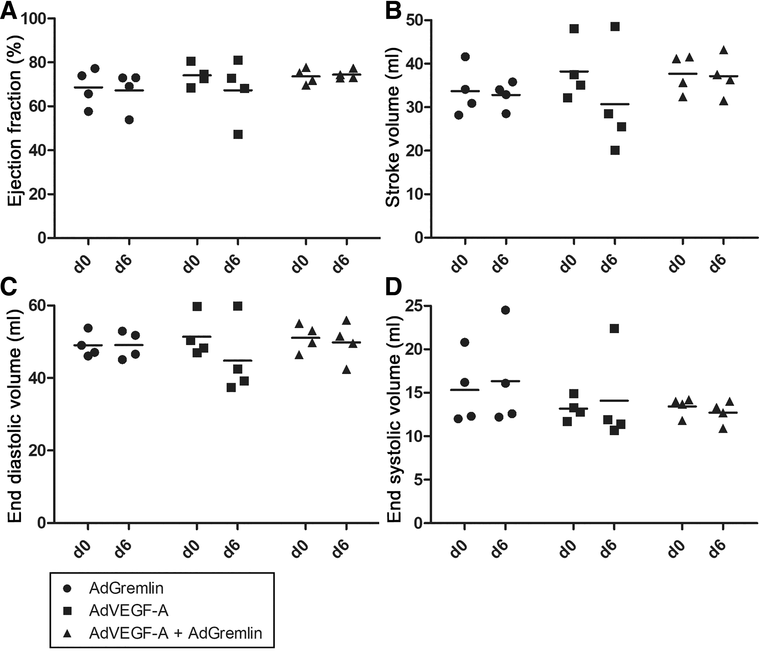
Gene transfers had no significant effect on cardiac function in healthy porcine hearts. Measurements were assessed using a left ventricular cine angiography. There were no significant differences in ejection fraction
Gene transfer had no effect on off-target tissues or blood parameters, except creatinine
In the collected safety tissues (lung, liver, spleen, kidney, and ovary) there were no inflammation or histological changes detected from hematoxylin-eosin stainings. There were no significant changes in blood parameters, except in VEGF-A with Gremlin group where creatinine values were increased (Fig. 4).
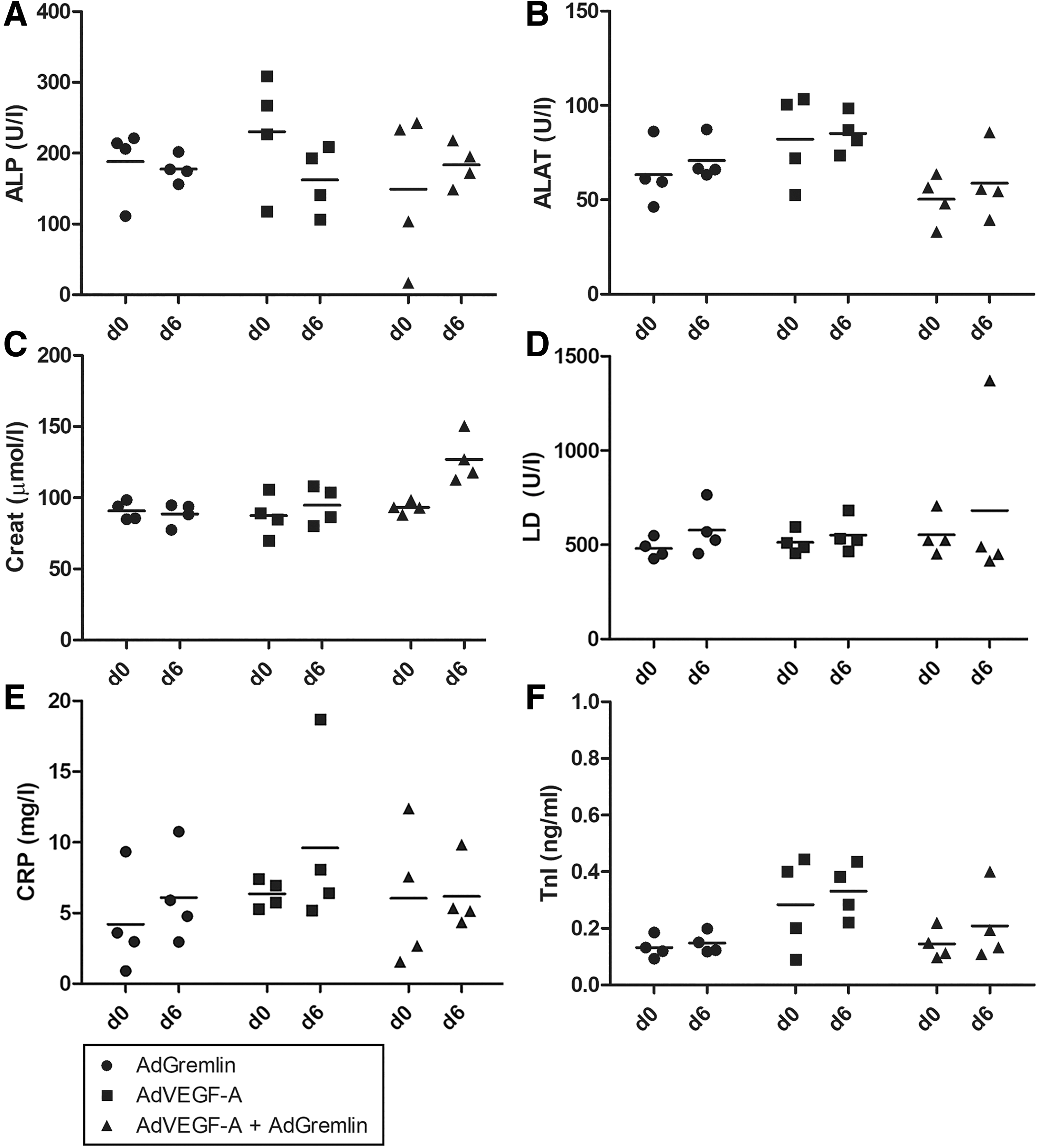
Adenoviral gene transfers had no effect on most of the tested blood parameters. ALP
Discussion
AdVEGF-A induces significant and even aberrant angiogenesis in the myocardium with a total dose of 5 × 10¹¹ vp. AdGremlin alone does not cause angiogenesis, but when combined with AdVEGF-A, it seems to modulate the angiogenic effect of VEGF-A. The aim of this study was to investigate the potential of Gremlin to modulate the effects of VEGF-A in a large animal model. The potent angiogenic effects of VEGFs have been previously modified with factors, such as dose, delivery method, transduction duration, and signaling pathway modulation. These signaling pathway modulations include Slit2 and EphrinB2/EphB4, 18,19 and Gremlin now adds to this list of modulators of angiogenic effects. Also, further studies are needed to investigate the molecular mechanisms behind these modulation effects.
VEGF-A is known not only for its angiogenic properties, but it has also notable side effects. Especially the increased permeability can cause pericardial fluid accumulation. AdGremlin seems to be a potential modulator of the angiogenic effect of VEGF-A that could be used to limit tissue edema and permeability related side effects of AdVEGF-A therapy. This could enable the use of larger doses of AdVEGF-A to increase the effective treatment area in the myocardium when using local intramyocardial injection delivery route.
In this study a 6-day time point was used to analyze the effects at the peak effect of adenoviral gene transfer, because the main target was to find out if Gremlin modulates the aberrant angiogenic effect induced by VEGF-A gene transfer. Even though both treatment groups had a similar pericyte coverage, both treatment groups accumulated a variable amount of pericardial fluid. This suggests that pericyte coverage 6 days after the gene transfer is either not yet fully developed, or not functionally capable of preventing the pericardial fluid accumulation. However, in the treatment group with only AdVEGF-A, but not in the other groups, three animals died before the 6-day end point. Deaths were due to the pericardial effusion causing a cardiac tamponade. Pericardial puncture and aspiration of fluid was done as a therapeutic procedure to prevent fatalities. By combining AdGremlin with the AdVEGF-A, no cardiac tamponade was observed, and no pericardial punctures were required. One shortcoming of the study was that due to the limited number of animals, the observations suggest, but do not prove, that AdGremlin could prevent the risk of fatal tamponade.
Adenoviral gene transfer did not cause inflammation or any histological changes in off-target organs. In AdVEGF-A with AdGremlin group there was a statistically significant change in kidney function parameter creatinine, but it was still close to the reference values for pigs (88–239 μM) 20 indicating a limited clinical significance. Small changes can also occur due to the use of contrast agent in the heart imaging.
The dual role of Gremlin could explain the possible mechanisms behind the modulator effects. It inhibits BMP signaling by blocking BMP2 and BMP4 ligand actions, which are known to induce angiogenesis. Gremlin also binds directly to VEGFR-216 and can compete with VEGF-A, reducing permeability and angiogenic effects of VEGF-A. Thus, Gremlin has potential to modulate the effects of therapeutic angiogenesis in vivo.
Footnotes
Acknowledgments
We thank animal caretakers Heikki Karhunen, Minna Törrönen, and Riikka Venäläinen for their assistance with the animal experiments.
Author Disclosure
No competing financial interests exist.
Funding Information
This study was supported by Finnish Academy Center of Excellence and National Virus Vector Laboratory for the production of viral vectors.