
Abstract

My lifetime goal in translational medicine began five decades ago, directed toward making a difference for patients with devastating neurological diseases. The terminology “translational medicine” as we use it today was not part of our vocabulary. Since the millennium, clinical gene therapy is better recognized as a subspecialty of translational medicine. A brief review of this innovation of gene delivery to patients makes us aware of how recently this was brought to the clinical world. The first successful gene therapy trial was ex vivo delivery for adenosine deaminase severe combined immunodeficiency (ADA-SCID) published in 1995. 1 The uniqueness of clinical gene therapy can be further appreciated, considering there were only two publications in the Journal of Translational Medicine in its inaugural year, 2003. The novelty and freshness of gene transfer as a tool for translational medicine is further emphasized by the small number of approved gene therapy products for commercial use at the time of this writing. These include Luxturna (voretigene neparvovec/AAV2-hRPE65v2; Spark Therapeutics, Philadelphia, PA) for autosomal recessive retinitis pigmentosa caused by mutations of the RPE65 gene, 2 and systemic delivery of Zolgensma (onasemnogene abeparvovec/AAV9.CB.SMN; Novartis, Basel, Switzerland) for the rapidly fatal disease, spinal muscular atrophy type 1 (SMA1). 3
My own career enabled an experience in bench to bedside medicine because of political events taking place in the 1960s. My first exposure to the translational world came at the expense of 60,000 American deaths and more than 3.3 million Vietnamese in one of the costliest wars in the history of the United States. Many of the young investigators reading this essay probably don't know that during the Vietnam war, the military draft was mandatory for every medical school graduate. The mandate for conscription (called the Doctor's Draft) suddenly changed one's destiny after internship, including the possibility of carrying a rifle under the overcast sky of Vietnam through green mountainous terrain or through hot and humid, insect-infested jungles. Circumventing this fate was a gift from heaven for the few selected to serve in the Public Health Service, in the branch that came to be known as “Yellow Beret” at the National Institutes of Health (NIH). Many of us who were selected had some research background in medical school (University of Texas Southwestern Medical Center). My further good fortune came through acceptance to the Medical Neurology Branch at the National Institute of Neurological Disorders and Stroke as a research fellow. This acceptance was a eureka moment for me and defined the remainder of my career, even up to the present. Chosen for this position was fate because I had only two formal years of neurology training (Columbia University) rather than the three “required.” This branch had a vivid history pioneering neuromuscular disease. Milton Shy, the first Director and his successor, W.K. Engel, clinically described new diseases and concepts in muscle diseases, including nemaline myopathy, central core disease, mitochondrial diseases, and myotubular and centronuclear myopathy. For young researchers like me, the traditional residency/fellowship regulatory guidelines that restrict research participation had not been established. My Branch Chief at the NIH strongly encouraged research involvement in a neuromuscular disease of interest to the individual.
At 27 years of age, I saw my first Duchenne muscular dystrophy (DMD) patient. He was 7 years old and on the threshold of losing ambulation; there was no treatment, and I was firmly committed to making a difference. Molecular-based tools were limited, and uncovering the genetic basis of the disease was not on the horizon. Insight was gained by understanding the pathological findings. This led to my first major translational paper published in Science Magazine in 19714 with confirmation in a subsequent report in Nature. 5 In the laboratory, we were able to reproduce the typical DMD focal areas of necrosis and regeneration by muscle ischemia. On the clinical side, this set the stage for clinical trials in DMD attempting to increase blood flow with aspirin and an ace-inhibitor, lisinopril. No clinical benefit was seen. The findings in the lab, however, showing compromised blood flow remain relevant almost 50 years later. This was not fully appreciated until the discovery of dystrophin by Lou Kunkel. 6 Normal skeletal-muscle neuronal nitric oxide synthase (nNOS) is localized to the cytosolic surface of the sarcolemma, and coincident with muscle contraction, nNOS produces NO. Given that NO is a potent vascular dilator, it provides a means of increasing metabolic requirements to the muscle. Overall, it validates the pathogenic contributions previously observed with pre-clinical gene therapy efforts. nNOS is a component of the dystrophin–glycoprotein complex. 7 In the absence of dystrophin, nNOS fails to localize at the sarcolemma, and consequently NO is deficient, resulting in focal ischemia. Under normal conditions, it is the spectrin repeats 16 and 17 of dystrophin that anchor nNOS to the muscle membrane and enhance exercise performance in the mdx mouse and dystrophic dog. 8 This has strongly encouraged some investigators currently involved in clinical gene therapy trials to treat patients with a micro-dystrophin cassette that includes repeats 16 and 17. The efficacy of this gene therapy approach is currently being tested.
In DMD, the next most relevant clinical contribution to date was the unequivocal proof that prednisone was effective for treatment. 9 Before this seminal publication in 1989, there were contrasting studies without a defining conclusion regarding the efficacy of steroids. The clinical trial was a collaboration between four centers, forming the first ever multicenter clinical collaborative investigative team exclusively devoted to muscular dystrophy. The group was established in 1978 and, for the first time, designed and published a protocol for DMD clinical trials. This was followed by one of the first attempts to define natural history of muscle strength, and function and establish power calculations. 10 The “Prednisone in DMD” clinical trial and publication 9 continues to impact current gene therapy trials. The strength of the study included enrolling a large cohort, 103 DMD boys, which was unparalleled in a study 30 years ago. Efficacy was established, and proof over decades shows that corticosteroids make a significant difference in prolonging ambulation. There were justifiably cautious comments made upon publication of the prednisone paper, considering risk of long-term use, but experience favors the risk–benefit ratio. Relevance to current gene transfer for DMD is valued, considering that criteria for enrollment in all current gene therapy trials includes a stable dose of prednisone for 3–6 months (Sarepta Therapeutics, Cambridge, MA; Pfizer, New York, NY; Solid Biosciences, Cambridge, MA). Corticosteroids also have importance related to suppression of adeno-associated virus (AAV) immunity and liver toxicity. The importance of immune-related adversity was shown in our spinal muscular atrophy clinical gene therapy trial. 3 Patient 1 in the SMA trial receiving a large dose of systemically delivered AAV9 by percutaneous infusion was found to have elevations in serum aminotransferase levels (31 times the upper limit of the normal range for alanine aminotransferase [ALT] and 14 times the upper limit for aspartate aminotransferase [AST] at 3 weeks post gene delivery without other liver-function abnormalities; i.e., total and indirect bilirubin and alkaline phosphatase). These elevations were attenuated by prednisolone treatment, which was subsequently administered in the remaining patients prophylactically. One patient in cohort 2 required additional prednisolone to attenuate elevated serum ALT and AST levels (35 times the upper limit of the normal range for ALT and 37 times for AST). The experience in the SMA trial (further discussion below) has been adopted for DMD gene therapy.
After the discovery of dystrophin and the molecular basis for DMD was fully established, treatment emphasis was reoriented toward correcting the molecular defect. Before gene therapy was a reality, gene correction through myoblast transfer was thought to be a favorable approach. 11 –13 Donor myoblasts injected into the muscles of affected patients were predicted to fuse with host muscle fibers, thus contributing their nuclei and potentially replacing dystrophin. Encouraging results in murine dystrophies had been reported and were tested in the clinical arena. A strategy we thought likely to be effective was to inject donor myoblasts once a month for 6 months to the biceps brachii muscle. 14 Over time, repeated episodes of necrosis and regeneration would incorporate satellite cells from the injected myoblasts to replace dystrophin deficient in endogenous muscle fibers. In the clinical trial, one extremity received myoblasts, and the opposite arm served as a sham-injected control. An incredible 110 million muscle cells were donated by fathers or brothers and injected each month. Six months after the final myoblast transfer, dystrophin was assessed using the peptide antibodies to the deleted exons of the dystrophin gene. There was no difference in muscle strength between sham- and myoblast-injected extremities. In one patient, 10.3% of muscle fibers expressed donor-derived dystrophin. Three others showed low-level dystrophin; eight had none. The findings showed the limitations of the method, but of more potential significance for future studies, exon-specific peptide antibodies permitted distinction between revertant fibers and dystrophin-positive fibers derived from donor DNA.
My formal entrée to the gene therapy world was inspired by the success of gene transfer in ADA-SCID with the full correction of the disease phenotype. DMD posed problems, but there was the potential for adenovirus (AD) gene transfer using the gutted virus that could package the 14 kb dystrophin cDNA. Several labs were working on this, including Wilson, 15 Karpati, 16 and Chamberlain. 17 My efforts concentrated on a collaboration with Jeff Chamberlain, using gutted AD carrying the full-length dystrophin gene to clinical trial. One of the initial disappointments was that this effort never came to trial because of the unfortunate experience with AD transfer for ornithine transcarbamylase deficiency.
On or about the same time as the direction shifted from AD, the investigators in translation were working toward introducing AAV to the clinic: Terry Flotte, AAV transfer for cystic fibrosis; 18 Roland Herzog, Jim Wilson, and Katherine High, breaking ground in hemophilia factor IX. 19 I was poised to join the gene therapy efforts based on my long history in clinical translation. The stars aligned for my entry when my academic colleague in pediatrics, Phil Johnson, invited me to join the team at Nationwide Children's Hospital (NCH; called Columbus Children's until 2007). I was on the Ohio State University Main Campus about 2 miles away and had been initiated into the AAV gene therapy world using AAV2 to transfer alpha-SG to the foot muscle of limb-girdle muscular dystrophy (LGMD) type 2D patients in 1999. This was chosen as a proof-of-concept (POC) clinical trial using hSGCA, a perfect fit for AAV. The vector was produced in the Wilson Lab at the University of Pennsylvania. Trial results favored gene expression in the extensor digitorum brevis muscle, a small foot muscle chosen for safety reasons. However, the study ended prematurely because of vector source. This experience was responsible for an invitation to join the Johnson Team at Columbus Children's. I was eager to work with Phil Johnson and his colleague K. Reed Clark, both experienced virologists. They were deeply invested in potential gene therapy for human immunodeficiency virus using a novel model delivering AAV carrying Fabs to generate neutralizing molecules targeting simian immunodeficiency virus. They were appreciative of what I was doing in monogenic muscle diseases such as DMD and LGMD. I could envision their help in multiple ways: viral production, delivery, and strategy for building cassettes. However, within months of my arrival, Phil Johnson came to my office with a big smile and announced that he had been recruited to the Children's Hospital of Philadelphia (CHOP) as their Chief Scientific Officer, and I would be named the Director of Gene Therapy at Columbus Children's. With some reluctance, I accepted the challenge and was blessed because Reed Clark elected to stay in Columbus for family reasons. I was given carte blanche to develop a neuromuscular disease (NMD) gene therapy program in my own vision.
As Reed and I discussed building the first independent gene therapy center for NMD, it quickly became apparent that we were going no place without a vector manufacturing core. Reed had contacts in AAV manufacturing, and discussed our options with Barrie Carter, who served as the Executive Vice President and Chief Scientific Officer of Targeted Genetics. The cost of AAV vectors was way beyond our budget, and we contemplated our options. My vision was still back at treating the 7-year-old DMD boy I met at the NIH (long gone by now). Reed advised to start with realistic expectations, and we went the CEO of NCH (Steve Allen), explaining the lack of vector, and he wrote a check for $2 million to build a one-unit vector manufacturing facility from an existing lab on our research floor. Reed engaged this like the pioneer in the field that he was and continues to be as he now works for Sarepta. This put the program squarely in my lap, and I recruited basic scientists with devoted interest in NMD. Brian Kaspar was well trained at the Salk Institute for Biological Studies, Paul Martin, Berkeley, Washington University; Scott Harper, University of Michigan; Doug McCarty and Haiyan Fu, University of North Carolina (hired right before I came by Phil). I knew the gene therapy turf, and in addition to vector needs, we needed a regulatory core to help with Investigational New Drug applications and a team to monitor outcome for clinical trials (recruited Linda Lowes and Lindsay Alfano). We also needed our own tissue lab for analyzing muscle biopsy results like what we had on the Ohio State University campus. This was critical item, and my long-time colleague, Zarife Sahenk, was recruited to NCH to direct the muscle lab and set up an independent research program. Another important player was a postdoctoral fellow who joined our center, Louise Rodino-Klapac (currently works for Sarepta). Our plan was to start with intramuscular POC AAV-mediated gene delivery; a renewed effort in SGCA gene delivery was successful. 20,21 We also delivered AAV-mediated mini-dystrophin (collaborated with Chris Walker, R. Jude Samulski, and Xiao Xiao), showing the potential of inducing transgene immunity when expressing the gene in a patient's deletion. 22 We also found that multiple intramuscular (i.m.) injections of AAV-mediated follistatin improved leg strength in Becker muscular dystrophy. 23 Determining preexisting AAV antibody titers was critical to achieve gene expression. With these successes and greater ability to produce vector at NCH, we attempted an isolated limb protocol for vector delivery with disappointing results with limited clinical impact. While these i.m. and isolated limb studies were progressing, AVI Therapeutics (later to become Sarepta Therapeutics) approached us to implement the exon skipping trial using antisense oligonucleotides (ASOs). This method had taken the DMD world by storm (highly touted in social media). We did a systematic study and showed that exon skipping was a viable, modest treatment option, although not curative. 24,25 Additional work has confirmed efficacy and the ASO has been approved and commercialized as Exondys 51 (eteplirsen). At the time of this writing, skipping exon 53 for DMD has now also been approved (Vyondys 53 [golodirsen]).
Dr. Mendell with SMA patient who is walking normally 4 years following gene therapy. Without treatment, children affected with SMA type 1 usually never sit up and often die by the age of 2.
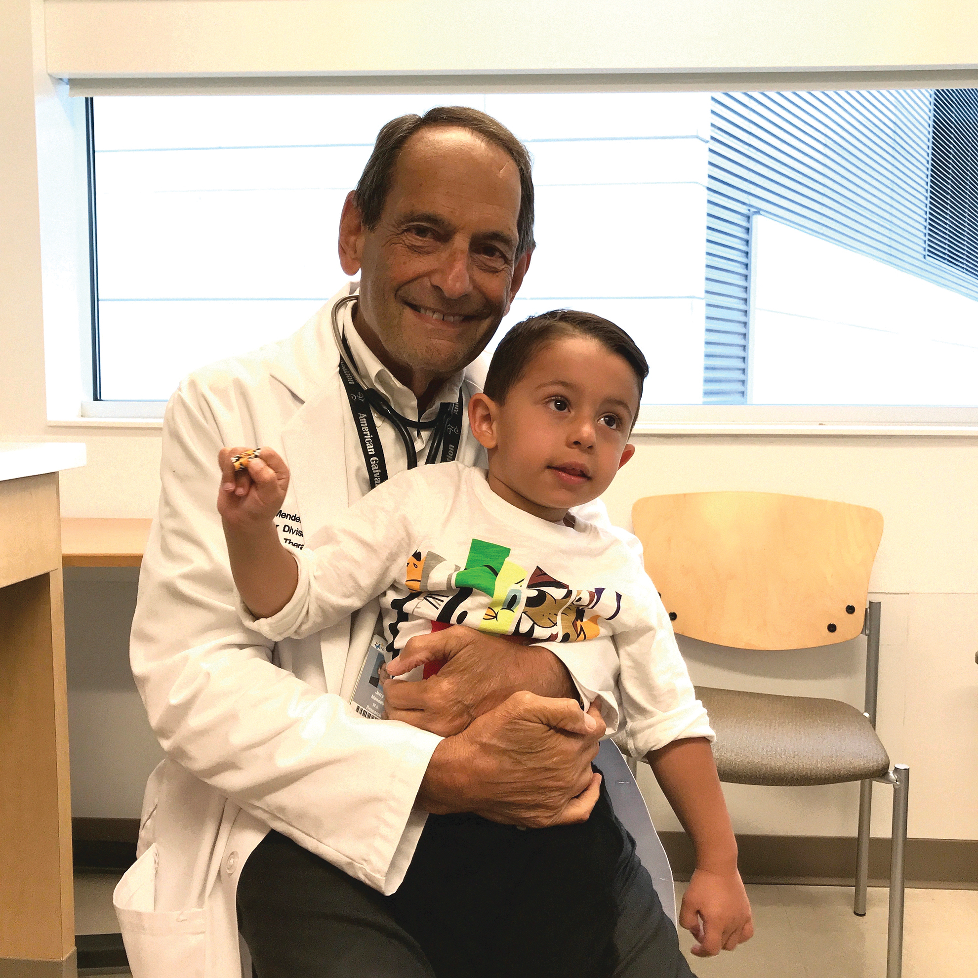
Dr. Mendell and a DMD patient who receives gene therapy alongside his teddy bear at Nationwide Children's Hospital, Columbus OH.

The next major clinical project was survival motor neuron (SMN) gene replacement. The success of this trial was teamwork. It began with preclinical studies by Arthur Burghes. I recruited Arthur to Neurology in 1987 from Ron Worton's lab in Toronto while they were working on dystrophin. Shortly after he came to OSU, he turned his focus toward SMA and developed the SMNΔ7 mouse model that simulated SMA type1. 26 This model enabled POC that set the stage for gene delivery. His lab showed that a transgenic mouse expressing SMN2 crossed into the severe SMA mouse model significantly extended life-span. 27 It was almost a year later that the Kaspar lab rescued the life and motor function of the SMNΔ7 mouse using scAAV9.CB.SMN by systemic delivery. 28 The third leg of the collaboration was my contribution leading the clinical trial, setting the stage for a major clinical breakthrough in gene therapy for SMA type 1. 3 The natural history predicts that only 8% survive by 20 months of age. The delivery of scAAV9.CB.SMN to infants at doses never given to patients in clinical gene transfer trials was a fortunate decision. Some experts advised against it, warning of the toxicity of this viral titer. Cohort 1 (n = 3) received 6.7 × 10¹³ vg/kg, and cohort 1 (n = 12) received 2.0 × 10¹⁴ vg/kg. It appeared at first that the naysayers might be correct. Upon starting the study, patient 1 in cohort 1 at low dose had elevated serum aminotransferase levels (ALT 31 times upper limit of normal [ULN] and AST 14 times ULN) in the third week post gene delivery. We aggressively treated the signs of liver toxicity, and we modified the protocol enabling prednisolone delivery to this subject and patients 2–15 (1 mg/kg/day) given for approximately 30 days, starting 24 hours before gene transfer. In the remainder of the study, only one additional subject had elevations that reached grade 4 level (serious adverse event). Of the total cohort, two other patients had elevation of ALT and AST, both <10 times ULN.
The efficacy in this study, sponsored by Avexis, went beyond expectations. By the conclusion at 2 years, all 15 patients were alive. At high dose, a rapid increase from baseline in the score on the CHOP INTEND scale followed gene delivery. The CHOP INTEND score increased by 9.8 points at 1 month and 15.4 points at 3 months compared with a decline in natural history. Of the 12 patients who had received the high dose, 11 sat unassisted, 9 rolled over, 11 fed orally and could speak, and 2 walked independently. The long-term implications were clear, and the Food and Drug Administration approved SMA gene therapy for children <2 years of age with SMA for what later became the Novartis product onasemnogene abeparvovec (Zolgensma®). In 2017, the clinical trial published in the New England Journal of Medicine 3 received the Science Breakthrough of the Year Award (People's Choice 2017). An important finding from the clinical trial was that the least-affected SMA patients treated at the earliest time did the best. This was translated to newborn screening for SMA now implemented in 18 States. So far, treating asymptomatic newborns without expression of SMN1 and two copies of SMN2 appears to prevent signs of disease.
After SMA, the passion for gene therapy has grown to new heights. DMD has been a life-long target that we have brought to clinical trial after preclinical studies looked very promising. Louise Rodino-Klapac has led efforts on the preclinical work. Almost everyone in the field knows the obstacles for achieving the same degree of success we had in SMA. The gene is too large for AAV, and we and others have tried to achieve full-length translation using dual vector delivery with homologous translation. This has had disappointing results. 29 We are forced to use a variant of a small dystrophin (micro-dystrophin gene transfer) to package in AAV. Obstacles for success relate to transduction efficiency and the amount of micro-dystrophin needed to reverse or protect against disease manifestations. We have used the principles learned from SMA, delivering 2 × 10¹⁴ vg/kg in the clinical trial. Patients with preexisting AAV antibody are excluded, and corticosteroids remain as a staple for gene transfer. Current data that have been presented at meetings on the first four subjects participating in an open-label trial look promising. The cassette includes hinges 1, 2, and 4 and spectrin-like repeats 1–3 and 24, adopted from the classic Chamberlain–Harper Study. 30 Preclinical experiments comparing other cassettes were not as effective. The gene is delivered through the systemic circulation, and at 12 weeks, the number of dystrophin-positive muscles showed a mean of 81.2% and an intensity of 96% in the first four subjects. Western blots supported high levels of micro-dystrophin and showed functional improvement at 1 year post gene delivery in the North Star Ambulatory Assessment, time to rise, climbing four stairs, and the 100 m run. Adverse events have been limited to increased liver enzymes in three subjects. DMD gene therapy looks promising, and studies supported by Sarepta have now been extended to a blinded-controlled trial and will move to Phase III within several months.
My life experiences have evolved through gratifying events promoted by early encounters with life-threatening conditions that provoked a passion to find ways to help. Gene therapy has offered a path, not fulfilled by any other means of translational medicine. The opportunity offered by Human Gene Therapy to share this life experience has required recalling deeply felt experiences for which I am grateful.
Author Disclosure
No competing financial interests exist. I have no investment in any product that I have ever brought to clinical trial, when serving as PI. I am a consultant for the following pharmaceuticals: Avexis, Inc, Sarepta Therapeutics, and Vertex Pharmaceuticals.