
Abstract
Recombinant adeno-associated viral (rAAV) vector-mediated gene therapy is being developed to treat X-linked retinitis pigmentosa (XLRP) in patients with mutations in the retinitis pigmentosa GTPase regulator (RPGR) gene. In preparation for a clinical gene therapy trial, we conducted dose range finding (DRF) studies with an AAV2 capsid with three surface tyrosine residues changed to phenylalanine (AAV2tYF) vector administered by subretinal injection in a naturally occurring RPGR-mutant canine model (XLPRA2) to compare two different human RPGR (hRPGR) transgenes and to establish a reasonable starting dose for a clinical trial. Different dose levels of two candidate vectors (0.15 mL at 1.2 × 10¹⁰–3.0 × 10¹² vg/mL of rAAV2tYF-GRK1-hRPGRco or 4 × 10¹⁰–3.0 × 10¹² vg/mL of rAAV2tYF-GRK1-hRPGRstb), 6.0 × 10¹¹ vg/mL rAAV5-GRK1-hRPGRco reference vector or Vehicle were subretinally administered, and the dogs were followed for 8 weeks postdose. Ophthalmic examinations, analyses of retinal structure by in vivo imaging using confocal scanning laser ophthalmoscopy (cSLO)/optical coherence tomography (OCT) in the Lower (4.0 × 10¹⁰ vg/mL) and Lowest (1.2 × 10¹⁰ vg/mL) Doses, immunological responses by cell based assays or enzyme-linked immunosorbent assay, RPGR transgene expression, and reversal of opsin mislocalization by immunohistochemistry were performed. No sustained signs of ocular discomfort or ophthalmic complications were noted in any of the injected eyes except some in the High Dose group (3.0 × 10¹² vg/mL), which showed signs of retinal detachment and inflammation. A change in fundus reflectivity suggestive of a rescue effect was seen in the High, Mid (6.0 × 10¹¹ vg/mL), and Low (1.2 × 10¹¹ vg/mL) Dose groups. cSLO/OCT demonstrated qualitative and quantitative evidence of rescue effect in eyes treated with the Lower Dose. Anti-hRPGR antibodies were absent, but neutralizing antibody titers against AAV2 were detected in all animals dosed with rAAV2tYF in an apparent dose-related pattern. RPGR expression was stronger for rAAV2tYF-GRK1-hRPGRco compared to rAAV2tYF-GRK1-hRPGRstb at all dose levels. Subretinal administration of rAAV2tYF-GRK1-hRPGRco and rAAV2tYF-GRK1-hRPGRstb both corrected rod and cone opsin mislocalization, two early markers of disease in the XLPRA2 canine model of RPGR-XLRP. These results support the selection and use of rAAV2tYF-GRK1-hRPGRco (AGTC-501) and guided the initial doses in clinical studies in patients with XLRP caused by RPGR mutations.
Introduction
X-
There are multiple alternatively spliced transcripts of the RPGR gene. An alternatively spliced transcript containing exons 1 to 15 and a large part of intron 15, RPGRex1-ORF15 , is the most abundant transcript in the retina of all species examined. 7 This transcript is localized in the connecting cilia of rod and cone photoreceptors and is likely involved in regulating transport through the photoreceptor cilium. 3,8 The highly repetitive purine-rich ORF15 region is a mutation hotspot, accounting for up to 80% of all reported RPGR mutations. 7,9 Naturally occurring frame-shift mutations in the ORF15 exon of mouse or canine RPGR result in RP that share important features with the human disease; in dogs this includes similar spatial distribution of retinal pathology and reduction of ERG function at an early age. 10 –14
Proof-of-concept studies first conducted in dog and subsequently in mouse models of XLRP have shown that subretinal delivery of recombinant adeno-associated virus (rAAV) vectors expressing a RPGRex1-ORF15 transgene can maintain photoreceptor structure and function. 15 –20 RPGR mutant XLPRA2 dogs have a two nucleotide microdeletion in ORF15 that causes early onset (4–5 weeks of age) retinal degeneration. 12,13 Gene therapy studies in XLPRA2 dogs, using an AAV2/5 vector carrying a “stabilized” human RPGR (hRPGRstb) cDNA, 21 showed stable rescue of photoreceptors from progressive degeneration for >2 years even when the treatment was initiated at late-stage disease. 15 –17
The hRPGRstb transgene is a “stabilized” 42 base-pair shorter version of hRPGR cDNA with 65 nucleotide substitutions; 15 amino acids deleted, 1 amino acid inserted, and 26 amino acids substituted. The function of this transgene's polypeptide appears not to be impaired. 17,21 A codon optimized full length hRPGR cDNA (hRPGRco) developed by rational design and codon optimization that maintains the original wild-type amino acid sequence has also been tested in XLRP dogs. Preliminary data using rAAV5 vector showed a similar level of structural and functional preservation of photoreceptors in XLPRA2 dogs with hRPGRco and hRPGRstb transgenes. 17
Toward development of an optimal gene therapy for RPGR-XLRP with the rAAV2tYF vector we conducted dose range finding (DRF) studies in RPGR-XLRP mutant (XLPRA2) dogs to compare efficacy and safety of two viral vector constructs: rAAV2tYF-GRK1-hRPGRco and rAAV2tYF-GRK1-hRPGRstb. The vectors were delivered using subretinal injection at a volume of 0.15 mL to XLPRA2 dogs during mid-stage disease (12 weeks of age) at a wide range of titers (1.2 × 10¹⁰–3.0 × 10¹² vg/mL). The results demonstrated the advantages of the rAAV2tYF-GRK1-hRPGRco vector and informed dose selection for the GLP safety study and clinical trials (NCT03314207). 22
Materials and Methods
Study design
Two DRF studies tested the efficacy of two vectors (rAAV2tYF-GRK1-hRPGRco and rAAV2tYF-GRK1-hRPGRstb) in XLPRA2 dogs 8 weeks after subretinal dosing (Table 1).
Design of dose range finding studies
DRF, dose range finding.
DRF study 1
A total of eight XLPRA2 dogs were used in this study. Six dogs were injected subretinally with 0.15 mL of rAAV2tYF-GRK1-hRPGRco in one eye and rAAV2tYF-GRK1-hRPGRstb in the contralateral eye at a vector concentration of 3.0 × 10¹² vg/mL (High Dose), 6.0 × 10¹¹ vg/mL (Mid Dose), or 1.2 × 10¹¹ vg/mL (Low Dose), and each dose was tested in two animals. rAAV5-GRK1-hRPGRco vector, previously reported to rescue photoreceptors in this dog model, 17 was used as a reference at the Mid Dose level (6.0 × 10¹¹ vg/mL) in both eyes of one dog. Both eyes of another dog were dosed with Vehicle (Alcon Balanced salt solution containing 0.014% [v/v] Tween-20, or BSST) and served as controls.
DRF study 2
This follow-up study was performed to evaluate two even lower dose levels. Two XLPRA2 dogs received a Lower Dose level (4.0 × 10¹⁰ vg/mL) of both rAAV2tYF vectors, one vector per eye. Finally, two additional dogs were dosed in one eye with the Lowest Dose level (1.2 × 10¹⁰ vg/mL) of rAAV2tYF-GRK1-hRPGRco, while the contralateral eyes received the Vehicle (BSST) and served as control.
AAV vector production
The rAAV2tYF-GRK1-hRPGRco and rAAV2tYF-GRK1-hRPGRstb vectors were produced using a proprietary Herpes simplex virus (rHSV) Assisted Vector Expansion (HAVE) system in suspension-cultured baby hamster kidney (sBHK) cells. 23 Two rHSV helper viruses, one containing the AAV2 rep and AAV2tYF cap genes and the other containing the hRPGRco or hRPGRstb expression cassette, were used to coinfect sBHK cells grown in serum-free medium. One day later the cells were lysed with Triton X-100 detergent, treated with Benzonase® (Merck), clarified by filtration, purified by AVB Sepharose® (GE Life Sciences) affinity chromatography followed by CIM SO3− (BIA Separations) cation-exchange chromatography, and eluted in 2.6 × BSST. The purified bulk was concentrated and buffer exchanged to 1 × BSST (drug substance) and sterile filtered (0.2 μm) to generate drug product. The drug product was further concentrated, as needed, using a 100 kDa MWCO Ultra centrifugal filter unit (EMD Millipore) and refiltered (0.2 μm) to generate drug product sub-lots of specific concentrations, which were stored at ≤ −65°C. The rAAV5-GRK1-hRPGRco vector was manufactured by double plasmid transfection method and purified by double iodixanol gradient centrifugation as previously described. 24 All vectors were formulated in 1 × BSST and assayed for vector genome concentration, purity, capsid and transgene identification, endotoxin levels, infectivity, and sterility as previously described. 25 Vector genome concentration was determined by quantitative real-time polymerase chain reaction with primers of SV40 polyadenylation sequence (polyA) region of the constructs.
Animals
Twelve XLPRA2 (RPGR-mutant) dogs 11,12 (five hemizygous males and seven homozygous females) were included in the studies (Table 1). Dogs were treated at 12 weeks of age, which corresponds to mid-stage disease (∼40% loss of photoreceptors at the time of treatment) 17 and observed for 8 weeks postdose. The dogs were bred and maintained at the University of Pennsylvania Retinal Disease Studies Facility (RDSF), and studies were carried out in strict accordance with the recommendations in the Guide for the Care and Use of Laboratory Animals of the National Institutes of Health in compliance with the USDAs' Animal Welfare Act, Animal Welfare Regulations, and the ARVO Statement for the Use of Animals in Ophthalmic and Vision Research. The study protocols were approved by the Institutional Animal Care and Use Committee of the University of Pennsylvania. Subretinal injections and all noninvasive imaging procedures were performed under general anesthesia, as previously described. 17 All efforts were made to improve animal welfare and minimize discomfort.
AAV vector administration
Subretinal injections of Vehicle (BSST) or AAV vectors were performed without prior vitrectomy under direct visualization through an operating microscope and a contact vitrectomy lens using a custom-modified subretinal cannula as previously reported. 16,17,26 The 0.15 mL volume injected aimed to produce a bleb that covers ∼12% of the retinal surface. The location and extent of the subretinal bleb were recorded by fundus photography (Retcam shuttle; Clarity Medical Systems, Pleasanton, CA) immediately after each dose (day 0) (Supplementary Figs. S1 and S2). On the second or third day postdose (day 1 or 2), retinal reattachment was confirmed by ophthalmic examination and fundus photography (Supplementary Figs. S3 and S4).
Ophthalmic examination
Dogs received an ocular examination 3–5 days predose, and on day 1–3, week 1, 2, 4, 7–8 postdose. Ocular comfort was assessed by evaluating the presence of blepharospasm, excessive discharge, excessive blinking, or redness when observing the dog in his/her normal environment and on the examination table. Pupils were dilated with tropicamide 1% solution (Akorn, Lake Forest, IL) 30 min before examination. A complete ophthalmic examination was performed using a Finoff Transilluminator (Welch Allyn, Skaneateles Falls, NY) and SL-17 Portable Slit Lamp (Kowa Company, Tokyo, Japan) for the anterior segment and anterior vitreous and binocular indirect ophthalmoscopy (All Pupil II; Keeler, Windsor, Berkshire, United Kingdom) using Volk 40D and 20D lenses (Classic BIO lenses; Volk Optical, Inc., Mentor, OH). Fundus photographs were taken following the examination. Intraocular pressure (IOP) was monitored at all time points, in conjunction with ophthalmic examinations, on nonsedated animals using a rebound tonometer (iCare vet tonometer; Tonovet, Salt Lake City, UT).
In life confocal scanning laser ophthalmoscopy and optical coherence tomography imaging and analyses
In DRF study 1 (High, Mid, and Low Dose) noninvasive retinal imaging by confocal scanning laser ophthalmoscopy (cSLO)/optical coherence tomography (OCT) (Spectralis HRA/OCT I, Heidelberg, Germany) was performed in anesthetized dogs before termination only if ocular examinations revealed obvious or suspicious retinal abnormalities. In DRF study 2 (Lower and Lowest Dose) detailed cSLO/OCT imaging and outer nuclear layer (ONL) thickness analysis were performed before termination as previously described 15,16 to assess potential rescue of photoreceptors within the treated areas.
Immunological analysis
Serum was collected from each dog at predose and at the time of termination (week 8) and stored at < −65°C. Neutralizing antibodies (NAbs) to AAV2 or AAV5 capsid were measured as previously described. 27 Antibodies to hRPGR were measured by enzyme-linked immunosorbent assay. Briefly, microtiter plates were coated overnight with a custom-made recombinant hRPGR protein (GenScript, Piscataway Township, NJ) or Dog IgG (ImmunoReagents, Raleigh, NC). Plates were then washed, blocked, and incubated with diluted dog test sera or mouse anti-hRPGR (custom-made monoclonal antibody, Lot#A215080168; GenScript) as a positive control. A mouse anti-hRPGR antibody positive control was used in the assay since a dog antibody was not available. Dog IgG in selected wells served as the control for dog secondary antibody. After sample incubation, a cocktail of horseradish peroxidase-conjugated anti-dog IgG and anti-mouse IgG was added to detect antibodies bound to hRPGR. Tetramethyl benzidine substrate was then added and absorbance measured spectrophotometrically.
Immunohistochemistry and histopathology
At 8 weeks postdose, the eyes were collected immediately after euthanasia with an intravenous injection of pentobarbital sodium and phenytoin sodium solution (Euthasol; Virbac, Fort Worth, TX). The ocular globes were fixed in 4% paraformaldehyde (PFA) for 3 h followed by 2% PFA for 24 h, trimmed, cryoprotected in 15–30% sucrose/phosphate-buffered saline solution, and embedded in optimal cutting temperature media as previously reported. 12 Cryosections were stained with hematoxylin and eosin (H&E) or used for fluorescence immunohistochemistry (IHC) using the primary antibodies listed in Supplementary Table S1, Hoechst 333421 nuclear stain, and imaged by widefield microscopy (Axioplan; Carl Zeiss Meditec) or confocal microscopy (Leica TCS SP5; Leica Microsystems). Digital images were taken, processed using the Leica Application suite program, and imported into a graphics program (Illustrator; Adobe). Quantitative assessment of RPGR expression was performed using MetaMorph software (Molecular Devices, San Jose, CA) by measuring integrated fluorescence in the inner and outer segments of the photoreceptor layer. Comparison of the treatment effects to mislocalization was done by an evaluator who was masked to the treatment. Retinal sections were H&E stained or labeled with each IHC marker to verify consistency of the results (pattern of labeling and intensity).
Results
Ophthalmic findings
Successful subretinal injections were achieved in all 12 dogs (24 eyes), with confirmed retinal reattachment on day 1 or 2 postdose in 23 of 24 eyes. In one eye dosed with Vehicle (Z544-OS), retinal reattachment could not be confirmed at day 1 due to preretinal and subretinal hemorrhages (Supplementary Fig. S2). Surgically induced focal hemorrhages were often seen at the retinotomy sites that gradually reduced in size or disappeared by the end of the study. No signs of ocular discomfort were observed during the postinjection period, except in one eye injected with Low Dose rAAV2tYF-GRK1-RPGRstb that had a mild reduction of the palpebral fissure 1 day after dosing. A low (<10 mm Hg) IOP was found in half of the eyes in the first few days following the procedure; most of them returned to within a normal range (10–25 mm Hg) and remained there at later time points until the end of the study. Ophthalmic findings are summarized in Supplementary Tables S2–S4.
Table 2 summarizes rescue and/or toxicity that was apparent in ocular examinations. A change in fundus reflectivity suggestive of a rescue effect (preserved reflectivity in the treated/bleb area in comparison to an increase in reflectivity in the untreated area) was seen in eyes injected with rAAV2tYF-GRK1-hRPGRco (OD) and rAAV2tYF-GRK1-hRPGRstb (OS) at High, Mid, and Low Doses as early as week 4 (12/12 eyes), Lower Dose at week 7 (4/4 eyes), and Lowest Dose at week 7 (1/2 eyes treated with rAAV2tYF-GRK1-hRPGRco). No signs of rescue were noted in the eyes treated with Vehicle.
Occurrence of funduscopic signs of retinal rescue and/or toxicity
Rescue = increased reflectivity in the untreated area in comparison to the treated/bleb area suggestive of a rescue effect from the treatment.
Toxicity = enlargement of the retinal vessels (perivascular cuffing), decreased reflectivity in the treated/bleb area suggestive of retinal detachment, or focal dark pigmentation suggestive of subretinal/retinal infiltration.
N/A = examination was not performed.
Grayed boxes: The eye was assigned to another treatment group, as indicated in the “Group” column.
Blank boxes: No rescue or toxicity was noted.
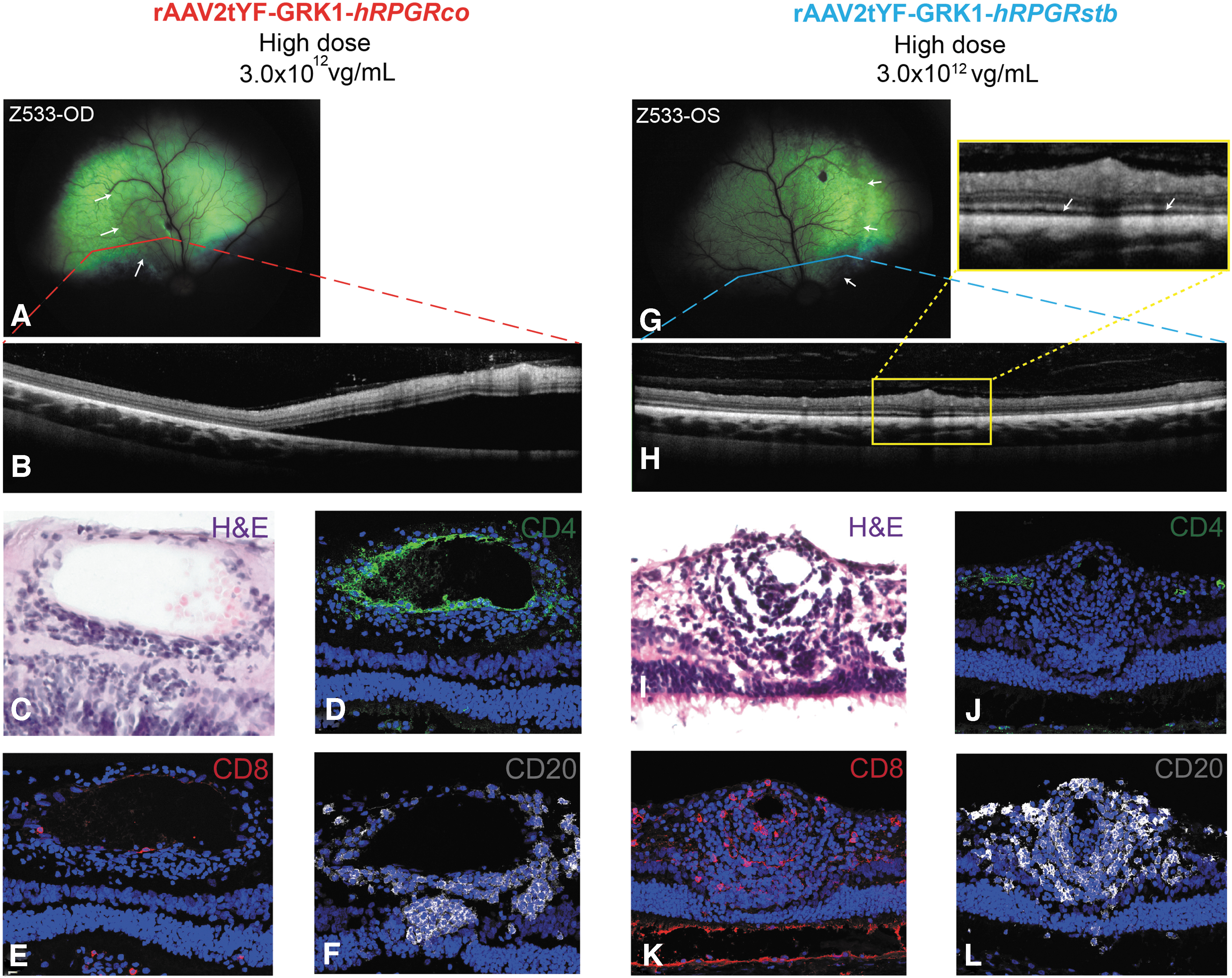
In all eyes that received the High Dose, retinal detachment was observed with both vectors at week 8. Enlargement of retinal vessels and perivascular cuffing were observed in one of two eyes dosed with either rAAV2tYF-GRK1-hRPGRco or rAAV2tYF-GRK1-hRPGRstb. Multifocal black pigmentation suggestive of chorioretinitis or retinochoroiditis was observed in one of two eyes treated with High Dose of rAAV2tYF-GRK1-hRPGRco and in one of two eyes treated with rAAV2tYF-GRK1-hRPGRstb (Table 2 and Supplementary Tables S2 and S3). A cSLO/OCT examination in these eyes confirmed the funduscopic lesions of retinal detachment (Fig. 1). No funduscopic signs of retinal toxicity were detected in the eyes treated with other lower doses (Mid, Low, Lower, or Lowest). In 6 of 10 eyes treated with the Lower Dose, Lowest Dose, and Vehicle control, a focal area of increased hyperreflectivity was seen for the first time at day 6 or week 2 postdose (Supplementary Table S2–S4). These areas of hyperreflectivity were likely lesions of phototoxic retinopathy caused by the light of the operating microscope at the time of the subretinal injection procedure, as it was found within or outside the treated retinal region.
Retinal toxicity in RPGR mutant dogs treated with the high dose of both rAAV2tYF-GRK1-hRPGRco and rAAV2tYF-GRK1-hRPGRstb.
Similar to the eyes treated with rAAV2tYF-GRK1-hRPGRco and rAAV2tYF-GRK1-hRPGRstb vectors, the two eyes dosed with the reference vector rAAV5-GRK1-hRPGRco (Mid Dose) showed reduced fundus reflectivity suggestive of retinal preservation as early as week 4 postdose, which remained stable until the end of the study with absence of retinal toxicity (Supplementary Table S4).
Rescue effect in eyes treated with rAAV2tYF-GRK1-hRPGRco or rAAV2tYF-GRK1-hRPGRstb vectors by in vivo cSLO/OCT
In vivo cSLO/OCT retinal imaging assessment was performed in DRF study 2 and demonstrated both qualitative and quantitative evidence of rescue effect in eyes treated with the Lower Dose of either rAAV2tYF-GRK1-hRPGRco (two of two eyes) or rAAV2tYF-GRK1-hRPGRstb (two of two eyes). Qualitatively evident was a region with ONL retention that was concentric to the injection bleb but larger (Fig. 2A, B, black arrows). Such “penumbral” expansion of treatment regions have been described previously. 17 Quantitative results in all four eyes in the Lower Dose groups showed ONL thickness sampled within the bleb area to be 39–47% of wild-type (WT), whereas the ONL samples in control region away from the injection was 29–38% of WT (Fig. 2D). The ONL thicknesses showed statistically significant differences between the regions (p < 0.0001, Student's unpaired t-test). Results in the two eyes injected with the Lowest Dose of rAAV2tYF-GRK1-hRPGRco showed the bleb area ONL thickness to be 50–52% of WT compared to 46% for the untreated control area (Fig. 2D). The difference between the regions was small but statistically significant (p < 0.0001, Student's unpaired t-test). In eyes treated with Vehicle, bleb area was 42–51% of WT compared to 41–47% of WT for the untreated control area (Fig. 2D); there was no statistically significant difference (p = 0.22, Student's unpaired t-test).
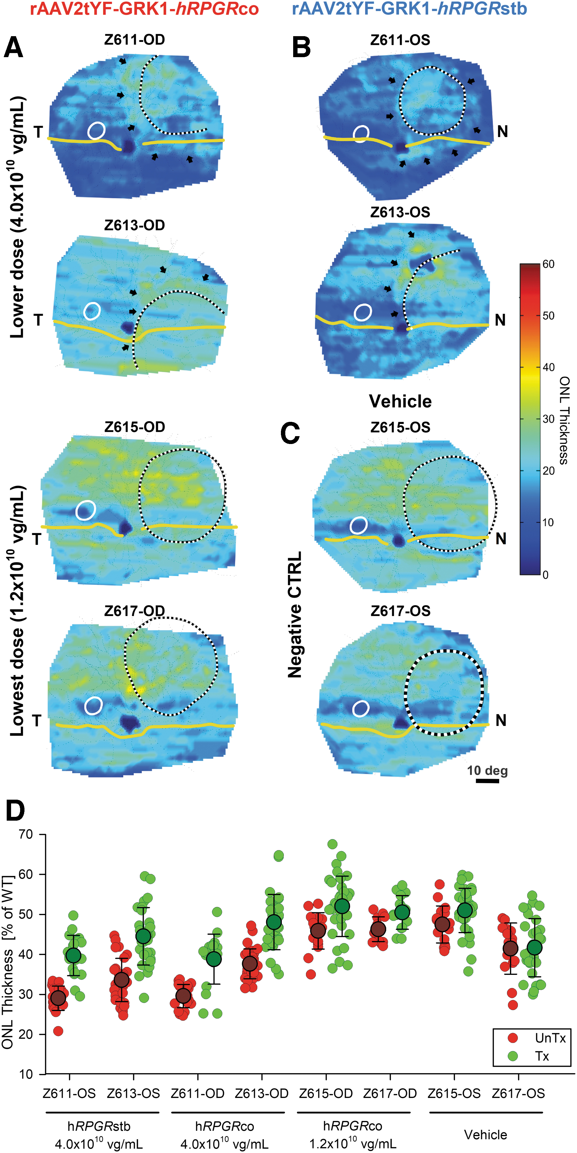
ONL thickness topography maps in eyes of RPGR mutant dogs injected with the Lower, Lowest doses, and Vehicle.
Immunological studies
To evaluate the immunological responses, we tested for both anti-hRPGR and NAbs to rAAV. Anti-hRPGR antibodies were absent in sera from XLPRA2 dogs dosed with either rAAV2tYF-GRK1-hRPGRco or rAAV2tYF-GRK1-hRPGRstb vector (data not shown). All animals had low but detectable predose titers of NAbs to AAV2 or AAV5 (Table 3). At termination (8 weeks postdose), NAb titers against AAV2 were detected in all animals dosed with rAAV2tYF in an apparent dose-related pattern. Both animals given High Dose, 1 of 2 animals given Mid Dose, and 1 of 2 Low Dose animals had high NAb titers (between 1:1,280 and 1:2,560). The other 2 animals, 1 given Mid and one given Low Dose of AAV2tYF-GRK1-hRPGR, had titers between 640 and 1,280 (Mid Dose) and 40 and 80 (Low Dose). All animals given Lower and Lowest Dose had very low NAb titers (1:10 to 1:40). The animal assigned to the Vehicle control group had very low NAb titers to AAV2 (1: 20 and 1:40). The animal given rAAV5-GRK1-hRPGRco (Mid Dose) had a titer between 1:80 and 1:160.
Titration of neutralizing antibody in treated retinitis pigmentosa GTPase regulator mutant dogs before and after vector administration
rAAV, recombinant adeno-associated virus.
Histological/immunohistochemical assessment
The levels of expression of the RPGR transgene, the structure of photoreceptors, and the correction of rod and cone opsin mislocalization were examined by IHC in the treated areas. The latter parameters are markers of disease that have been shown to be corrected upon efficient RPGR gene augmentation. 15 –17 Results for eyes dosed with rAAV2tYF-GRK1-hRPGRco or rAAV2tYF-GRK1-hRPGRstb are summarized in Table 4.
Summary of histological and immunohistochemical findings
Toxicity = retinal perivascular infiltration consisting primarily of T cytotoxic (CD8+) and B lymphocytes (CD20+), with T helpers (CD4+) in the treated area.
Qualitative assessment of RPGR immunolabeling: +++ intense, ++ strong, + moderate, +/− rare, − absent.
Semiquantitative assessment of number of cells in which mislocalization was observed: Rare <25%, Moderate 25–75%, Frequent >75%.
Blank boxes: No abnormal findings were noted.
Gray boxes: The eye was assigned to another treatment group, as indicated in the “Group” column.
Black boxes: Not performed.
CIS, cone inner segment; H&E, hematoxylin and eosin; RPGR, retinitis pigmentosa GTPase regulator.
RPGR expression and H&E staining
Assessment of the pattern of RPGR immunolabeling on cryosections that were stained and imaged under similar conditions revealed that RPGR expression was exclusively located in the inner segments (IS) and ONL of the photoreceptors located within the treated areas of all eyes dosed with rAAV2tYF-GRK1-hRPGRco or rAAV2tYF-GRK1-hRPGRstb vector (Fig. 3). In eyes treated with rAAV2tYF-GRK1-hRPGRco, RPGR expression was found exclusively in the treated area in the IS and the ONL, at all doses (Fig. 3, A1–A5). The same localization of RPGR transgene expression has been previously observed in retinas that have undergone PFA fixation, unlike an immunolabeling pattern restricted to the connecting cilium area that is seen in unfixed retinas. 17,18
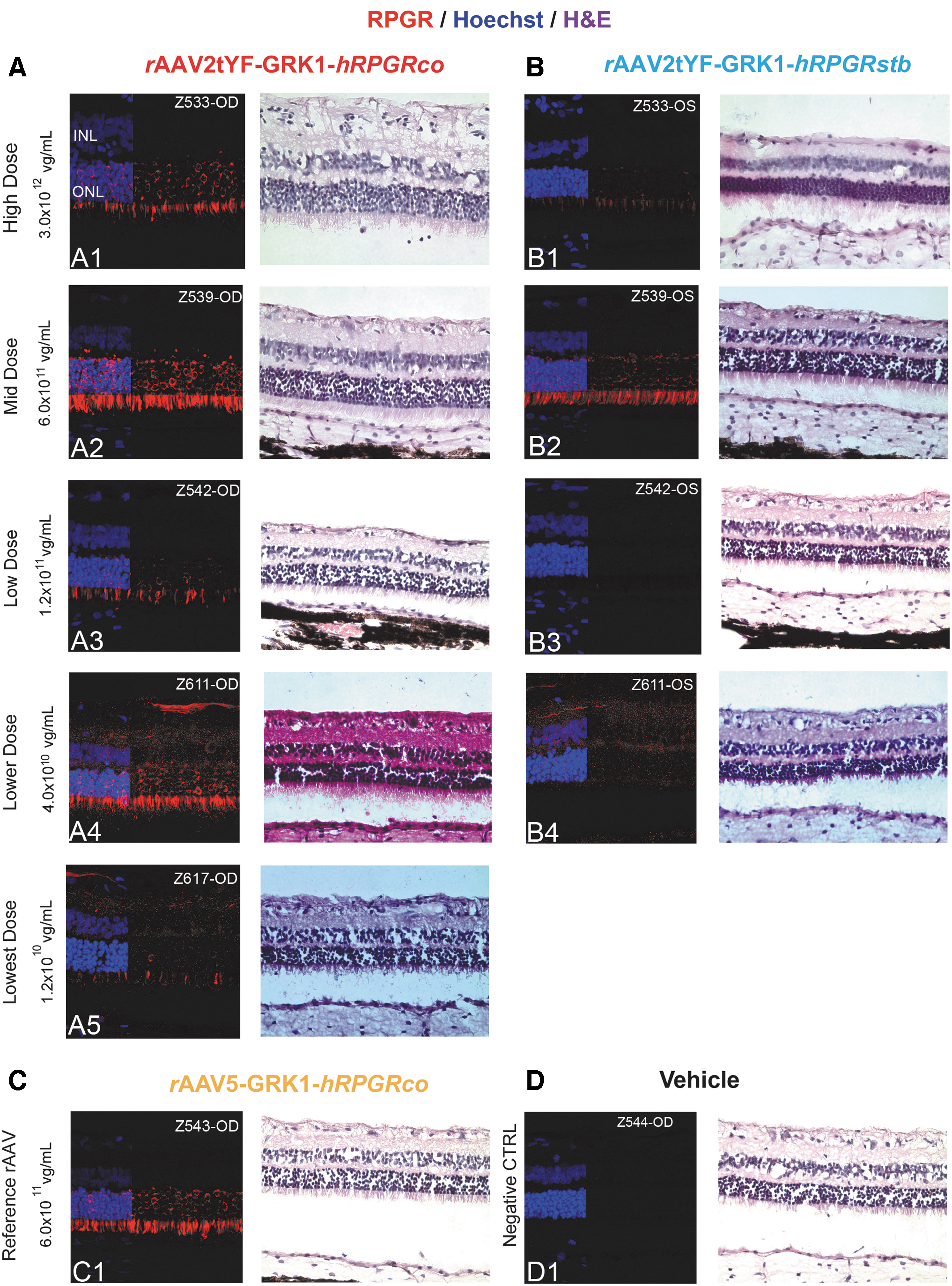
RPGR transgene expression in RPGR mutant dogs. Photomicrographs of RPGR-immunolabeled retinal cryosections from the treated areas of eyes injected with different doses of:
Quantitative assessment of RPGR immunostaining found that the expression level of RPGR was higher in the eyes treated with rAAV2tYF-GRK1-hRPGRco than with rAAV2tYF-GRK1-hRPGRstb at all tested doses (Fig. 4). Intensity of RPGR immunolabeling varied without a clear dose–response effect, although a trend was apparent. RPGR labeling was barely detectable in the Low Dose group and absent in the Lower Dose group treated with rAAV2tYF-GRK1-hRPGRstb. The level of RPGR expression in the eyes treated with reference vector rAAV5-GRK1-hRPGRco was lower than the expression in the eyes treated with the rAAV2tYF-GRK1-hRPGRco at the same dose level (Mid Dose). RPGR protein was not detected in Vehicle dosed retinas.
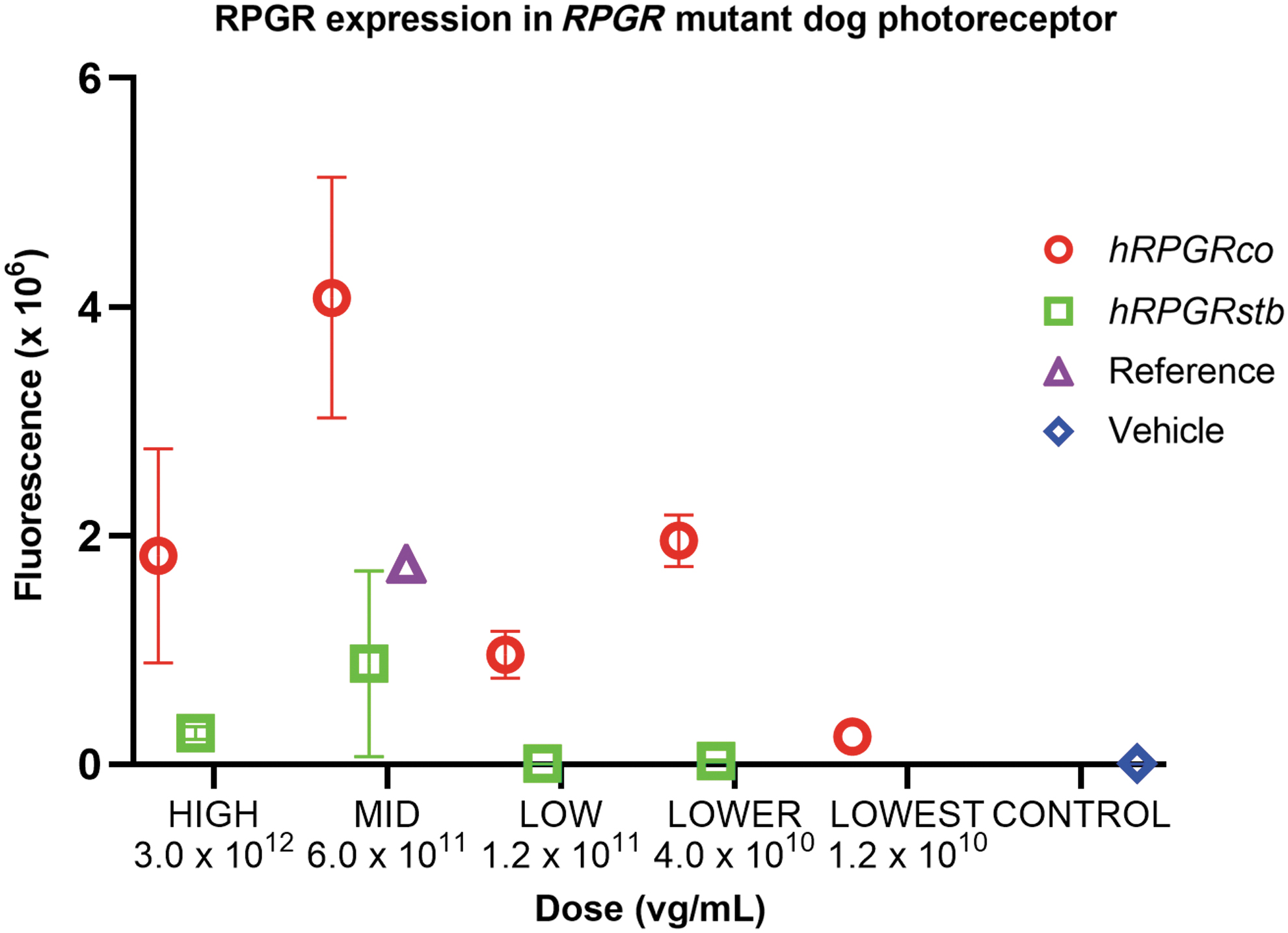
Quantification of RPGR immunolabeling in photoreceptors of RPGR mutant dogs. rAAV2tYF-GRK1-hRPGRco shows higher RPGR expression at all dose levels compared to rAAV2tYF-GRK1-hRPGRstb and AAV5-GRK1-hRPGRco.
Cone photoreceptor morphology
Human cone arrestin immunolabeling on cryosections that were stained and imaged under the same conditions revealed cone labeling in all eyes (including Vehicle dosed eyes) consistent with the persistence of cone photoreceptors at 20 weeks of age in XLPRA2 mutant dogs (Fig. 5). In High and Mid Doses of both vectors and reference vector treated eyes, the cone IS were elongated and normal in appearance (Fig. 5, A1, A2, B1, B2, and C1). The eyes treated with Low, Lower, and Lowest Dose vector and Vehicle had slightly shorter cone IS (Fig. 5, A3–A5, B3–B5), which are consistent with the XLPRA2 disease progression. In several eyes, the cone outer segments were disrupted due to retinal detachment (observed clinically and confirmed by OCT imaging in the High Dose treatment group) or due to artifactual retinal separation during tissue processing and sectioning.
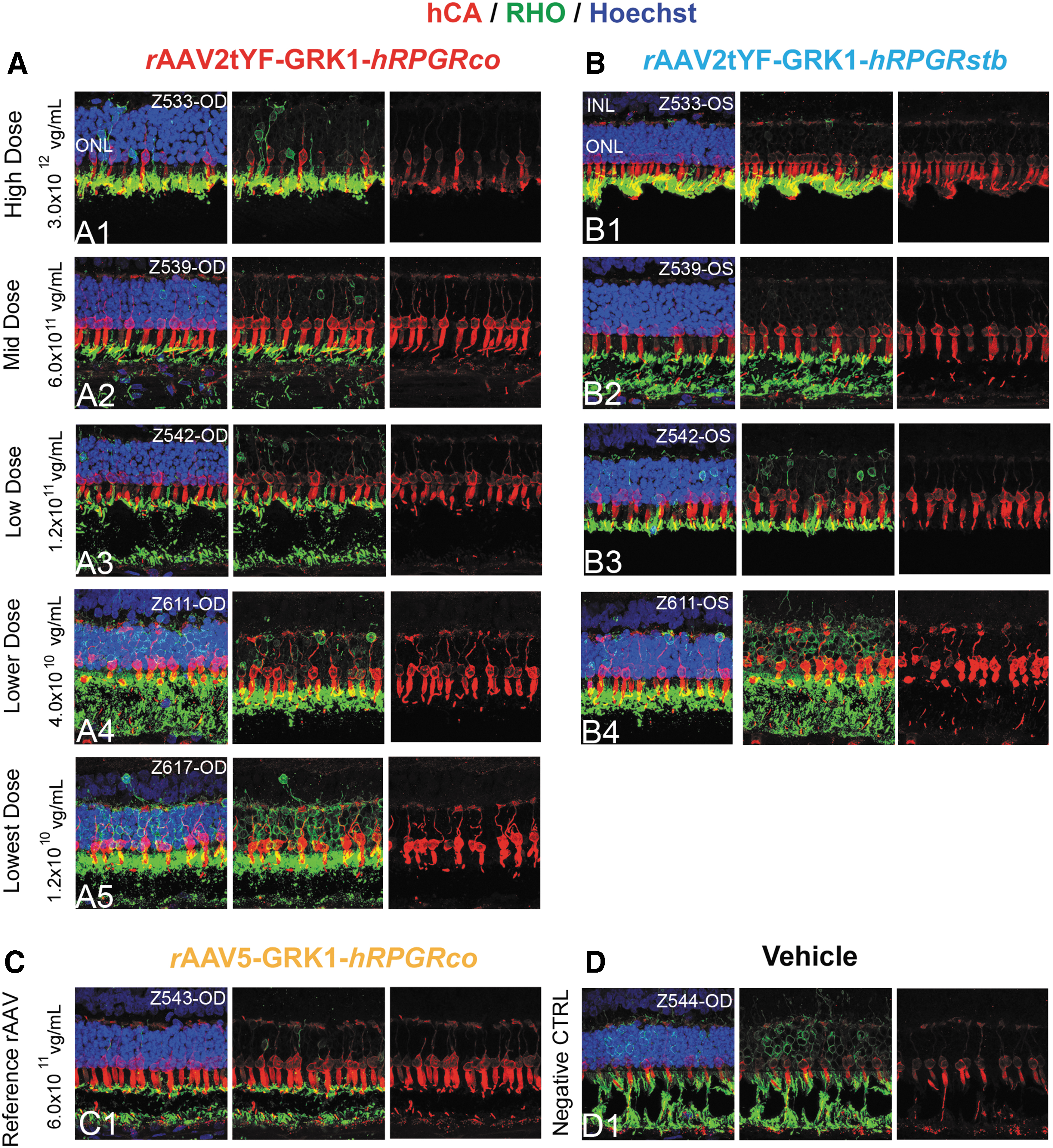
hCA and RHO immunolabeling in RPGR mutant dogs. Photomicrographs of hCA (red) and RHO (green) co-immunolabeled retinal cryosections from the treated areas of eyes injected with different doses of:
Correction of rod opsin mislocalization
Rod opsin (RHO) immunolabeling on cryosections (Fig. 5) revealed variable degrees of correction of RHO mislocalization in the High, Mid, and Low Dose of the rAAV2tYF-GRK1-hRPGRco or rAAV2tYF-GRK1-hRPGRstb-treated eyes. With the exception of one dog (Z534 not shown), immunolabeling of RHO in High Dose treated animals was primarily located in the rod outer segment compared to the Vehicle treated eyes that showed RHO labeling throughout the entire rod photoreceptor cell (rod outer and inner segments and ONL) (Fig. 5D). The Lower and Lowest Doses did not reveal correction of RHO mislocalization in any of the rAAV2tYF-GRK1-hRPGRco or rAAV2tYF-GRK1-hRPGRstb-treated eyes (Fig. 5, A4, A5, B4). Similar to Vehicle treated eyes, RHO was localized throughout the entire rod photoreceptor cells.
Correction of M/L cone opsin mislocalization
M/L cone opsin immunolabeling on cryosections (Fig. 6) revealed correction of M/L opsin mislocalization in all eyes treated with High, Mid, and Low and in half of the eyes treated with Lower Dose of rAAV2tYF-GRK1-hRPGRco or rAAV2tYF-GRK1-hRPGRstb, with labeling found exclusively in the cone outer segments. In eyes treated with the Lowest Dose of rAAV2tYF-GRK1-hRPGRco, correction of M/L opsin mislocalization was reduced and only seen partially in cones. All Vehicle dosed eyes showed no correction of M/L cone mislocalization, which was indicated by labeling throughout the cone photoreceptor cell (Fig. 6D).
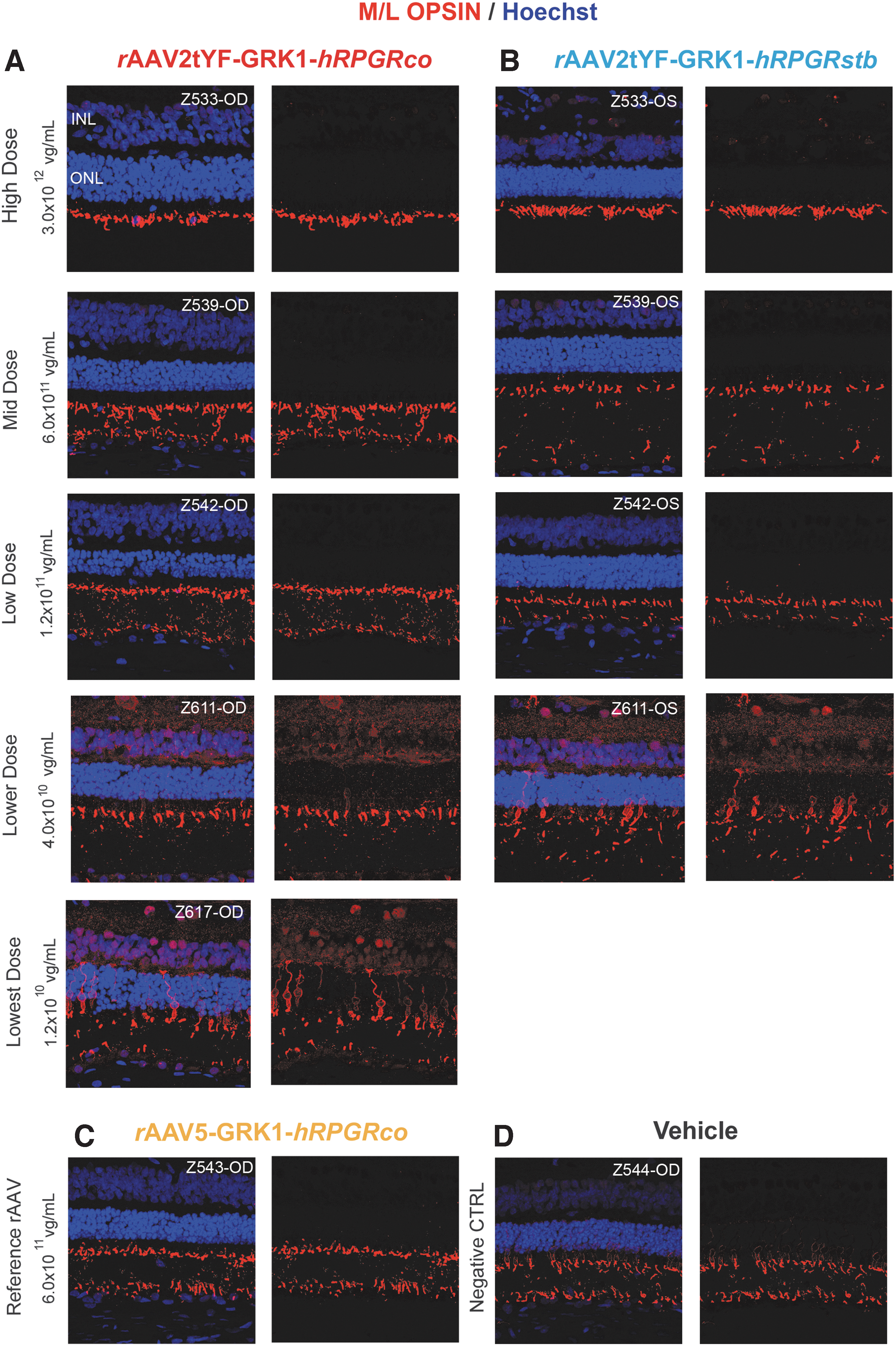
M/L cone opsin immunolabeling in RPGR mutant dogs. Photomicrographs of M/L opsin (red) immunolabeled retinal cryosections from the treated area of eyes injected with different doses of:
Correction of S cone opsin mislocalization
Despite nonspecific extensive punctate background labeling with the S cone opsin antibody, immunolabeling on cryosections (Supplementary Fig. S5) revealed persistent mislocalization of S opsin in all the rAAV2tYF-GRK1-hRPGRco or rAAV2tYF-GRK1-hRPGRstb-treated eyes, with labeling throughout the cone photoreceptor cell. Although correction was incomplete, the High and Mid Dose rAAV2tYF-GRK1-hRPGRco showed partial correction of mislocalization (Table 4). Correction of mislocalization of S cone opsin was absent in the eye injected with reference vector (rAAV5-GRK1-hRPGRco) or Vehicle.
Histopathological signs of retinal toxicity
Retinal perivascular infiltration was observed in treated areas of all eyes dosed with the High Dose of both rAAV2tYF-GRK1-hRPGRco and rAAV2tYF-GRK1-hRPGRstb vectors (Fig. 1C, I). Perivascular infiltration was also observed in one of two eyes dosed with a Mid Dose of rAAVtYF-GRK1-hRPGRco (Supplementary Fig. S6B). Cellular infiltration around blood vessels was observed within the treated area and expanded into the inner retina in areas devoid of blood vessels. IHC with cell-specific markers showed that these infiltrates consisted primarily of cytotoxic T (CD8+), B (CD20+), and helper T (CD4+) lymphocytes (Fig. 1D–F, J–L and Supplementary Fig. S6C–E, H–J). No signs of cellular inflammation were observed at any other dose of either vector.
Discussion
To develop an effective AAV-based gene therapy product for RPGR-XLRP treatment, each component of the therapeutic vector, including capsid, promoter, transgene, and other regulatory elements, needs to be optimized. Previously we reported GRK1 to be an efficient promoter in NHP rod and cone photoreceptor cells. 17 In addition, we reported comparable preservation of photoreceptors in canine eyes treated with GRK1 driven hRPGRstb or hRPGRco delivered by AAV5 at a single dose (7.2 × 10¹¹ vg/mL). 17 In this study we expanded comparison of potency and efficacy of the two transgenes packaged in rAAV2tYF vector under control of the GRK1 promotor in a dose–response (0.15 mL at 1.2 × 10¹⁰–3 × 10¹² vg/mL) study in XLPRA2 dogs. We used the outcome to inform the selection of construct and dose levels to move forward to GLP safety study and a human clinical trial (NCT03314207). 22
Comparison of two hRPGR transgenes for efficacy and gene expression in RPGR-mutant dogs
hRPGRco is a stabilized full length hRPGR cDNA maintaining the wild-type amino acid sequence of the endogenous hRPGR protein. In contrast, hRPGRstb is a shortened version of hRPGR cDNA with several amino acid changes. 21 Earlier data comparing the hRPGRstb and hRPGRco transgenes delivered by an AAV5 vector at a single dose level in RPGR-mutant dogs showed a similar level of efficacy for both transgenes (ONL rescue, correction of mislocalization, and structural preservation of rods and cones). 17 In this study we performed a direct comparison of efficacy using rAAV2tYF-hRPGR at 5 dose levels, ranging from Lowest Dose (0.15 mL at 1.2 × 10¹⁰ vg/mL) to High Dose (0.15 mL at 3.0 × 10¹² vg/mL) (Tables 2 and 4). In these two DRF studies, we found the hRPGRco and hRPGRstb transgenes to show similar levels of rescue by fundus reflectivity, ONL thickness, correction of opsin mislocalization, and length of cone IS at all dose levels with some exceptions where rAAV2tYF-GRK1-hRPGRco showed stronger correction.
Rescue assessed by fundus reflectivity was noted in all eyes treated with either rAAV2tYF-GRK1-hRPGRco or rAAV2tYF-GRK1-hRPGRstb at Mid, Low, Lower Doses and in one of two eyes treated with the Lowest Dose with hRPGRco. Eyes administered High Dose revealed rescue in ocular examinations up to week 4 postdose. However, rescue was masked by signs of toxicity at week 8. Quantitative analysis of ONL thickness in treated versus untreated retina showed rescue of the ONL in treated retina at the Lower (39–47% of WT) and Lowest Doses (50–52% of WT, Fig. 2). In vehicle treated or untreated retinas, ∼60% of the ONL is lost over 20 weeks of XLRP progression (40% of WT). 12,17
Partial or complete correction of both rod opsin mislocalization was observed in all animals in High, Mid, and Low Dose groups. Partial or complete correction of both rod and M/L cone opsin mislocalization was observed in all animals in all dose groups. Both AAV2tYF constructs showed better correction than the AAV5 reference construct. Partial correction of S cone opsin mislocalization was seen in the High and Mid Dose rAAV2tYF-GRK1-hRPGRco groups and in one animal in the High Dose hRPGRstb group. Incomplete correction could be due to the inefficient expression of the transgene in canine S cones driven by the human GRK1 promoter. Although the AAV2tYF capsid was effective in transducing S cones in NHP and canine retinas, 24,28 and a human GRK1 promoter was effective in NHP foveal cones, the activity of the GRK1 promoter in canine cones might be less optimal in agreement with earlier published work, 29 where GFP expression was not detected in cones of wild-type dogs dosed with rAAV5-GRK1-GFP. This finding was consistent with Weiss et al., 30 which showed absence of protein expression driven by GRK1 promoter in canine cones. However, the GRK1 promoter driving GFP or therapeutic transgene in dogs has shown expression in cones and corrected some S cone opsin mislocalization at higher dose of 7.2 × 10¹¹ vg/mL. 17 Taken together, these results suggest that the GRK1 promoter is able to drive expression in canine S cones but less effectively than in rods and M/L-cones. Both AAV2tYF constructs showed better correction than the AAV5 reference construct.
RPGR expression levels as assessed by IHC were higher in eyes treated with rAAV2tYF-GRK1-hRPGRco than with rAAV2tYF-GRK1-hRPGRstb at all dose levels (Table 4 and Figs. 3 and 4). RPGRco was expressed at all dose levels, while RPGRstb was expressed at only High and Mid Doses. There was no RPGRstb expression at the Low and Lower Dose Levels. AAV2tYF-RPGRco expression was higher than AAV5-RPGRco expression at the same Mid Dose level.
Safety analysis
Subretinal dosing of either rAAV2tYF-GRK1-hRPGRco or rAAV2tYF-GRK1-hRPGRstb vectors did not result in discomfort with the exception of one animal that had a narrowed palpebral fissure (day after dosing, thus likely due to surgery). Retinal detachment and inflammation in the treated area were found only in the High Dose group for both vector constructs (0.15 mL at 3 × 10¹² vg/mL). Minimal histological signs of inflammation (without retinal detachment) were noted in the Mid Dose group treated with rAAV2tYF-GRK1-hRPGRco (Supplementary Fig. S4). No clinical signs of inflammation or retinal detachment were observed in any other dose groups.
In summary, we compared two rAAV vector constructs with different transgenes in extensive DRF studies in a canine model of RPGRORF15 -XLRP and showed that the rAAV2tYF-GRK1-hRPGRco construct results in higher levels of RPGR transgene expression than that achieved with rAAV2tYF-GRK1-hRPGRstb or AAV5-GRK1-hRPGRco. In addition, we also showed that while the Mid Dose (0.15 mL at 6.0 × 10¹¹ vg/mL) of the vector led to optimal correction of disease attributes, structural and functional rescue of photoreceptors was also achieved when treating at mid-stage disease with rAAV2tYF-GRK1-hRPGRco at the Lowest Dose (1.2 × 10¹⁰ vg/mL), thus significantly expanding its therapeutic index and guiding a starting dose for a clinical trial. Overall, results of these studies support the selection of rAAV2tYF-GRK1-hRPGRco for further clinical development.
Footnotes
Author Disclosure
C.S., G.Y., J.A.N., A.M.T., P.M.R., D.R.K., J.D.C., and M.S.S. are or were employees and shareholders of AGTC and have a conflict of interest to the extent that this work potentially increases their financial interests. C.S., G.Y., and J.D.C. coauthors have left AGTC.
Funding Information
This work was supported by a Sponsored Research Agreement from AGTC, Inc. to the University of Pennsylvania and research grants from NIH/National Eye Institute R01-EY06855, P30-EY001583 and from the Foundation Fighting Blindness.
Supplementary Material
Supplementary Figure S1
Supplementary Figure S2
Supplementary Figure S3
Supplementary Figure S4
Supplementary Figure S5
Supplementary Figure S6
Supplementary Table S1
Supplementary Table S2
Supplementary Table S3
Supplementary Table S4
References
Supplementary Material
Please find the following supplemental material available below.
For Open Access articles published under a Creative Commons License, all supplemental material carries the same license as the article it is associated with.
For non-Open Access articles published, all supplemental material carries a non-exclusive license, and permission requests for re-use of supplemental material or any part of supplemental material shall be sent directly to the copyright owner as specified in the copyright notice associated with the article.