
Abstract
Virus-targeted therapy for tumors can effectively prolong the survival rate of patients and has become a new trend for cancer biotherapy. Oncolytic adenovirus (OAd) can specifically replicate in tumor cells, allowing the therapeutic genes carried to be rapidly copied. As known, solid tumors are always hypoxic, and researchers often overlook a key point, the replication of OAd depends not only on its own activity but also on the cellular hypoxic environment in which the virus replicates. In this study, we constructed an OAd carrying Decorin, HRE-Ki67-Decorin, combining the Ki67 promoter upstreamed with hypoxia-response element (HRE) sequences to drive adenoviral E1A. The OAd HRE-Ki67-Decorin had better replication ability under hypoxic conditions, downregulated cellular immunosuppressed growth factor TGF-β. In addition, HRE-Ki67-Decorin was potent in suppressing tumor growth and participated in the assembly of tumor extracellular matrix by expressing Decorin in subcutaneous renal cancer cell tumor models. Tumor sections from HRE-Ki67-Decorin-treated tissues had less collagen fibers and more spread of virus among tumor tissues. These results indicated that chimeric HRE-Ki67 promoter-regulated OAd carrying Decorin might be an effective anticancer treatment strategy.
Introduction
Renal cell carcinoma, which originated from renal parenchymal tubular epithelial cells, is a common malignant tumor of the urinary system, accounting for 3–5% of new cancer cases per year. 1 Due to the overexpression of multiple drug resistance genes in renal cell carcinoma, conventional chemotherapy is not effective, and the median survival time of patients with terminal cancer is only 12 months after chemotherapy, which is not sensitive to radiotherapy. At present, high doses of interleukin-2 (IL-2) or alpha interferon (IFN-α) are first-line drugs in the treatment of nonspecific immunotherapy for renal cell carcinoma, but their clinical application results show low remission rate, only a few patients obviously benefit. 2
Recently, oncolytic virus immunotherapy is a class of therapeutic agent that has recently received more attention since the FDA approval in 2015 of a genetically modified herpes simplex virus, type 1 talimogene laherparepvec (T-VEC; Imlygic). 3 Oncolytic adenovirus (OAd) is currently one of the most widely used carriers. OAd has been genetically engineered to selectively replicate in tumor cells, lyses tumor cells, and releases progeny viruses to infect other cancer cells. 4 In 1996, Onyx reported that the adenovirus Onyx-015, which deleted the E1B55KD protein-encoding gene, replicated only in tumor cells without affecting normal cells. 5 To further improve anticancer activity of OAd, many researchers had used the tumor-specific promoter to regulate the adenovirus E1A gene to develop novel OAds. 6 E1A is the first gene transcribed in OAd-infected cells and plays a key role in viral replication. Using the tumor-specific promoter to regulate E1A expression, OAd can be controlled to proliferate in tumor cells and had high safety. 7
Our previous research showed that a 253-bp-length Ki67 core promoter was a highly specific promoter, with low activity in normal cells and high activity in tumor cells. 8 We found that Ki67 promoter-regulated OAd had good effect on inhibiting tumor cells in vitro, but was less efficient in animal experiments. It had been reported that adenoviral replication was not only determined by the activity of the promoter itself but also by the activity of the promoter in the cellular environment while the virus replicated in Hedjran et al. 9
Although mammalian tissues exist at 2% to 9% oxygen (O2) conditions, tumor environment is known to be hypoxic, exhibiting a median O2 level of 1.3%. Hypoxia is a common feature of solid tumors, including renal carcinoma. To augment the activity of the promoter integrated into OAd, several tumor-selective control elements were incorporated. Hypoxia-inducible factors (HIFs) bound to hypoxia-responsive elements (HRE) to modulate target gene transcription in tumor cells. 10 Kwon et al. integrated HRE into the alpha-fetoprotein promoter to regulate E1A transcription and improved the ability of the virus to replicate in liver cancer cells. 11
Decorin is a leucine-rich proteoglycan that was first discovered in collagen fibers and performs multiple functions on stromal and epithelial cells. 12 There is increasing evidence that Decorin is downregulated in a variety of human tumors and plays a wide range of biological roles in tumor cells, including proliferation, metastasis, and angiogenesis. Recent studies have shown that Decorin may affect the proliferation of A549 and promote the expression of p53 and p21 by decreasing the expression of transforming growth factor-β1 (TGF-β1) and cyclin D1. 13 However, few articles have reported the effects of Decorin on renal cell carcinoma cells, and the mechanism by which Decorin genes affect metastasis remains unknown.
In this study, we introduced a replication-competent oncolytic adenovirus regulated by chimeric HRE and 253-bp long Ki67 core promoter and expressing Decorin, HRE-Ki67-Decorin. Our study suggested that this genetically engineered adenovirus promoted virus replication under hypoxia and participated in the assembly of tumor extracellular matrix (ECM) by expressing Decorin in subcutaneous renal cancer cell tumor models. Ultimately, HRE-Ki67-Decorin mediated a markedly enhanced antitumor activity for renal cancer cells.
Materials and Methods
Cell culture
Renal epithelial cell 293, human normal renal tubular epithelial cell HK-2, and human renal cell adenocarcinoma ACHN cells (Chinese Academy of Sciences Shanghai Branch Cell Bank) were cultured in Dulbecco's modified Eagle's medium supplemented with 10% (v/v) fetal bovine serum (FBS) and 1% (v/v) penicillin/streptomycin at 37°C in a 5% CO2 incubator. Clear cell renal cell carcinoma OSRC-2 cells were cultured with RPMI-1640 containing 10% FBS and 1% (v/v) penicillin/streptomycin at 37°C in a 5% CO2 incubator. Hypoxia was achieved using prewarmed aluminum hypoxic chambers evacuated through the use of 94% N2/5% CO2. Oxygen concentration was controlled to the desired level of 1%.
Adenoviruses construction
The constructed shuttle plasmids pCA13-Decorin, pKi67-EGFP, pHRE-Ki67-EGFP pKi67-Decorin, and pHRE-Ki67-Decorin were co-transfected into HEK-293 cells with adenoviral cytoskeleton plasmid pBHGE3 (Microbix Biosystems Inc.) through Lipofectamine 2000 (Invitrogen) according to manufacturer's instructions. HEK293 cells were infected with adenovirus Ad-Decorin, Ki67-EGFP, HRE-Ki67-EGFP Ki67-Decorin, and HRE-Ki67-Decorin with a density of about 80%; when half of the cells were floated, the virus was collected and the titer was measured by tissue culture infection dose 50 (TCID50) methods.
Fluorescence microscope and flow cytometry to detect the expression of fluorescent proteins
Normal renal epithelial cell HK-2 and renal cell carcinoma cells OSRC-2 and ACHN were infected with Ki67-EGFP and HRE-Ki67-EGFP under normoxic condition (21% O2) and hypoxic condition (1% O2) for 24 h at multiplicity of infection (MOI) = 20. Cells were checked under fluorescence microscope. The three different cells infected with Ki67-EGFP or HRE-Ki67-EGFP under normoxia and hypoxia were also collected for flow cytometry.
RNA isolation, reverse transcription, and quantitative real-time PCR
HK-2, OSRC-2, or ACHN cells were plated in six-well plates and for different experiments. Total cellular RNA was extracted using 1 mL of TRIzol reagent (Invitrogen) per well in six-well plates. Five hundred nanograms RNA was used for cDNA synthesis with HiScript® Reverse Transcriptase reagent Kit (Vazyme biotech, China). Samples were analyzed in triplicate by the Applied Biosystems 7500 PCR System. The targeted genes and endogenous housekeeping gene GAPDH were used as normalizing controls and amplified, using the PrimeScript™ RT reagent Kit (TaKaRa). The cycle number (Ct) was calculated and the fold changes of gene expression were determined using the double ΔCt (2−ΔΔCT) method.
Western blot
Proteins were extracted from the cells. An aliquot of 100 μg proteins were separated by 10% SDS-PAGE gel and transferred onto a PVDF membrane. Membranes were incubated with blocking buffer (5% nonfat milk in TBS containing 0.1% Tween-20) for 2 h at room temperature. Thereafter, samples were probed overnight at 4°C with primary antibodies against HIF-1α, vascular endothelial growth factor (VEGF), p53, GAPDH (Santa Cruz), or E1A (Merk millipore). After extensive washing, the membrane was incubated with secondary antibody IgG (Zhongshan, Beijing) for 1h at room temperature. The blotting membranes were visualized by Bio-Rad chemiluminometer.
CCK-8 proliferation experiment
Inoculated cells were infected with Ad-Decorin, Ki67-Decorin, and HRE-Ki67-Decorin at MOI = 1, 10, 20, or 100. Four days after infection, cell proliferation was analyzed by recording the absorbance of the sample at 450 nm using the CCK-8 assay. The survival rate was calculated as follows: survival rate = (experimental well − blank well)/(control well − blank well) × 100%. The final data were the average of three independent experiments.
Virus replication assay
Logarithmically growing renal cancer cells (105 cells/well) were cultured in six-well plates for 24 h. Later, cells were infected with Ki67-Decorin or HRE-Ki67-Decorin at an MOI of 20 under normoxia or hypoxia, respectively, for another 48 h. Then, cells were lysed through three cycles of freeze and thaw. The titers of viral progenies were quantified on HEK-293 cells with the TCID50 method. The replication multiples of adenovirus in the tested cancer cells were equal to viral progeny titers at 48 h, normalized with that at the beginning of infection.
ELISA experiment
OSRC-2 and ACHN cells were infected with Ad-Decorin, Ki67-EGFP, Ki67-Decorin, HRE-Ki67-EGFP, or HRE-Ki67-Decorin at MOI = 20 for 24 h. The culture medium was removed, and the 600 μL serum-free culture medium was added for 24 h. Then, the supernatant of each well collected was centrifuged at 4,000 rpm for 5 min to take the supernatant as samples. TGF-β1 or Decorin expression was determined by ELISA according to the manufacturer's protocol for TGF-β ELISA kits (Dakewe, China) or Decorin (Abcam).
Animal experiment
Thirty female 4-week-old BALB/c nude mice were purchased from Beijing Vital River Laboratory Animal Technology. Animal welfare and laboratory procedures were performed in strict accordance with the Guide for the Care and Use of Laboratory Animals. In addition, all animal experiments were approved by the Animal Care Committee of the Xuzhou Medical University, Xuzhou, China. ACHN-luciferase renal cell carcinoma cells (5 × 10⁶/mouse) were subcutaneously injected into the right side of the nude mice. When the tumor diameter reached 3–5 mm, 1 × 10⁹ pfu of Ad-Decorin, Ki67-EGFP, Ki67-Decorin, HRE-Ki67-EGFP, or HRE-Ki67-Decorin, or an equal volume of PBS was injected into the tumor. Tumors were measured once a week and their volume was calculated. Three mice from different groups were sacrificed on day 7 after administration and genomic DNA was extracted from tumor tissues and livers; real-time PCR was used to detect the content of viral genome.
Immunohistochemistry staining analysis
The tumor tissue slides were fixed in 10% formalin, embedded in paraffin, and cut into 3-mm sections. Deparaffinized tumor sections were treated with primary antibody for Decorin, hexon, or Collagen Type I. After incubation with a secondary antibody, tissue sections were then counterstained with hematoxylin. Masson's trichrome staining was used to analyze the distribution of collagen fibers (stained blue color). Representative sections were stained with Masson's trichrome and then observed by light microscopy. The collagen fiber area was analyzed with Image-Pro Plus software.
Statistical analysis
Data are represented by the mean ± standard deviation. Experimental data were processed by SPSS16.0 statistical software. Statistical significance was defined as a p value <0.05.
Results
OAd driven by HRE suppressed renal cell carcinoma under hypoxic conditions
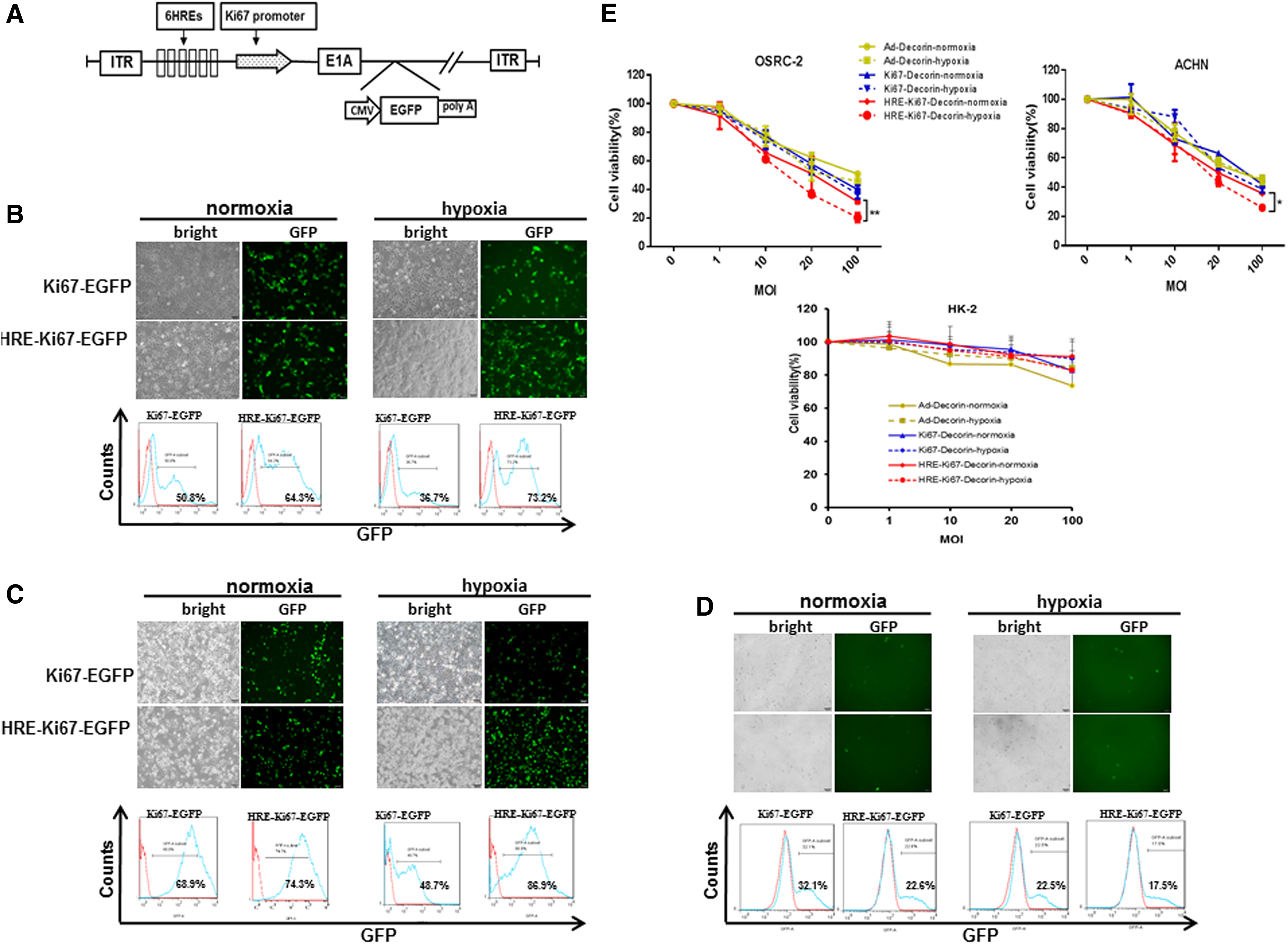
To investigate whether the OAd driven by a chimeric promoter combined HRE with Ki67 promoter has a therapeutic effect on renal cell carcinomas, we constructed a replication-competent type V adenovirus HRE-Ki67-EGFP with six additional HRE sequences based on the previous constructed OAd Ki67-EGFP with a reporter gene EGFP (Fig. 1A).
Oncolytic adenovirus driven by HRE suppressed renal cell carcinoma under hypoxic conditions.
Two renal cancer cells ACHN and OSRC-2 and normal renal tubular epithelial cell HK-2 were infected with Ki67-EGFP or HRE-Ki67-EGFP under normoxia and hypoxia, respectively. The infection efficiency of the virus for cells was analyzed under fluorescence microscope observation and flow cytometry by detecting the expression of EGFP. As shown in Fig. 1B–D, there was no significant difference between ACHN and OSRC-2 under normoxic condition. Under hypoxic condition, both renal cancer cells had higher EGFP expression in the HRE-Ki67-EGFP group than that in the Ki67-EGFP group. Moreover, Ki67-EGFP had less EGFP expression under hypoxia treatment than that under normoxia. The expression of EGFP in normal renal tubular epithelial HK-2 was significantly lower than that in renal cancer cells under normoxia and hypoxia. It is suggested that HRE can enhance the expression of EGFP under hypoxic conditions in the two renal cancer cells, but had less efficiency in normal cell HK-2.
Excessive accumulation of tumor ECM is one of main contributors to drug resistance in renal cell carcinomas. To overcome desmoplasia of renal cancer cells, we utilized three different adenoviruses expressing Decorin (a nonreplicative adenovirus Ad-Decorin and two replication-competent adenoviruses Ki67-Decorin and HRE-Ki67-Decorin, which were constructed through replacing EGFP with Decorin in Ki67-EGFP or HRE-Ki67-EGFP plasmid). Next, we examined the inhibitory effect of these adenoviruses on renal cancer cells under normoxic and hypoxic conditions. As shown in Fig. 1E, oncolytic capabilities of Ki67-Decorin and HRE-Ki67-Decorin were confirmed in two renal cancer cells (ACHN and OSRC-2) and were dose dependent. Ad-Decorin only had modest cytotoxic effect at high MOI (MOI, 100) on renal cancer cells. In addition, HRE-Ki67-Decorin showed better inhibition efficiency under hypoxic conditions than that under normoxic conditions. However, survival rate did not change significantly in HK-2 cells infected with three viruses under normoxic or hypoxic conditions. These data suggest that the oncolytic HRE-Ki67-Decorin induced a potent antitumor response for renal cancer cells under hypoxic conditions.
Hypoxia regulated the expression of cellular HIF-1α, VEGF, and p53
Under hypoxic conditions, the HIF-1α binds to the hypoxia-response element (HRE), which induces the expression of downstream target genes, VEGF and p53. 14 As showed above, HRE-Ki67-Decorin showed obvious inhibition effect in tumor cells, but not in normal cells. Then, we aimed to detect the expression of hypoxia-related proteins HIF-1α and VEGF in renal cancer cells ACHN and OSRC-2 and normal HK-2 cells. These cells were treated under hypoxia for 0, 0.5, or 4 h, and quantitative PCR was used to detect the mRNA expression of HIF-1α and VEGF. The results showed that HIF-1α and VEGF mRNA level were significantly downregulated compared with the control cells (Fig. 2A upper). Cellular HIF-1α and VEGF mRNA level under normoxia were also detected; as shown in Fig. 2B lower, HIF-1α and VEGF mRNA expression level had no significant difference with increasing incubation time. To detect the post-transcriptional changes of hypoxia-related genes, ACHN, OSRC-2, and HK-2 were treated under hypoxia for 0, 0.5, 1, 2, and 4 h, and Western blotting assay was performed to detect the expression of HIF-1α and its downstream target genes VEGF and p53. Interestingly, the results showed that HIF-1α, VEGF, and p53 protein were all upregulated time dependently (Fig. 2B). These data showed that the mRNA of HIF-1α and VEGF was reduced, but the proteins were increased under hypoxia. Our data also indicated that hypoxia can modulate the related protein expression both in tumor cells and normal cells. The absence of HRE-Ki67-Decorin-mediated oncolytic capabilities in normal cells was due to the chimeric adjuster with hypoxia-response element (HRE) and Ki67 promoter.

Hypoxia regulated the expression of cellular HIF-1α, VEGF, and p53.
HRE enhanced the replication ability of OAd under hypoxic condition in renal cancer cells
The E1A gene is the first viral gene to be transcribed after adenovirus transfecting into tumor cells, and the E1A protein plays a key role in viral replication. 15 Ad-Decorin, Ki67-Decorin, or HRE-Ki67-Decorin was added to infect ACHN or OSRC-2 with MOI = 20 under hypoxia for 0.5, 1, or 4 h, respectively. The quantitative PCR assay was performed and the results showed that E1A mRNA was significantly upregulated in renal cancer cells treated with HRE-Ki67-Decorin or Ki67-Decorin compared to Ad-Decorin, in which there was no E1A expression (Fig. 3A). ACHN, OSRC-2, and HK-2 cells were treated the same method as mentioned above. Total cell lysates were harvested for Western blot assay. The results showed that E1A protein was increased largely in HRE-Ki67-Decorin-infected renal cancer cells compared with Ad-Decorin and Ki67-Decorin, whereas in HK-2 cells, the expression of E1A protein was little or even absent (Fig. 3B). Next, TCID50 assay was used to determine the virus particles with infectivity. HK-2, ACHN, and OSRC-2 were infected with Ki67-Decorin or HRE-Ki67-Decorin with MOI = 20 under normoxia and hypoxia, respectively, for 48 h. The cells were lysed to release virus and the supernatant was collected for the next assay. As shown in Fig. 3C, there was no significant difference in the replication ability of Ki67-Decorin and HRE-Ki67-Decorin in renal cancer cells under normoxic conditions. However, under hypoxia, HRE-Ki67-Decorin has significantly improved replication ability compared to Ki67-Decorin. In addition, there was no significant difference in replication of these two adenoviruses in HK-2 cells under normoxia or hypoxia. The above results indicated that the recombinant adenovirus can selectively replicate in renal cancer cells without affecting normal cells, and HRE can enhance the replication ability of the oncolytic virus under hypoxia. These results indicated that HRE, as a hypoxic reaction element, enhanced the ability of the virus to replicate under hypoxic conditions.

HRE enhanced the replication ability of oncolytic adenovirus under hypoxic conditions in renal cancer cells.
HRE-regulated recombinant adenovirus carrying Decorin downregulated intracellular TGF-β
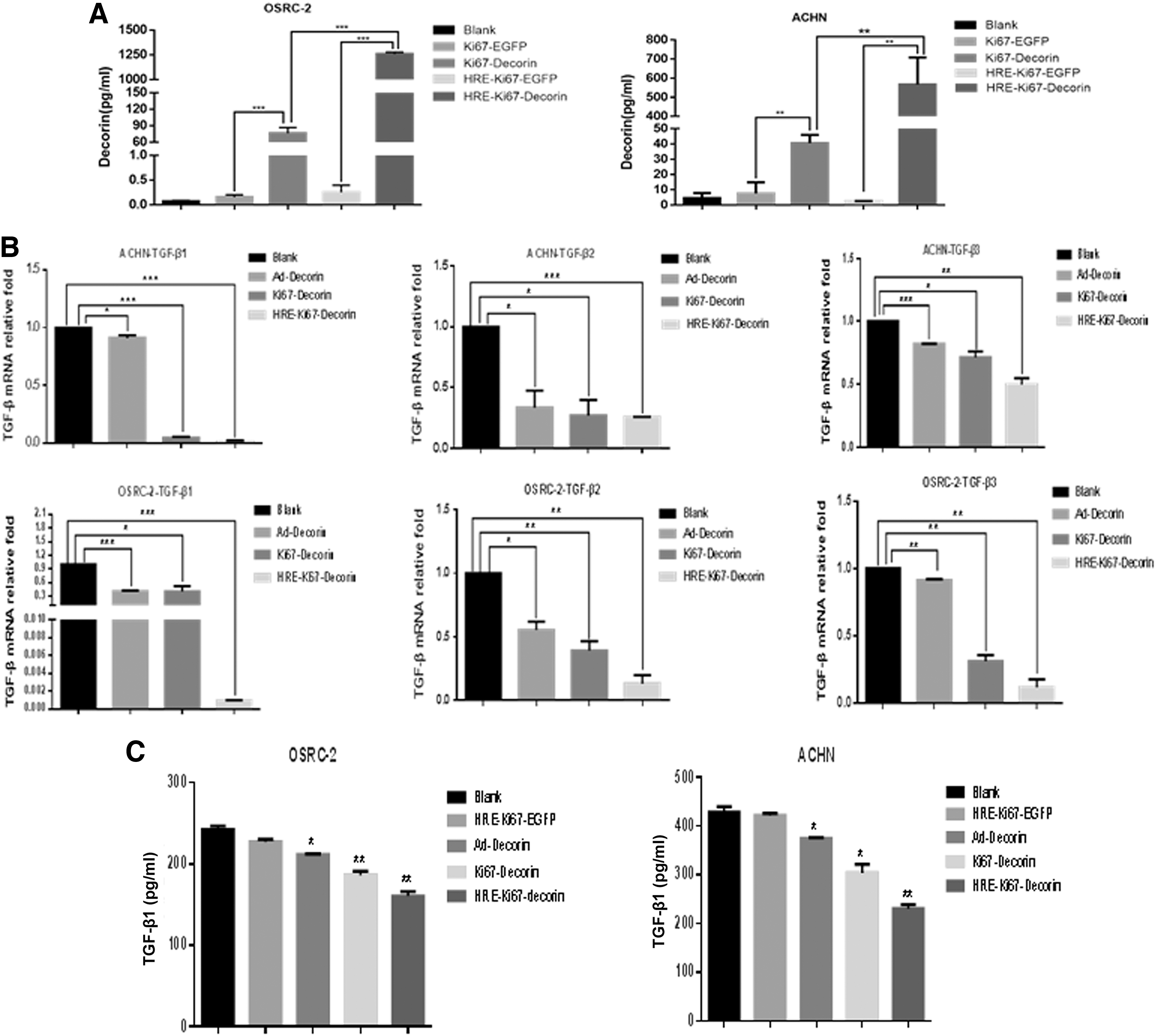
Decorin is a low molecular proteoglycan rich in leucine. It is widely distributed in tissues and can bind to various growth factors and cytokines to regulate cell growth and tissue remodeling. We determined higher secreted Decorin level in renal cancer cells infected with HRE-Ki67-Decorin and Ki67-Decorin than those cells infected with other control viruses under hypoxic conditions. The results showed that Ki67 promoter-mediated OAd expressed Decorin validly and HRE motifs chimericing upstream of Ki67 promoter enhanced viral replication and translation of Decorin gene under hypoxic conditions (Fig. 4A).
HRE-regulated recombinant adenovirus carrying Decorin downregulated intracellular TGF-β.
TGF-β is the first growth factor known to interact with Decorin. 16 To verify Decorin function mediated by HRE-regulated recombinant adenovirus, ACHN and OSRC-2 were infected with Ad-Decorin, Ki67-Decorin, and HRE-Ki67-Decorin under hypoxic conditions with MOI = 20. TGF-β expression was detected by quantitative real-time PCR and ELISA, respectively. The results showed that Decorin mediated by three viruses inhibited the expression of TGF-β1, TGF-β2, and TGF-β3 mRNA in ACHN and OSRC-2. However, HRE-Ki67-Decorin showed a more significant decrease in TGF-β mRNA expression compared to the other two groups (Fig. 4B). We also examined cellular TGF-β protein by ELISA. As shown in Fig. 4C, it showed that in the renal cancer cells, TGF-β was not significantly different between HRE-Ki67-EGFP and control group, while there was less TGF-β protein in cells infected with Ad-Decorin, Ki67-Decorin, and HRE-Ki67-Decorin. Comparing with the Ad-Decorin and Ki67-Decorin groups, HRE-Ki67-Decorin inhibited TGF-β more effectively. These results suggested that HRE-Ki67-Decorin has a significant inhibitory effect on TGF-β under hypoxic conditions.
HRE-regulated recombinant adenovirus carrying Decorin inhibited renal cancer cell growth in vivo
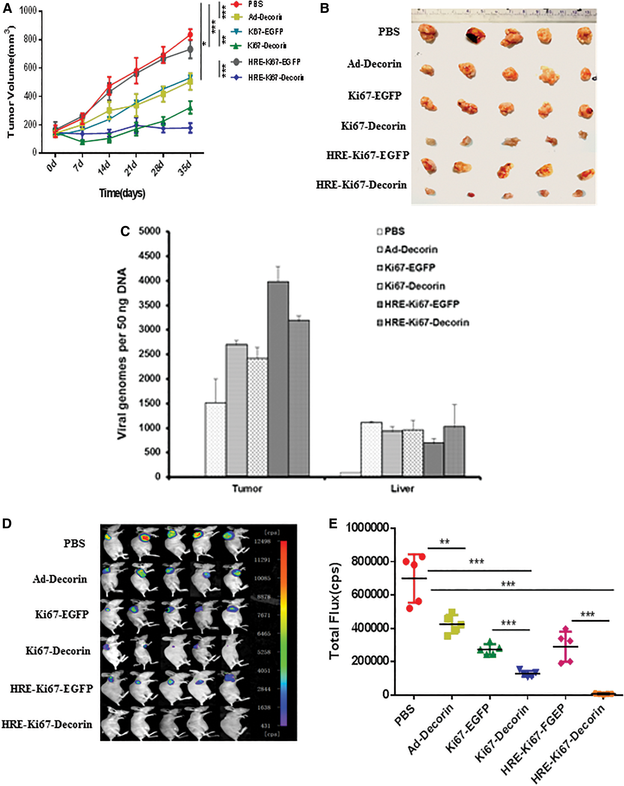
To evaluate the activities of oncolyitc adenovirus carrying Decorin in vivo, we used ACHN-luciferase cells to establish a xenograft model in nude mice. Approximately, 3 × 10⁶ cells were injected subcutaneously in the right flank of the nude mice. We randomly divided 30 tumor-bearing mice into 6 groups. Each mouse was injected with 1 × 10⁹ pfu of Ad-Decorin, Ki67-Decorin, Ki67-EGFP, HRE-Ki67-EGFP, or HRE-Ki67-Decorin. The same volume of saline (PBS) was also used as a control. Compared with the group treated with saline, intratumoral injection of Ad-Decorin, Ki67-Decorin, Ki67-EGFP, HRE-Ki67-EGFP, or HRE-Ki67-Decorin reduced the tumor burden, and HRE-Ki67-Decorin caused a significant decrease in tumor volume (Fig. 5A, B). Moreover, three mice were sacrificed from each group on day 7 after administration and genomic DNA was extracted from tumor tissues and livers; real-time PCR was used to detect the content of viral genome. As shown in Fig. 5C, tumor treated with Ad-Decorin, Ki67-Decorin, Ki67-EGFP, HRE-Ki67-EGFP, or HRE-Ki67-Decorin indeed had high-level viral expression, although there was a modicum viruses that existed in the liver. The fluorescence analysis detected by in vivo imaging system also showed that the tumor volume of the Ad-Decorin, Ki67-Decorin, or HRE-Ki67-Decorin mice was smaller compared with the Ki67-EGFP, HRE-Ki67-EGFP, and PBS groups (Fig. 5D, E). These data indicated that HRE-Ki67-Decorin could induce a sufficient antitumor effect in renal cell carcinoma in vivo.
HRE-regulated recombinant adenovirus carrying Decorin inhibited renal cancer cell growth in vivo.
Recombinant adenovirus carrying Decorin inhibits the expression of collagen fibers in renal cell carcinoma
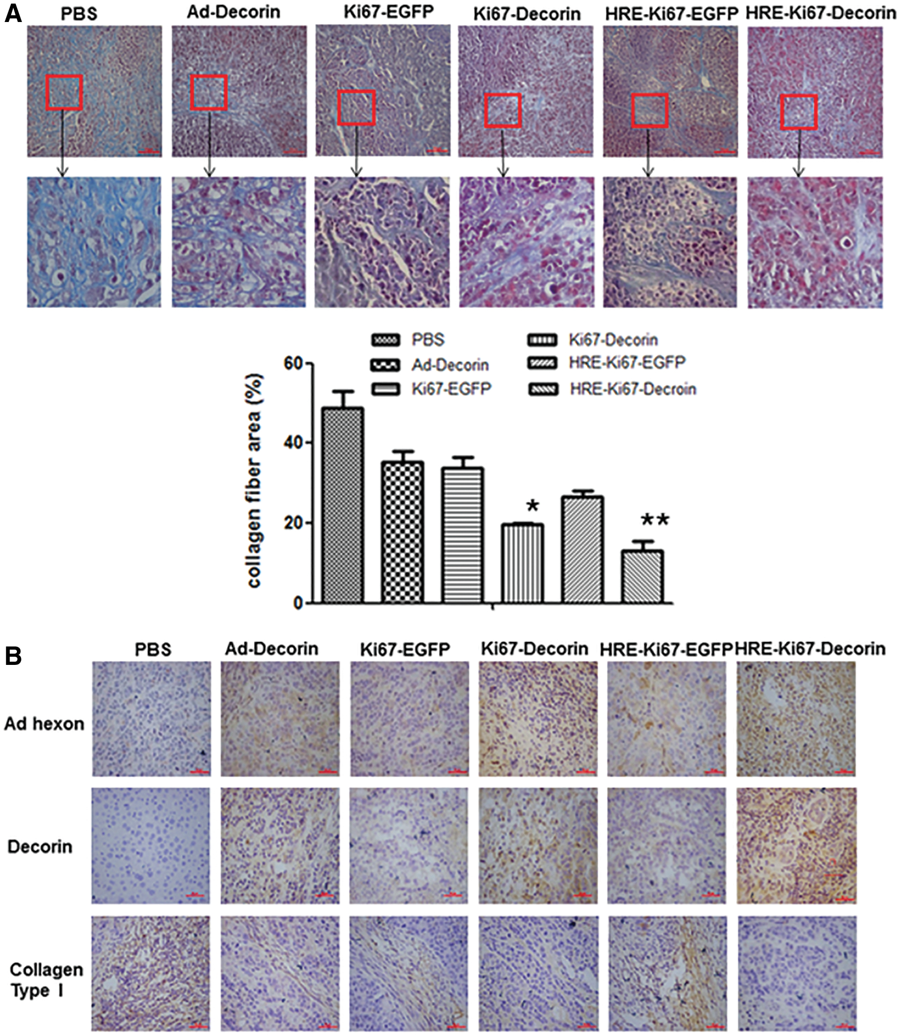
Finally, we aimed to elucidate whether HRE-Ki67-Decorin also effectively remodels the ECM in renal cell carcinoma. At the end of in vivo experiment, the mice were sacrificed and the tumors were removed, fixed in 4% paraformaldehyde, and paraffin embedded to make paraffin sections. The expression of collagen fibers in the ECM was measured by Masson staining. As shown in Fig. 6A, the number of collagen fibers in tumor samples of the Ad-Decorin, Ki67-Decorin, and HRE-Ki67-Decorin treatment groups was significantly reduced compared with the PBS and Ki67-EGFP and HRE-Ki67-EGFP groups, and the collagen fibers became thinner and shorter than the control group (the blue striped substance in the figure was collagen fiber-specific painted). The collagen fiber area was also analyzed with Image-Pro Plus software. Compared to the PBS group, relative collagen fiber area was less in Ki67-Decorin or HRE-Ki67-Decorin group. In addition, we also detected the expression of tumor tissue adenovirus hexon (adenovirus hexon protein), Decorin, and Collagen Type I (type I collagen fibers) by immunohistochemical staining. The results showed that Decorin-positive staining was significantly increased in the tumor tissues of the Ad-Decorin, Ki67-Decorin, and HRE-Ki67-Decorin treatment groups compared with the PBS, Ki67-EGFP, and HRE-Ki67-EGFP groups, and the expression of Collagen Type I is significantly reduced. Meanwhile, the expression of hexon in Ad-Decorin, Ki67-Decorin, and HRE-Ki67-Decorin group was significantly higher than that in the control groups Ki67-EGFP and HRE-Ki67-EGFP, and no positive hexon was observed in the PBS group (Fig. 6B). These data suggested that the replication ability of adenovirus HRE-Ki67-Decorin was better in tumor tissues, and Decorin could downregulate the expression of collagen fibers in tumor tissues and promote the spread of virus in tumor tissues.
Recombinant adenovirus carrying Decorin inhibited the expression of collagen fibers in renal cell carcinoma.
Discussion
It is well known that hypoxia is one of the common features of solid tumors and plays an important role in the development of tumors. 17 Numerous studies had shown that hypoxia can stimulate tumor cell proliferation, induce angiogenesis, accelerate invasion and metastasis, 18 and is important for tumor treatment tolerance. 19,20 The production of the HIF-1α is critical during tumor hypoxia. HIF-1α induces the expression of downstream hypoxia-responsive genes by binding to the HRE, including heme oxygenase and glycolytic enzymes and their related enzyme-glucose transporters, VEGF, erythropoietin, p53, 21 and platelet-derived growth factor β. 22 Therefore, HRE used to regulate target gene expression is an ideal tumor treatment strategy. 23,24 Interestingly, our data showed that HIF-1α and VEGF mRNA were reduced, but the proteins were increased under hypoxia. Similar phenomenon was also reported in other literature. 25 The author explained this paradox might be due to gene allelic copy number loss, and mRNA levels were high in G1 and low in proliferating cells. Moreover, high protein levels protect tumor cells from stress-induced cell death. So eukaryotic translation is not fully coupled with transcription.
The occurrence and development of tumors is a complex process that requires sustained proliferation signals and can activate invasion and distant metastasis. 26 Decorin is a member of the leucine-rich small proteoglycan family and is widely distributed in extracellular matrices and epithelial cells. The increasing evidence showed that Decorin participates in cell growth, differentiation, proliferation, adhesion, and metastasis by interacting with multiple signaling molecules 27 –29 — Decorin is a negative regulator of tumor growth, whether it induces Decorin overexpression in vivo or as a recombinant protein, it can inhibit the proliferation, metastasis, and tumor angiogenesis of tumor cells from different tissues, suggesting that Decorin is likely a tumor suppressor that may become a new antitumor drug. The more detailed mechanism of Decorin is being discovered gradually.
In this study, HRE was integrated as an enhancer into the Ki67 promoter to regulate the transcription of adenoviral E1A gene and Decorin was inserted into the viral genome, and we constructed a novel OAd HRE-Ki67-Decorin. We also used HRE-Ki67-EGFP, which carrying report gene EGFP, to determine the activity of OAd in normal cells and renal cell carcinoma cells under normoxia and hypoxia (Fig. 1). The results showed that under hypoxic conditions, HRE can enhance the replication ability of virus in renal cell carcinoma cells and has lower infection efficiency to normal cells. The CCK-8 assay results showed that Ad-Decorin, Ki67-Decorin and HRE-Ki67-Decorin inhibited renal cancer cells growth, and HRE-Ki67-Decorin had a higher antitumor activity for renal cancer cells under hypoxic conditions (Fig. 1E). Quantitative PCR and Western Blot assay determined the higher level E1A expression under hypoxic conditions, and TCID50 method further confirmed that replication of HRE-Ki67-Decorin was enhanced under hypoxic conditions (Fig. 3). These data suggested that HRE as a hypoxia response element enhanced the replication ability of HRE-Ki67-Decorin in renal carcinoma cells.
TGF-β has a wide range of biological activities as a multipotent cytokine. 30 When TGF-β is released into the ECM, it is recognized by the TGF-β receptor and binds to the corresponding proteins. TGF-β acts as a cellular immunosuppressor among tumor, and the mechanism may be to promote apoptosis and growth arrest of immune cells, and enhance the immune evasion ability of tumor cells. TGF-β can downregulate the expression of MHC II antigens and cause changes in the shape of immune cells and some cytokines, weaken the ability of immune surveillance of tumors, and also mediate the resistance of tumor cells to antitumor drugs and promote the growth of tumor blood vessels. TGF-β has become a common target for researches of tumor inhibition. 31 TGF-β is also the first growth factor to interact with Decorin, 16 and Decorin significantly blocked TGF-β-induced tumor cell metastasis and proliferation. 32 Decorin reduced TGF-β-induced tissue fiber regeneration and inflammation in different animal models. 33,34 Therefore, inhibition of TGF-β by Decorin might be an effective and logical antitumor strategy. In this study, we also found that Decorin overexpression mediated by HRE-Ki67-Decorin could significantly inhibit TGF-β (TGF-β1, TGF-β2, and TGF-β3) expression under hypoxic conditions (Fig. 4).
Solid tumor is characterized by excessive deposition of ECM. 35,36 Excessive accumulation of tumor ECM caused drug resistance in renal cell carcinomas. Major ECM components, such as several types of collagen and fibronectin, have been reported to increase the resistance of cancer cells to apoptosis and chemotherapeutic agent. In this study, we found that HRE-Ki67-Decorin showed potent inhibition of tumor growth in nude mice. Masson staining and immunohistochemical staining of tumor sections from subcutaneous xenografts showed that the expression of Decorin decreased. The amount of collagen fibers was significantly decreased in the Ad-Decorin, Ki67-Decorin, and HRE-Ki67-Decorin treatment groups compared with the PBS and control viruses.
In summary, this study confirmed that HRE-Ki67-Decorin can increase the expression of Decorin gene in renal cell carcinoma cells, and HRE can enhance the targeting, transcriptional activity, and replication ability of the virus under hypoxic conditions, and in vivo, HRE-Ki67-Decorin had improved therapeutic effect for renal cancer through enhancing viral distribution and remodeling tumor ECM. It is expected to play an important role in the treatment of anti-renal cell carcinoma.
Footnotes
Author Disclosure
No competing financial interests exist.
Funding Information
This project is supported by grants from the National Key Research and Development Plan (2018YFA0900900), the National Natural Science Foundation of China (No. 81872488), the Natural Science Foundation of Jiangsu Province (Grants Nos. BK20161156, BK20161157), the Education Department of Jiangsu Province (No. 13KJB320028), Social Development Key Project of Jiangsu Province (No. BE2016643), Jiangsu Provincial Medical Youth Talent (QNRC2016774, QNRC2016773, CXTDA2017034, The Project of Invigorating Health Care through Science, Technology and Education), and Jiangsu Provincial Key Medical Discipline, The Project of Invigorating Health Care through Science, Technology and Education (No. ZDXKA2016014).