
Abstract
Hypopharyngeal carcinoma is one of the most aggressive subtypes of squamous cell carcinoma of the head and neck. Although significant progress has been made in surgical techniques, radiotherapy, and chemotherapy, the prognosis is still poor. Mesenchymal stem cells (MSCs) have attracted substantial attention as tumor-targeted cellular carriers for cancer gene therapy. We have previously shown that recombinant baculovirus-adeno-associated vectors (BV-AAV) possessed high efficiency for multi-gene coexpression in human bone marrow MSCs (BMSCs) and BV-AAV-engineered BMSCs could effectively target hypopharyngeal cancer tissues in vivo. However, it was not clear whether BV-AAV-engineered BMSCs as cellular vehicles, mediating the expression of the sodium iodide symporter (NIS), would be effective in controlling the growth of hypopharyngeal carcinoma by radioiodine therapy. We constructed a hybrid BV-AAV containing the Luc-P2A-eGFP fusion or NIS sequence to modify BMSCs (BMSCs-Bac-Luc-P2A-eGFP or BMSCs-Bac-NIS). The 125I uptake of BMSCs-Bac-NIS was analyzed by an automatic gamma counter in vitro and micro-single-photon emission computed tomography (SPECT)/computed tomography (CT) imaging in vivo. The value of radioiodine therapy for hypopharyngeal carcinoma was evaluated by measuring tumor volume, glucose metabolism (via 2-deoxy-2-[18F] glucose [18F-FDG] positron emission tomography/CT), and proliferation of tumor cells. We demonstrated that 125I uptake of BMSCs-Bac-NIS persists over long-term in vitro (at least 8 h). Radioactive uptake could be detected by SPECT/CT 1 h after 125I injection in the BMSCs-Bac-NIS group, showing that this strategy allows for the tracking of real-time migration and transgene expression of BMSCs. Radioiodine therapy resulted in a significant reduction in tumor growth (386.93 ± 249.23 mm3 vs 816.56 ± 213.87 mm3 in controls), increased survival, and decreased SUVmax of 18F-FDG. The hybrid BV-AAV that can provide a variety of genes and regulatory elements, as a novel gene therapy strategy opens the prospect of NIS-mediated radionuclide therapy of hypopharyngeal carcinoma after MSC-mediated gene delivery.
Introduction
Most patients with hypopharyngeal cancer have a poor prognosis because they present with advanced disease, poor general health, and have severe nutritional problems. At present, the treatment of hypopharyngeal cancer includes surgery, chemotherapy, and radiotherapy. However, the survival rate of advanced cancer has barely improved. At the same time, although targeted therapy has shown promising results in the treatment of breast, colon, and lung cancers, its success rate in head and neck cancer is relatively low. 1,2 As such, there is an urgent need to find new treatment options. Mesenchymal stem cells (MSCs) are regarded as ideal carriers for targeting tumors in vivo; however, the safe and efficient genetic modification of MSCs remains a key technical challenge. Viral vectors are widely used in gene delivery, but their immunogenicity, insertion mutation potential, and carcinogenicity limit their clinical application. 3 Baculovirus vectors are ideal vectors, because they have an extensive capacity for foreign DNA, lack the risk of DNA integration into the host cell genome, and are easy to prepare. 4 –6 However, many studies have demonstrated that the transduction efficiency of baculovirus vectors is still too low for clinical application and needs to be further improved, especially when a multi-gene expression cassette is inserted. 7 Therefore, to overcome these limitations, we designed and constructed a baculovirus–adeno-associated viral vector (BV-AAV), which was flanked by two AAV inverted terminal repeats (ITRs) to improve the infection efficiency. Our previous studies have demonstrated that BV-AAV vectors represent a novel, highly efficient, and noncytotoxic method for the functionalization of human bone marrow MSCs (BMSCs). BMSCs functionalized by BV-AAV can effectively locate to hypopharyngeal cancer tissues; we dynamically imaged BMSC migration in real time to confirm cancer targeting and biodistribution of BMSCs in a hypopharyngeal carcinoma model. 8
The sodium iodide symporter (NIS) is becoming the counterpart for human studies of green fluorescent protein and luciferase, which have been used extensively in cells and other organisms. NIS expression and activity correlate to cell viability because only living cells can accumulate iodide (I−). NIS also offers higher detection sensitivity, because it actively transports its substrates rather than simply binding a substrate stoichiometrically. 9
As there have been higher success rates of radioiodine therapy in thyroid cancer, even in advanced metastatic disease, this strategy has been regarded as the most effective targeted internal radiation cancer treatment available with far fewer side effects than other treatments. 10 The introduction of NIS gene expression in nonthyroidal tumor tissue provides the possibility to induce the accumulation of radioiodine in tumor tissues, affording radioiodine imaging and therapy. However, normal thyroid tissue should be strictly protected from radioiodine uptake, which affects the feasibility of the clinical application of this strategy.
The aim of this study was to assess whether intravenously injected BMSCs modified by BV-AAV could be fully implanted into tumors based on the bystander effect of NIS-expressed BMSCs, to achieve the effect of tumor radioiodine therapy.
Materials and Methods
Culture and differentiation of human BMSCs
Human BMSCs were provided by the Chinese Academy of Sciences Cell Bank (Shanghai, China) and cultured in Human Bone Marrow Mesenchymal Stem Cell Growth Medium (Cat. HUXMA-90011; Cyagen Biosciences, Inc., China) at 37°C in 5% CO2. Cells of passage 4 through 6 were used for experiments. The differentiation ability of the BMSCs was corroborated by adipogenic and osteogenic differentiation. 11,12
Recombinant BV-AAV construction and transduction
The baculoviral plasmid pFBGFPR was provided by the School of Biomedical Sciences (Faculty of Medicine, The Chinese University of Hong Kong, Hong Kong, China), and pFBNIS was obtained from the laboratory of Rui Jin Hospital. Recombinant BV-AAV carrying Luc-P2A-eGFP or NIS (Bac-CMV-Luc-P2A-eGFP-ITR and Bac-CMV-NIS-ITR) were constructed and prepared as described previously. 8 Viral vectors were amplified by infecting Sf9 cells; upon subsequent ultracentrifugation (100,000g, 45 min), the viral titer (PFU/mL) was determined using the end-point dilution method 13 and quantitative real-time PCR. 14
As per the optimal infection conditions determined from previous experiments, 8 BMSCs were infected with Bac-CMV-Luc-P2A-eGFP-ITR and Bac-CMV-NIS-ITR at multiplicity of infection (MOI) = 400 (BMSCs-Bac-Luc-P2A-eGFP and BMSCs-Bac-NIS, respectively) in phosphate-buffered saline (PBS) at 27°C for 4 h with mild shaking. Uninfected BMSCs were used as the control group. Expression of eGFP was viewed under a fluorescence microscope (Carl Zeiss, Inc., NY). Transduction efficiency was quantified by detecting eGFP through flow cytometry (Beckman Coulter, Miami, FL).
125I uptake studies
BMSCs (2 × 10⁴ cells/well) were seeded into 24-well plates and incubated for 24 h before being infected with Bac-CMV-NIS-ITR at MOI = 400 in PBS at 27°C for 4 h with mild shaking. Control BMSCs were incubated with PBS under the same experimental conditions. After 4 h, the virus was removed and cells incubated for 24 h at 37°C in complete medium. BMSCs-Bac-NIS were seeded into 24-well plates and incubated in a radioiodine working solution containing 500 μL Dulbecco's modified Eagle's medium, 3.7 kBq Na125I (Shanghai GMS Pharmaceutical Co., Ltd., China) and 10 μM sodium iodide (NaI). Fetal bovine serum (FBS) plays an important role in maintaining the cell activity of stem cells. Our previous experiments have confirmed that high levels of 125I uptake were observed at ∼15 min, and peaked at ∼60 min. Therefore, the iodide uptake was allowed to proceed for 1 h to reach a stable accumulation level. To investigate the influence of cell state on iodine uptake, 1 h later, BMSCs-Bac-NIS were divided into three groups, and incubated with 0%, 5%, and 10% FBS, respectively, in the working solution; these groups were named BMSCs-NIS +0%FBS, BMSCs-NIS +5%FBS, and BMSCs-NIS +10%FBS, respectively. The radioiodine working solution was aspirated at different time points (0.5, 1, 2, 4, 6, 8, and 24 h). In the uptake inhibition assay, NaClO4 (30 mM) was added to the radioiodine working solution (inhibition group). BMSCs were washed three times with ice-cold PBS, lysed with NaOH (1 M) for 15 min, and radiation was measured, in counts per minute (CPM), using an automatic gamma counter (Shanghai Hesuo Rihuan Photoelectric Instrument Co., Ltd., China).
Animals and the hypopharyngeal carcinoma mouse model
All animal experimental protocols were approved by the ethical principles of animal experimentation under the approval of the Institutional Animal Care and Use Committee of Ruijin Hospital. Female nude mice at 5–6 weeks old were used (BALB/c nu/nu; Jie Si Jie Laboratory Animal Corp., China). Mice were given 1% NaI drinking water to reduce the thyroid uptake of radionuclides. After mice were given NaI water for 2 weeks, FaDu cells (5 × 10⁶ cells in 100 μL of PBS) were injected into the left axilla of the mice. When the length of the tumor reached 5 mm, the tumor-bearing mice were selected for subsequent experiments.
Micro-single-photon emission computed tomography/computed tomography imaging and immunofluorescence analysis
A BMSCs-Bac-NIS PBS suspension (100 μL PBS containing 2 × 10⁵ cells) was prepared. On days 0 and 2, the suspension was injected into the tail veins of the hypopharyngeal carcinoma model mice (n = 3). As a control group, nude mice were injected with 2 × 10⁵ uninfected BMSCs (n = 3) through the tail vein in 100 μL of PBS. Five days later, 125I (74 MBq) was injected into the nude mice through the tail vein. One hour later, the experimental nude mice were anesthetized with isoflurane. Micro-single-photon emission computed tomography (SPECT)/computed tomography (CT) scans were used to obtain axial, coronal, and sagittal images, and images were reconstructed using InVivoScopeH 1.43 software (Bioscan) for observation and analysis.
The nude mice exposed to micro-SPECT/CT imaging were killed with an excess of isoflurane, and the tumors were removed and rapidly frozen for immunofluorescence staining. Tumor tissue slides were incubated with polyclonal rabbit anti-NIS antibody (Proteintech; 1:200) overnight at 4°C, and then incubated with rhodamine (TRITC) anti-rabbit secondary antibody for 1 h (YEASEN Biology, China; 1:100). Finally, nuclear staining of the sample was performed using 4′,6-diamidino-2-phenylindole, and the tissues were observed under a fluorescence confocal microscope (Carl Zeiss, Inc.).
In vivo 131I therapy
Two experimental groups (BMSCs-Bac-NIS and BMSCs groups) were established, each with five hypopharyngeal carcinoma model mice. In the BMSCs-Bac-NIS group, 100 μL PBS containing 2 × 10⁵ BMSCs-Bac-NIS was injected into the tail vein three times (days 0, 2, and 4). In the BMSCs group, 100 μL PBS containing 2 × 10⁵ BMSCs was injected into the tail vein at the same time points. According to the biological distribution data obtained in previous experiments, 8 on the seventh day, 18.5 Mbq (0.5 mCi) 131I was injected into the tail veins of the two groups of mice. Tumor volumes were measured weekly using an electronic vernier caliper for 8 weeks, and estimated using the formula: tumor volume (mm3) = length × width × height × 0.5.
Histology and immunohistochemistry
Two weeks after 131I administration the hypopharyngeal carcinoma model mice were killed with an excess of isoflurane. Tumors were removed, fixed with 4% paraformaldehyde, and dehydrated. A portion of the sections were stained with hematoxylin and eosin (H&E), and other sections were placed in a repair kit of ethylenediaminetetraacetic acid antigen repair buffer (pH 9.0) for immunohistochemical staining. Incubation of the sections in 3% hydrogen peroxide solution blocked endogenous peroxidase activity. After blocking the serum, the sections were incubated with a monoclonal antibody targeted to the proliferative marker proliferation cell nuclear antigen (PCNA), (1:2,000) overnight (4°C). Next, horseradish peroxidase–goat anti-rabbit universal secondary antibody (1:1) was added and sections were incubated for 50 min. The slides were washed three times and diaminobezidin was added. Finally, the nuclei were counterstained with hematoxylin.
Micro-positron emission tomography/CT imaging
Hypopharyngeal carcinoma model mice were randomly divided into two groups, BMSCs-Bac-NIS group or BMSCs group, with three mice in each group. Micro-positron emission tomography (PET)/CT scans were performed 2 weeks after administration of 131I. Mice were fasted for 6 h before imaging, and tumor volume and weight were measured. After isoflurane-inhalation anesthesia, the corresponding dose of 2-deoxy-2-[ 18 F] glucose ( 18 F-FDG) was injected into the tail veins of the mice. Imaging began after 30 min, and mice continuously inhaled anesthesia throughout the procedure. Axial, coronal, and sagittal images were scanned by micro-PET, and image analysis was performed using Inveon Research Workplace 3.0 software (Siemens); two nuclear medicine doctors independently assessed the images, outlined the region of interest (ROI), and calculated the maximum standardized uptake values (SUVmax) using the software. SUVmax was estimated by dividing the relevant ROI concentration by the ratio of injection activity to body weight.
Statistical analysis
Data were expressed as mean ± standard deviation of at least three independent experiments. Statistical differences between groups were determined by unpaired t-tests using GraphPad Prism 7.00 (GraphPad Software, Inc.). p < 0.05 was considered statistically significant.
Results
Recombinant BV-AAV construction and infection
We used a hybrid baculovirus vector containing the Luc-P2A-eGFP fusion or NIS sequence under the control of the cytomegalovirus promoter. To enhance the transfection efficiency, baculovirus vectors (Bac-CMV-Luc-P2A-eGFP-ITR and Bac-CMV-NIS-ITR) were flanked by ITRs, which are key elements of adeno-associated viruses. The virus vectors have been constructed in the previous study. 8 A schematic representation of the plasmid is given in Fig. 1A. The titer of BV-AAV was 109 PFU/mL. Our previous research found that the optimal infection condition of BV-AAV carrying Luc-P2A-eGFP for BMSCs was at an MOI of 400, and the infection efficiency was as high as 92.84 ± 1.14% with no obvious toxic effects (Fig. 1B). As illustrated in Fig. 1B, the BMSCs were efficiently infected with Bac-CMV-Luc-P2A-eGFP-ITR at an MOI of 400.
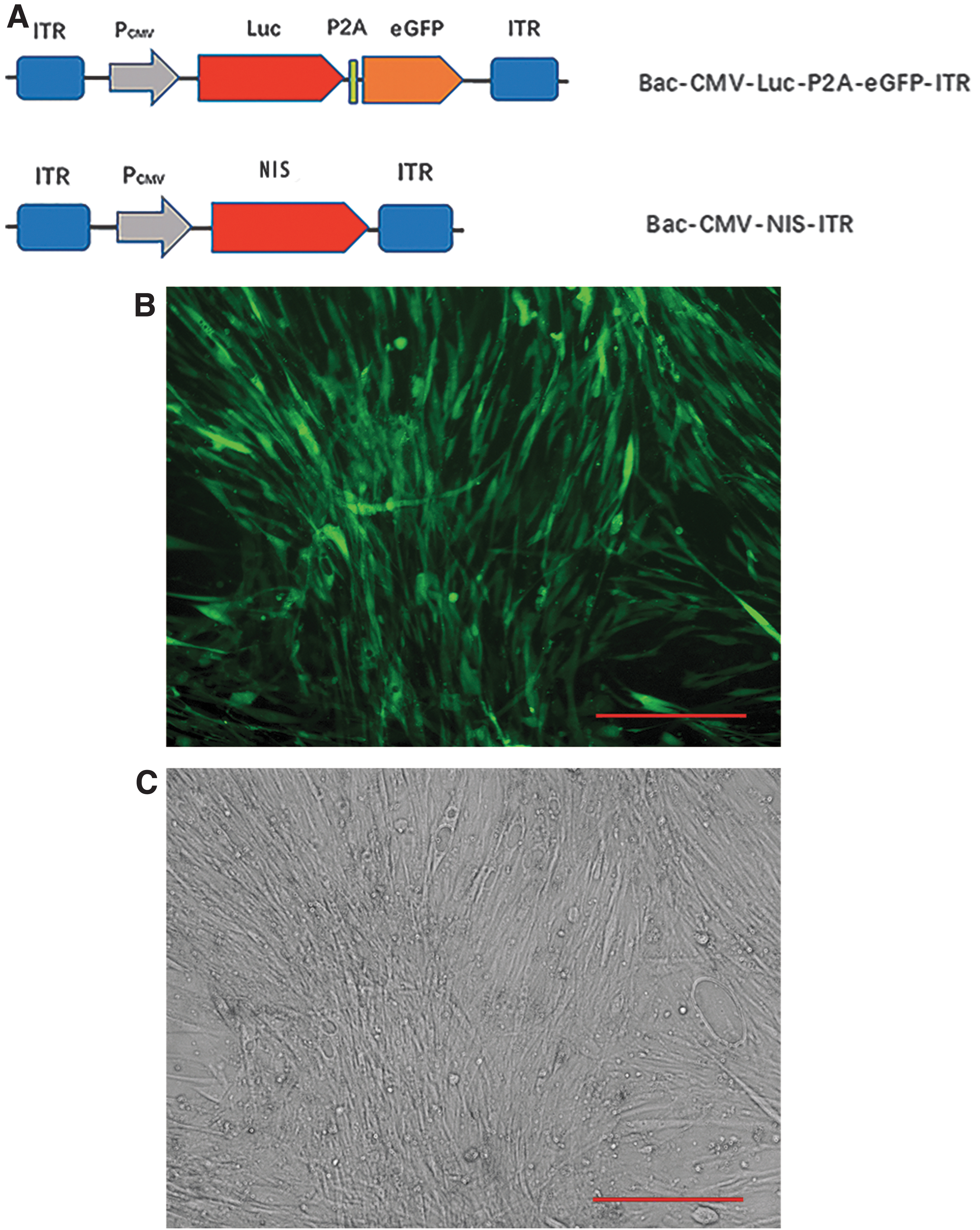
Schematic representation of the BV-AAV vectors constructs and optimal MOI.
Iodide uptake by BMSCs-Bac-NIS
The uptake of 125I by BMSCs-Bac-NIS varied with the incubation time, reaching a peak at ∼4,500 CPM at 1 h, and up to 15-fold higher iodide uptake was detected in BMSCs-Bac-NIS compared with the BMSCs and the inhibition group. One hour later, different concentrations of FBS were added to the working solution of BMSCs-Bac-NIS. As illustrated in Fig. 2A, at 2 h, the iodine levels of the three groups remained high. At 4 h, the uptake level of 125I in the BMSCs-NIS +0%FBS group was significantly lower than that at 1 h, whereas the level of 125I uptake in BMSCs-NIS +5%FBS and BMSCs-NIS +10%FBS was not significantly different from that of the previous group at 1 h. After 8-h 125I incubation, the radioactivity in the BMSCs-NIS +10%FBS group plateaued, implying a steady state. There was no significant difference in iodine intake levels between BMSCs-NIS +0%FBS group at 1 h and the BMSCs-NIS +10%FBS group at 8 h. The uptake level of 125I in the BMSCs-NIS +0%FBS group and the BMSCs-NIS +5%FBS group significantly decreased over time, and the BMSCs-NIS +0%FBS group decreased more significantly. The iodine intake levels of all three groups at 24 h were significantly decreased, but significantly higher than that of the control group.
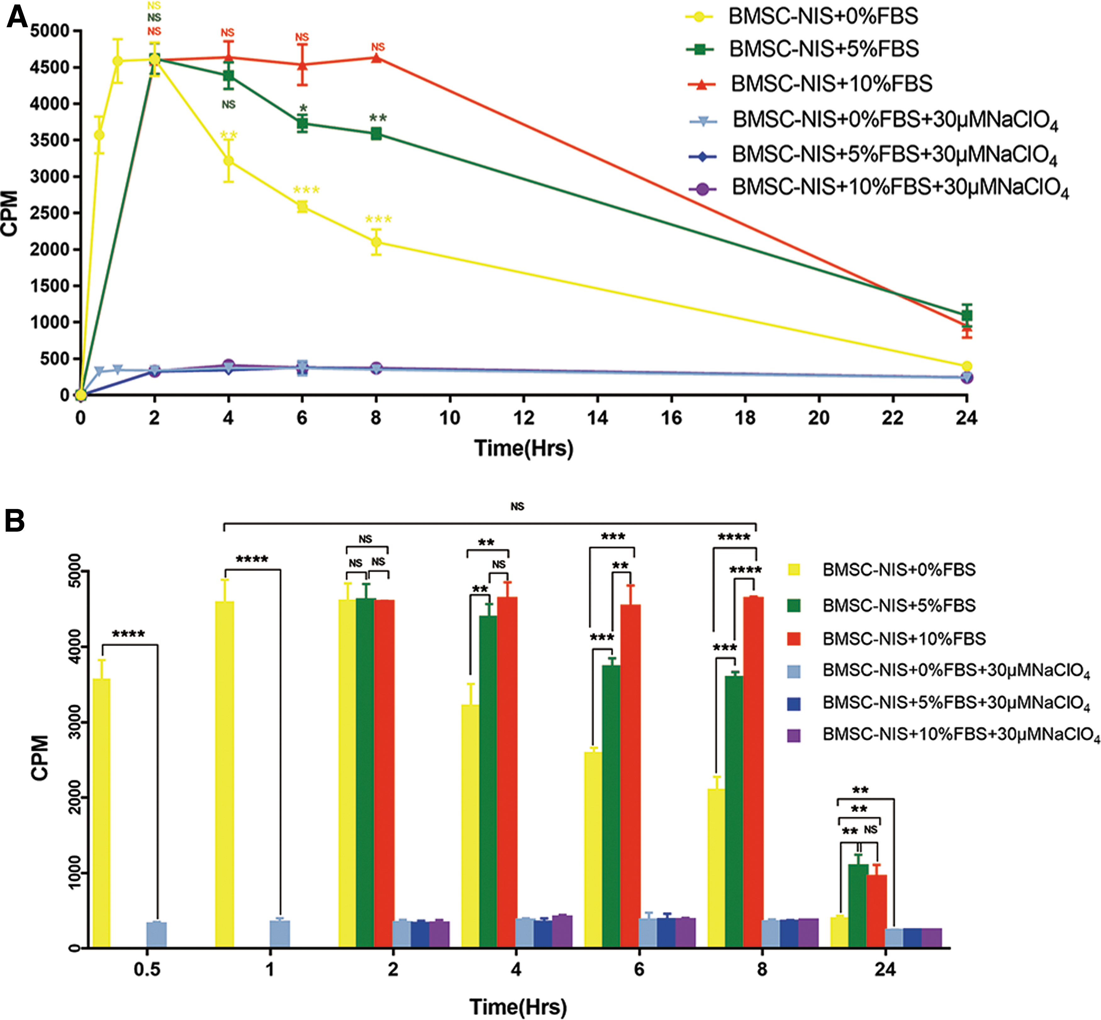
Time course of iodide uptake by BMSCs-Bac-NIS.
As given in Fig. 2B, there was no significant difference in the iodine intake level among the three groups at 2 h. At 4 h, there was no significant difference in the uptake level between the BMSCs-NIS +10%FBS and the BMSCs-NIS +5%FBS groups. The iodine uptake level of the BMSCs-NIS +0%FBS group was significantly lower than that of the former two groups. The iodine levels in the three groups at 6 and 8 h were significantly different. At 24 h, there was no significant difference in the intake levels of the BMSCs-NIS +10%FBS and BMSCs-NIS +5%FBS groups, but it was still significantly higher than the BMSCs-NIS +0%FBS group.
Micro-SPECT/CT imaging and immunofluorescence analysis of BMSCs-Bac-NIS in a mouse model of hypopharyngeal carcinoma
Three days after the last injection of cells, 74 MBq 125I was injected and radioiodine biodistribution was tracked using a micro-SPECT/CT in nude mice. No significant accumulation of iodine was detected in the tumor site after administration of BMSCs (Fig. 3A), whereas significant iodide uptake was observed in tumors after BMSCs-Bac-NIS application (Fig. 3B). Iodide uptake associated with native NIS expression was observed in the thyroid gland and stomach. To further determine NIS expression and BMSC biodistribution in vivo, tumor slides were incubated with NIS-specific antibodies. NIS-specific immunoreactivity was detected on the membranes of BMSCs-Bac-NIS in tumors of mice (Fig. 3C).
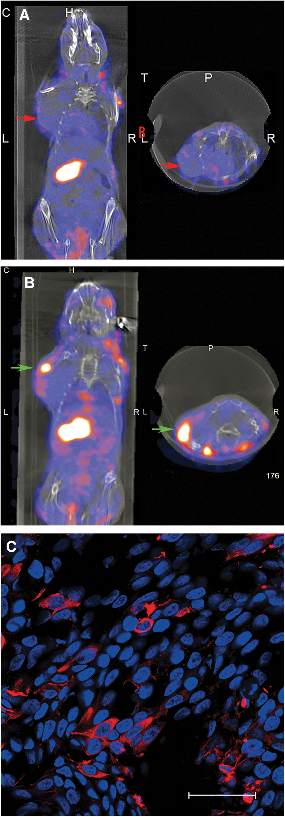
Biological distribution of radioiodine in vivo by micro-SPECT/CT and immunofluorescence staining. 125I micro-SPECT/CT imaging of mice harboring hypopharyngeal carcinoma (FaDu cells) after BMSC-mediated NIS gene delivery 1 h post-125I administration.
Therapeutic effects of 131I therapy in vivo
Mice were injected with 100 μL PBS containing 2 × 10⁵ BMSCs-Bac-NIS or BMSCs three times through their tail veins, and 18.5 MBq (0.5 mCi) 131I was injected intravenously 3 days after the last cell transplant. There was no significant difference in tumor volume between the two groups at each time point before 131I administration. Similarly, there was no significant difference in tumor volume between the two groups 1 week after 131I administration. However, at week 2, tumor growth rate and volume were significantly reduced in the BMSCs-Bac-NIS group, and the difference between tumor volumes in the BMSCs-Bac-NIS group (386.93 ± 249.23 mm3) and BMSCs group (816.56 ± 213.87 mm3) was statistically significant (p = 0.02, p < 0.05). At week 3, the difference in tumor volume between the two groups became more obvious (p = 0.008, p < 0.01). At week 4, only two nude mice survived in the BMSCs group. During the fifth week after cell transplantation, all five experimental mice in the BMSCs group died, whereas all five nude mice in the BMSCs-Bac-NIS group survived. With the extension of observation time, the tumor volume of the Bac-NIS group was also increased. After 4 weeks, the tumor volume increased faster in the BMSCs-Bac-NIS group. At week 6, two nude mice died in the BMSCs-Bac-NIS group, and the rest three died at week 7. The results suggested that radioiodine treatment after BMSCs-Bac-NIS transplantation through the tail vein could delay the tumor growth and prolong the life of the tumor model mice (Fig. 4).

In vivo 131I therapy after BMSC-mediated NIS gene transfer. Two weeks previously, FaDu cells were injected in the left axilla of the mice. One week later, mice were divided randomly into two groups receiving an intravenous injection of BMSCs-BAC-NIS (n = 5) or BMSCs (n = 5) in 2-day intervals, before receiving 131I (18.5 MBq) 72 h after the last injection of cells. **p < 0.01, *p < 0.05.
Histochemical and immunohistochemical analysis
Tumors were removed from experimental nude mice treated with 131I for 2 weeks. Tumor tissue slides were incubated with H&E and immunohistochemical staining with a PCNA antibody was performed. H&E staining showed that after 2 weeks of 131I treatment, the tumor tissue of the BMSCs-Bac-NIS group showed extensive necrotic areas (Fig. 5B), which were not visible in the BMSCs group (Fig. 5A). Immunohistochemical staining of tumor tissue revealed that most of the nuclei in the tumor tissue of the BMSCs group were positive for PCNA (a large number of yellow particles), indicating the presence of actively proliferating cells (Fig. 5C). There were fewer yellow particles in the BMSCs-Bac-NIS group than the BMSCs group (Fig. 5C, D).
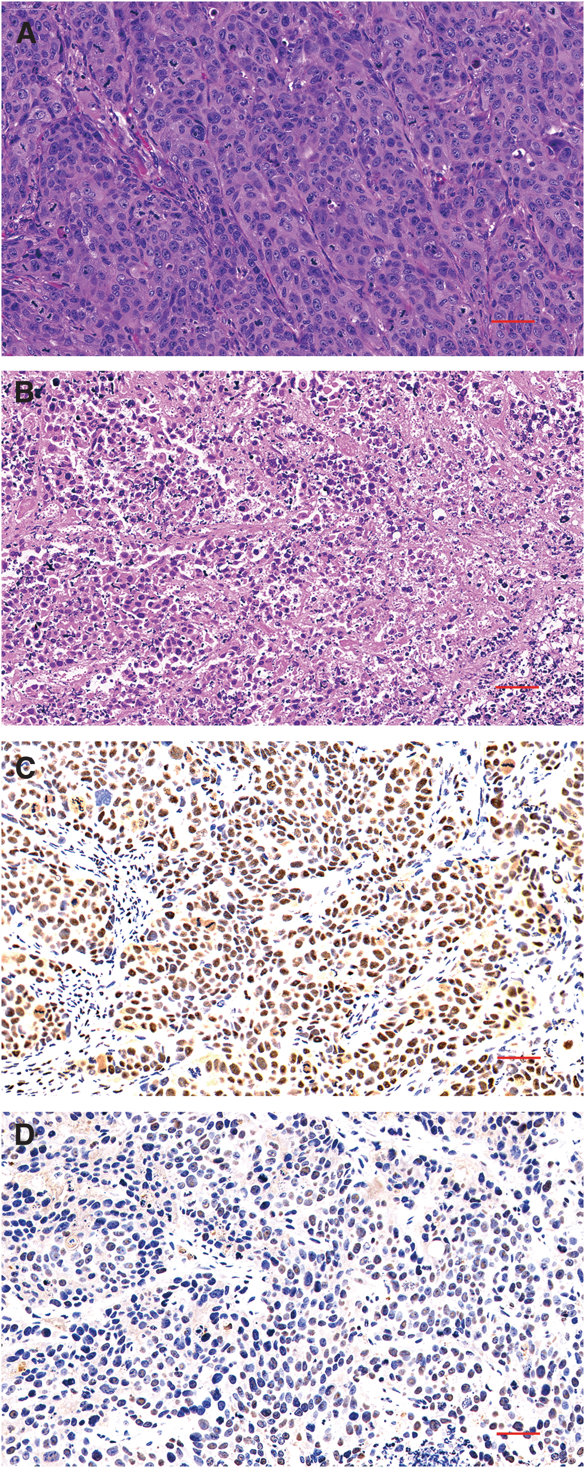
Histological and immunohistochemical study of tumor tissue in mice after 2 weeks of radioiodine treatment.
Effect of 131I therapy on 18 F-FDG micro-PET of xenografts
To estimate whether 131I therapy of intravenously transplanted BMSCs-Bac-NIS decrease glucose uptake in tumors, tumor-bearing mice were analyzed by 18 F-FDG micro-PET in vivo. The SUVmax of the BMSCs group (Fig. 6A) was 0.95 ± 0.08 (n = 3); the SUVmax of the BMSCs-Bac-NIS group (Fig. 6B) was 0.73 ± 0.03 (n = 3); and the SUVmax of 18 F-FDG PET in the BMSCs group was significantly higher than that in the BMSCs-Bac-NIS group (p < 0.05) (Fig. 6C).
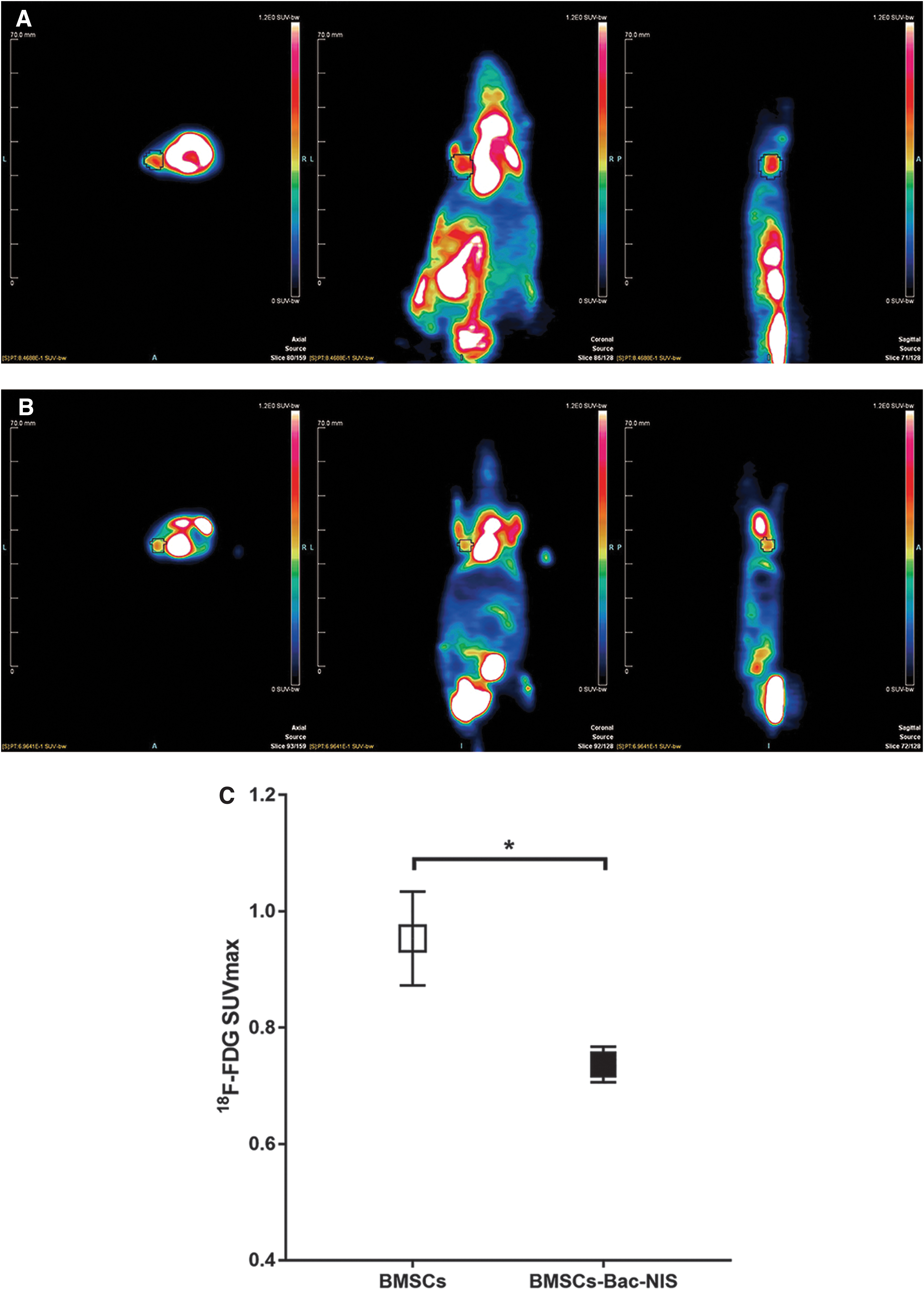
The effect of 131I on
18
F-FDG micro-PET of xenografts. Axial, coronal, and sagittal micro-PET imaging of xenograft tumors in
Discussion
Head and neck tumors are one of the most common tumors in developing countries 15,16 ; these tumors are the sixth highest incidence tumor type in the world, 17 accounting for ∼6% of all cases, and it is estimated that there are 650,000 head and neck tumor cases and 350,000 deaths every year worldwide. 18 Hypopharyngeal cancer is the worst prognosis pathological subtype of head and neck tumors. Most patients with hypopharyngeal cancer were found to be in advanced stages because of the abundant lymphoid supply and soft tissue boundary of the oropharynx and laryngopharynx. In the absence of distant metastasis, chemoradiotherapy is currently considered the most important treatment. 19 However, the current treatment results are still not ideal, especially when the disease is found in the late stage. In addition, as many as 26% of the patients with complete clinical remission after radiotherapy and chemotherapy still have residual regional lymph node metastasis. 20 It is clear there is an urgent need to find new treatment options.
The unique expression of NIS in the thyroid gland and no other important organs mitigates potential adverse effects of ionizing radiation on other organs. Therefore, thyroid cancer, even advanced metastatic diseases, can get better therapeutic effects through radioiodine therapy. The introduction of hNIS gene expression in nonthyroidal tumor tissue provides a mechanism to induce the accumulation of radioiodine in tumor tissues, allowing for imaging and therapy in nonthyroidal tumors. But the protection of normal thyroid tissue is particularly important. In clinical application, normal thyroid tissue should be protected from radioiodine uptake; this has been a limitation for radioiodine treatment of nonthyroidal tumors. However, in hypopharyngeal cancer patients, radioiodine treatment could be an option; according to the NCCN clinical practice guidelines in Oncology: Head and Neck Cancers (2019.V1), patients requiring (amenable to) a laryngectomy (T3, N0; Most T3, N1–N3; T4a, N0–N3) should simultaneously undergo a thyroidectomy. Therefore, radioiodine therapy has great clinical value in patients with advanced hypopharyngeal carcinoma.
Optimal expression of NIS in hypopharyngeal carcinoma is a key component in this strategy. Owing to the intrinsic tumor-homing capabilities of stem cells, they have been used as smart vehicles for targeted gene therapy. A recent study by our group 8 demonstrated that BMSCs modified by BV-AAV could target the hypopharyngeal tumor site with high efficiency and proliferate in the cancer microenvironment, serving as ideal vectors for targeted gene delivery to hypopharyngeal tumors. Here, the aim was to study BMSCs modified by BV-AAV as promising cellular carriers for targeted delivery of NIS to hypopharyngeal tumors, providing the basis for targeted radioiodine therapy of hypopharyngeal cancer.
BV-AAV was constructed to modify mammalian cells, including stem cells, which have shown high efficiency, no cytotoxicity, and maintained MSCs tumor-homing properties, demonstrating its potential for improving gene therapy. Moreover, this vector has great application value in delivering large multigene DNA constructs into most mammalian cells, including stem cells. Because of the very large foreign DNA cargo capacity, this multi-gene delivery vector can provide a variety of genes and regulatory elements, offering unlimited possibilities for the development of molecular biology research.
Using MSCs as the cell carrier of delivering NIS to laryngopharyngeal cancer, supporting the function of dynamic radionuclide imaging and radionuclide therapy, it is necessary to express NIS correctly on the targeted cell membrane and maximize the cellular retention time of iodide to have the greatest therapeutic effect. 21 Unfortunately, previous experiments have confirmed that when cells are incubated with solutions of I− working solution for 30 min, the peak value is reached and then 125I uptake decreases over time. 22 –24
The expression and activity of NIS are related to cell viability, because only living cells can accumulate iodine. 9 Thus, we speculate that the decrease in iodine uptake of stem cells modified by NIS may be because of the absence of FBS in the working solution, resulting in the decrease of cell activity. We have confirmed, through experiments adding FBS into working solutions with 125I to maintain BMSCs in good condition, that the I− uptake into BMSCs-Bac-NIS persists relatively long-term (at least 8 h) in vitro. hNIS-expressing nonthyroid cells do not organify iodide, 25 so maintaining the concentration of intracellular iodide should keep not only BMSCs activity, but also the high concentration of iodine in extracellular fluid. It is suggested that radioiodine should be given many times after BMSCs-Bac-NIS transplantation to maintain the accumulation of iodine in BMSCs. A previous study demonstrated that cells coinfected with vectors for hNIS and hTPO (human thyroperoxidase, an important enzyme in thyroid hormone synthesis) prolonged the retention of radioiodide. 26
The biological distribution of BMSCs-Bac-NIS after whole-body infusion through the tail vein showed that 125I signal was detected at tumor sites and native NIS-expressing organs. This suggests that the number of BMSCs expressing NIS was high enough to be used as tracers to produce signals over background. These data reflect the importance of real-time tracking of the migration of BMSCs, and provided experimental support for tumor-targeted BMSCs-Bac-NIS implantation for administration of 131I.
Many studies have shown that MSCs differentiate into peripheral cells or tumor-related fibroblasts after accumulation in tumor tissue, which could secrete vascular endothelial growth factor, interleukin-8, transforming growth factor β, epidermal growth factor, and platelet-derived growth factor, 27 –29 thus forming a microenvironment to support tumor growth. Moreover, it has been shown that MSCs can also induce the activation of Akt and ERK in endothelial cells, thus increasing its recruitment and angiogenesis potential. 28 In an in vitro coculture experiment, MSCs stimulated the invasion and proliferation of breast cancer cells. 29 These data limit the application of MSC as a gene delivery targeting vector.
The therapeutic gene used in this experiment is NIS, which is treated with radioiodine by taking 131I. During the decay process of radioactive 131I, β-rays (99%) and γ-rays (1%) are released; as the effective range of β-rays is only 2 mm, it can selectively destroy tumor tissue without affecting adjacent tissue. After the tumor is destroyed, it will gradually undergo necrosis and achieve the purpose of treatment. This treatment approach leads to the death of transgenic expression cells, which mitigates the promoting effect of MSCs on tumor growth and increases the safety of this treatment.
18 F-FDG is a kind of molecular probe that has been widely used and shown to be effective in tumor detection and evaluation of therapeutic efficacy. 30,31 18 F-FDG can enter the cell through the same glucose transporter-1 (GLUT-1) as glucose, and then be phosphorylated by hexokinase and remain in the cell. Many studies have found that the concentration of FDG is related to some tumor pathological characteristics, including tumor size, 32,33 tumor malignancy, 34 pathological type, 35,36 tumor differentiation, 37 and micro growth mode. 38 The increase of glycolysis or energy metabolism in tumor cells results in the increase of 18 F-FDG uptake and accumulation, which is an indicator of tumor growth and development. 39,40 SUVmax, as a commonly used semi-quantitative indicator, helps to quantify the metabolic changes in tumors. In this study, we evaluated the role of BMSCs expressing NIS, mediated by BV-AAV, as a cell carrier target in the treatment of hypopharyngeal cancer by radioiodine. We not only measured the change in tumor volume and the life span of the experimental mice, but also evaluated the SUVmax.
According to a study of FDG uptake in a subcutaneous-transplanted tumor animal model of lung cancer, 41 FDG was mainly concentrated in the hypoxic tumor cells of the starvation group, but the uptake of nonhypoxic tumor cells and tumor stromal cells was relatively low; in the nonstarvation group the result was the opposite. Therefore, in this study, mice were fasted for 6 h before micro-PET/CT scans; BMSCs-BAC-NIS (treatment group) or BMSCs (control group) transplantation, 131I was injected intravenously and PET-CT was performed 2 weeks later. The results showed that the SUVmax in the treatment group was significantly lower than that in the control group (p < 0.05), indicating that tumor glucose metabolism was decreased after treatment, indicating that tumor growth was inhibited.
Many clinicopathological indexes are considered to be related to tumor proliferation, invasion, and prognosis, such as Ki-67, PCNA, cyclin D1, and nm23. 42 –44 Tumor cells have strong proliferative activity, and PCNA is one of the most commonly used evaluation indexes to study tumor proliferative activity. In this study, monoclonal antibodies targeting PCNA were used for immunohistochemical analysis of tumor tissue sections. The results showed that the number of PCNA-positive granules in the treatment group was significantly reduced, suggesting that tumor proliferation was significantly inhibited, which was in line with the tumor volume and SUV value of 18 F-FDG PET results.
In the sixth week, the growth rate of tumor volume in the treatment group was significantly faster than before. We infer that it may be owing to the decrease of BMSCs and 131I in vivo, suggesting that the effect of radioiodine therapy can be further improved by repeated doses of BMSCs-Bac-NIS and radioiodine injection. Many studies have shown that tumor radiation stimulation can increase the infiltration of MSCs into tumor tissues, 45 –48 which is encouraging for patients with tumor recurrence and tumor metastasis.
Footnotes
Author Disclosure
No competing financial interests exist.
Funding Information
This research was supported by the Project of Shanghai Municipal Commission of Health and Family Planning (Grant No. 201540387).