
Abstract
Hematopoietic stem cell gene therapy has become a successful therapeutic strategy for some inherited genetic disorders. Pre-clinical toxicity studies performed to support the human clinical trials using viral-mediated gene transfer and autologous hematopoietic stem and progenitor cell (HSPC) transplantation are complex and the use of mouse models of human diseases makes interpretation of the results challenging. In addition, they rely on the use of conditioning agents that must induce enough myeloablation to allow engraftment of transduced and transplanted HSPC. Busulfan and total body irradiation (TBI) are the most commonly used conditioning regimens in the mouse. Lenticular degeneration and atrophy of reproductive organs are expected histopathological changes. Proliferative and nonproliferative lesions can be observed with different incidence and distribution across strains and mouse models of diseases. The occurrence of these lesions can interfere with the interpretation of pre-clinical toxicity and tumorigenicity studies performed to support the human clinical studies. As such, it is important to be aware of the background incidence of lesions induced by different conditioning regimens. We review the histopathology results from seven long-term studies, five using TBI and two using busulfan.
Introduction
Rare diseases are defined as life-threatening or chronically debilitating conditions that have a prevalence threshold in the range of 40 to 50 cases out of 100,000. 1 There are ∼7,000 reported diseases, many of which are caused by single gene mutation. Gene therapy has shown clinical promises as a strategy for treating some of these diseases, yet remains facing challenges around safe, consistent, and durable gene delivery to targeted tissues. So far, most gene therapies under development are aimed to treat rare diseases for which no effective alternative treatment exists. 2,3 Among several promising gene therapy medicinal product (GTMP) under development, gene therapy performed with ex vivo corrected hematopoietic stem and progenitor cell (HSPC) has progressed to successful clinical testing for several immunodeficiencies, blood and storage disorders, with two therapies already registered for the pharmaceutical market and several more likely to join in the next few years.
Before administration of an investigational GTMP in a clinical trial, the sponsor must provide adequate information about the pharmacological and toxicological studies that demonstrated that it is reasonably safe to conduct the proposed clinical investigations. 4 The pre-clinical program should have a satisfactory scientific rationale to mimic and support the design of the clinical trial, providing characterization of potential toxicity and guiding clinical risk assessment and monitoring.
Pre-clinical toxicity studies performed to support the human clinical studies using autologous genetically engineered HSPC transplantation are complex. For better reconstitution of the hematopoietic system, murine HSPC and often vectors expressing the mouse homolog of the therapeutic transgene must be used. These studies also use mouse models of human diseases, which can make interpretation of the results difficult. In addition, ex vivo gene therapy relies on the use of conditioning agents to achieve stable engraftment of transduced HSPC. Animals undergoing transplantation of congenic or allogenic HSPC receive a chemotherapy or radiotherapy treatment that depletes proliferating hematopoietic progenitors in the bone marrow (myeloablation), creating space for the engraftment of gene-modified HSPC.
Therefore, a profound knowledge of the mouse disease model-specific biological background as well as the effects of conditioning regimens administered is required. Conditioning treatment should be tailored to reach good levels of engraftment, while minimizing toxicity. Considerations need to be given not only to the type and intensity of conditioning regimens but also to the mouse sensitivity to them that can be different between strains or models.
Two types of conditioning regimen are commonly used, total body irradiation (TBI) and chemotherapy (with compounds such as busulfan). Both treatments induce histopathological changes, nonproliferative and proliferative, which could interfere with the toxicological evaluation of the HSPC therapy.
TBI as a conditioning regimen
Ablation of hematopoietic stem cells together with an effective level of immune suppression are desirable properties of a preparative regimen for bone marrow transplantation and TBI (using gamma rays) meets these requirements in clinical and pre-clinical settings. 5 TBI induces excess double-strand breaks in DNA that lead to cell death, particularly in actively dividing cells such as hematopoietic progenitor cells. TBI of mice is a widely used research tool and there are international guidelines (Association for Assessment and Accreditation of Laboratory Animal Care: AAALAC-accredited facilities) for humane care and use of animals that remain immunocompromised for a period after HSPC transplantation. 6
The irradiation protocols are tailored to induce adequate bone marrow cell death such that it results in animal lethality if the recipient animal does not achieve adequate donor cell engraftment from bone marrow transplantation (so called “lethal irradiation”). 7 Pathological processes of irradiation injury begin immediately after exposure, but clinical and histological features may not become apparent for weeks or months to years. 8 As such, radiation injuries are classified as acute or late effects. Tumorigenesis is an important consequence of radiation exposure.
Busulfan as a conditioning regimen
Busulfan (1,4-butanediol dimethanesulfonate), a cell cycle phase-nonspecific alkylating agent, has myeloablative properties targeting dividing marrow cells and minimally immunosuppressive properties that help to overcome graft rejection of donor stem cells. 9 Cytotoxicity to highly proliferating tissues is an early expected change following administration of busulfan, especially hematopoietic precursor and stem cells in the bone marrow. 9 Busulfan can be safely administered to mice by intraperitoneal injection at a dose between 60 and 100 mg/kg and does not require the specialized facilities and equipment necessary to perform TBI. Importantly, at these doses, myeloablation is not complete and as such mice are able to survive without receiving support bone marrow cells. 9
Busulfan has been shown to be mutagenic to microorganisms, mammalian cells, Drosophila, and rodents and is also considered carcinogenic to humans. At high dose, the hematopoietic cell depletion can lead to aplastic anemia and there is different strain susceptibility among mice exposed to treatment with busulfan. 10 Other toxic changes might be observed such as lenticular degeneration and atrophy of reproductive organs.
Busulfan's clinical use has been well established in the treatment of hematological malignancies such as chronic myeloid leukemia and other myeloproliferative syndromes and it has been extensively used in human allogeneic hematopoietic stem cell transplantation. 11 It is also commonly used with or without cyclophosphamide as a conditioning regimen for hematopoietic stem cell transplantation in GTMP trials. 11 It helps to create space in the bone marrow for new blood stem cells to grow. Eight weeks after transplantation with HSPC, all leukocyte subsets are measurable in blood and tissues. 12
Pre-clinically, busulfan has been used in various species, 9,13 –15 including C57BL/6 mice, rhesus monkey, and NOD/SCID mice, to allow engraftment of modified HSPC.
We describe the experience gained from six pre-clinical studies using mouse transplanted with HSPC conducted in the GLP SR-TIGET Test Facility (Milan, Italy) using either TBI or busulfan (Table 1). Results from an investigative time-course study using busulfan performed at GSK (Ware, United Kingdom) were included to gain insight on acute lesions (0 to 4 weeks of the study) that can be expected in gene therapy studies and to better understand the proliferative changes of the hemolymphoid system. Among strains and disease models, we observed differences in timing and incidence of lesions, supporting the importance of collecting historical data and having concurrent control animals to perform a weight of evidence assessment of pathology findings observed in the study.
List of the long-term toxicity and tumorigenicity studies
CGD, chronic granulomatous disease; MLD, metachromatic leukodystrophy; MPS1, type 1 mucopolysaccharidosis; TBI, total body irradiation.
The aim of this review is to share histopathological data related to conditioning regimens that we have collected from several pre-clinical safety studies using mouse model of human diseases performed in a GLP accredited facility (GLP SR-TIGET), conducted according to a comprehensive and consistent study plan and standardized procedures.
Materials and Methods
Animals and husbandry
Mouse strains were purchased from Jackson Laboratory or from Charles River. Mice were maintained at San Raffaele Scientific Institute SPF Animal Facility under specific pathogen-free conditions. Animals were kept in ventilated cages, had prophylactic gentamicin in drinking water (gentamicin sulfate 80 mg/250 mL for 2 weeks, starting from the day of irradiation), were housed in sterile cages, and received sterile food (2918 Teklad Global Diet; Harlan Laboratories) and water.
All experimental procedures were approved by the Animal Care and Use Committee of the Fondazione San Raffaele del Monte Tabor and communicated to the Ministry of Health and local authorities in accordance with the Italian legislation and carried out in accordance with Animals (Scientific Procedures) Act 1986 and the GSK Policy on the Care, Welfare, and Treatment of Animals.
Mortality and morbidity were checked at least five times a week and animals were individually observed for the recording of clinical changes (e.g., reduced activity, hunched posture, and stained fur) and body weight on a weekly basis. Hematology and clinical chemistry samples were collected and evaluated at different time points after transplantation and at termination (data not shown). To guarantee quality, robustness, and traceability, studies were designed and performed following the Guidelines for GLP (Good Laboratory Practice) in compliance with the Organization for Economic Co-operation and Development (OECD) Principles of GLP [as revised in 1997, ENV/MC/CHEM(98)17].
Gene therapy studies
TBI studies
Three long-term toxicity and tumorigenicity studies using mouse models of human diseases (MPS1—type 1 mucopolysaccharidosis, MLD 16 —metachromatic leukodystrophy, and CGD—chronic granulomatous disease) and two using wild-type C57BL/6 were conditioned by TBI.
Seven- to 9-week-old mice were irradiated using a Rad Source RS-2000 Irradiator with a total of 750–900 cGy divided into two administrations with an interval of 2 to 3 h.
Procedures used were fully compliant with national and international regulations and were defined in Standard Operating Procedures. The dosing regimen consisted of a lethal irradiation at different dose depending on strain susceptibility and fractioned into two consecutive doses (Table 1).
Busulfan study
Busulfan was the conditioning regimen used in one long-term toxicity and tumorigenicity study using Th3/+ mice (mouse model of human beta-thalassemia).
Seven- to 9-week-old mice were given four intraperitoneal injection of busulfan (in acetone: peanut oil 1:10 v/v; Sigma-Aldrich, St. Louis, MO) at the dose of 25 mg/kg/administration for 4 days (Table 1). 17
Investigative busulfan study
In the time-course study, B6C3F1 mice between 10 and 12 weeks of age received four intraperitoneal injections of busulfan at the dose of 40 mg/kg (Busilvex, Pierre Fabre volume of 10 mL/kg diluted in water: acetone) on day 0, 14, 28, and 42. Control mice were injected with water: acetone alone. 18
Transplantation of murine hematopoietic progenitors
Following conditioning (4 h after irradiation or 24 h after last busulfan injection), mice received mock-transduced murine HSPC (isogenic Lin− progenitors purified by antibody-mediated depletion of cells expressing lineage differentiation markers and cultured in absence of lentiviral vector) by intravenous injection in the tail vein according to standard transplantation procedures for mouse. The transplanted cell dose ranged between 4 × 10⁵ cells (Th3/+) and 1 × 10⁶ cells (MLD). In the time-course study using busulfan, the animals did not receive murine HSPC.
Evaluation of the engraftment levels
To monitor the level of transplant engraftment following transplantation, the donor/recipient allelic mismatch for CD45 pan-leukocyte antigen was used. Chimerism and cell origin were monitored using antibodies specific for CD45.1 or CD45.2 surface antigen. In addition, clinical pathology (hematology) data gave information on the bone marrow repopulation, and, when possible, the reversibility of the disease phenotype was evaluated by transgene expression or enzyme activity measurement (e.g., Alpha-
Histopathology
A full necropsy with macroscopic examination was performed on all animals (including intercurrent deaths). Tissues taken at necropsy were fixed using industry-standard procedures in 10% neutral buffered formalin. They were then paraffin-embedded and sectioned at 4 μm according to local standard operative procedures. A routine hematoxylin and eosin stain was used to produce the final slides.
Although the tissue list examined microscopically differed slightly between studies, the following organs were systematically examined: adrenal gland, aorta, brain, heart, kidneys, large intestine (cecum, colon, and rectum), liver, lungs with bronchi, lymph nodes (mesenteric and mandibular), pancreas, skeletal muscle, skin, small intestine (duodenum, ileum, and jejunum), spleen, sternum and femur with bone marrow, stomach with forestomach, thymus, and reproductive organs. The time-course study using busulfan focused on the hematopoietic system with the addition of heart, jejunum, lungs, liver, kidneys, and eyes.
Histopathological findings observed were graded by a pathologist using the industry standard semiquantitative approach as follows: Grade 1—Minimal: Finding affecting <10% of the organ Grade 2—Mild: Finding affecting between 10% and 30% of the organ Grade 3—Moderate: Finding affecting between 30% and 50% of the organ Grade 4—Marked: Finding affecting between 50% and 80% of the organ Grade 5—Severe: Finding affecting between >80% of the organ
Immunostaining with anti-CD3 (16669, working concentration of 10 mg/mL; Abcam) and anti-CD45R (MCA1258G, working concentration of 0.5 mg/mL; Serotec) antibodies was performed on all lymphoid hyperplasia and lymphomas from the busulfan time-course study.
A pathology peer review was performed on all animals with significant histological findings, including all neoplastic findings.
Results
In general, similar values of engraftment were observed between the reviewed studies and persisted until termination. For example, the percentage of engraftment in MLD mice was at 95% in females and 92% in males. In CGD male mice, the level was 80%. In Th3/+ mice,
17
there was difference in engraftment values between males and females, which was maintained throughout the study. The average was 60%, with males at 84% and females at 52%. Engraftment percentages did not significantly change with time in males, while females presented higher variability and some animals had lower percentages at termination. The engraftment level in MPS1 mice was evaluated indirectly by the alpha-
It should be noted that no correlation was observed between individual engraftment level and mortality in the studies reviewed.
Because of their intrinsic toxicity (myeloablative properties), conditioning regimens can be the cause of early unscheduled death. Depending on the susceptibility of the mouse strain, this occurred in our experience between 2 and 4 weeks after conditioning regimen administration in up to 20% of the animals receiving HSPC. Animals affected by this acute toxicity were not included in the evaluation. However, microscopic observations in these animals were similar to those described in the time-course study using busulfan, where no animal has been excluded from the analysis aimed to characterize the follow-up of acute changes (characterized by severe decreased cellularity in the hematopoietic tissues).
In addition, it should be noted that the phenotype of the model can also be the cause of the unscheduled cull. For example, CGD mice can present extensive granulomatous inflammation in the skin with secondary abscess formation, which was the cause of the animals' removal and was considered out of scope for this review.
TBI: unscheduled deaths
Moderate to marked glomerulonephropathy (Supplementary Fig. S1) was considered the main cause of death and morbidity of irradiated animals. Pleural effusions and depletion of lymphoid cells in hematolymphoid tissues (lymph nodes, spleen, thymus, and bone marrow) were also frequently observed. These deaths occurred from the 40th week post-treatment onward. The strain and sex of the mouse played an important role in the incidence of the findings. For example, in a preliminary study (data not shown), CGD female mice appeared to be much more susceptible than males to glomerulonephropathy. Females developed the disease earlier and at a higher severity (with mortality) than males. That was the reason for including only males in the 52-week study.
In details, 13 (1 male and 12 females) out of 17 deaths in the 52-week study using MLD mice and 4 out of 9 animals in the 52-week study using CGD mice were attributed to glomerulonephropathy. Only one C57BL/6 mouse unscheduled death, amid the two studies, was attributed to glomerulonephropathy. It should be noted that no MPS1 mouse had findings in kidneys; however, the microscopic evaluation was limited to phase I animals (26 weeks after transplantation).
The other causes of death were attributed to proliferative changes, such as generalized lymphomas, adenocarcinomas of the gastrointestinal tract, or cutaneous sarcomas. The incidence of nonhematopoietic tumors was low, usually being the cause of death of only one animal per study, but still considered to be related to irradiation (Supplementary Table S1). Lymphomas were generalized, that is, involving multiple organs such as thymus, spleen, lung, liver, mandibular/mesenteric lymph nodes, heart/aorta, kidney, brain, and spinal cord (Fig. 1a). The histotype of lymphomas observed in TBI animals were of the lymphoblastic type, characterized by a quite uniform population of medium-sized round cells with scant cytoplasm, medium-sized nucleoli, and starry sky macrophages (Fig. 1d). 19,20
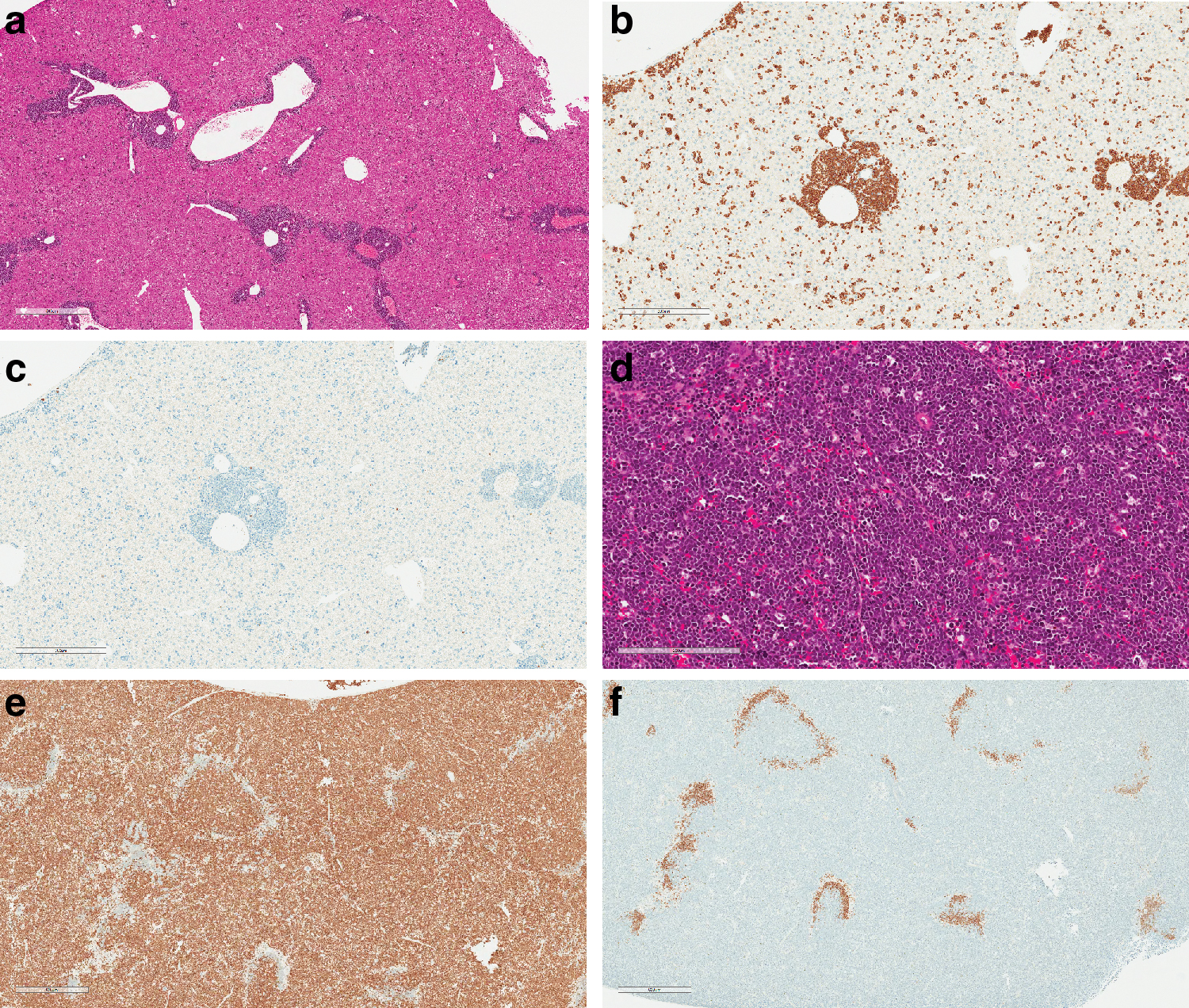
Lymphoblastic lymphoma generalized, from the busulfan time-course study showing the typical uniform population of medium-sized round cells. B6C3F1 mouse terminally killed at week 36.
Additional lesions attributed to TBI were observed in the reproductive system and the lens. Lenticular degeneration was observed up to a marked severity in most animals from all studies (Supplementary Fig. S2). This finding was characterized by fiber fragmentation, cleft formation, and the presence of Morgagnian globules. 21 Similarly, ovarian atrophy characterized by a reduced number of healthy antral follicles and no corpora lutea 22 and testicular atrophy, characterized by the absence of most or all germ cells from affected tubules lined only by Sertoli cells, 23 was observed generally at a marked level in most unscheduled deaths using TBI as the conditioning regimen (Supplementary Fig. S3). All studies combined, these changes were observed first around 20 weeks post-treatment in the unscheduled deaths. However, the incidence and severity of histopathological changes showed differences across various strains (Supplementary Table S2).
TBI: terminal sacrifice. Nonproliferative lesions
At the end of the studies, irradiated animals from all studies had high incidences of lenticular degeneration with virtually every animal affected. There was no sex or strain difference and the severity was up to marked. Ovarian and seminiferous tubules atrophy were similarly observed in virtually all the animals with no strain difference, usually at a marked severity. In the hematopoietic organs, findings considered to be related to irradiation were observed in the spleen and the thymus and consisted of a mild decreased cellularity when compared to controls. Finally, glomerulonephropathy was observed in most mice exposed to the irradiation.
TBI: terminal sacrifice. Proliferative lesions
Irradiated animals had, at the end of the studies, relatively few proliferative findings that are summarized in Table 2. However, the incidence of certain tumors or hyperplasia was higher in the irradiated groups in comparison with the concurrent controls. Specifically, one gastrointestinal tract adenoma (duodenum) and three adenocarcinomas (duodenum, jejunum, or colon) were observed in the 52-week study using C57BL/6 mice (N = 30 treated animals). One adenocarcinoma (ileum) was observed in the 52-week study using MLD mice when these tumors were not present in the control groups (N = 32 treated animals). CGD mice, however, had a similar incidence of these tumors between control and irradiated groups. Harderian gland adenomas were observed in the MLD mice, with a higher incidence in irradiated animals. It should be noted that these glands have not been evaluated in the other studies.
Busulfan time-course study
Incidence and rounded percentages of lymphoid proliferative changes in treated animals. No changes were observed in terminally killed control animals at any time point.
Another irradiated mouse-specific finding was observed in the adrenal glands. Cortical hyperplasia was present in 5 out of 22 remaining animals from the 52-week study using C57BL/6 mice, 13 out of 15 remaining animals from the 52-week study using MLD mice and 6 out of 12 remaining animals from the 52-week study using CGD mice. Only one control animal (CGD) had this hyperplasia (the adrenals were not read for the other two studies).
At the end of the studies, the incidence of lymphomas was low, generally one per study, and occurred only in control animals (Table 3). These lymphomas in control mice not exposed to TBI were of the pleomorphic type where neoplastic cells may be predominantly large, small, or composed of a mixture of large and small cells. 19,20
Summary of lymphomas observed in the studies (incidences and rounded percentages) as observed in unscheduled death and in terminal killed animals
Localized lymphomas were observed in one organ (usually spleen or thymus), whereas generalized lymphomas were observed in multiple tissues such as liver or kidney, in addition to the lymphoid organs.
Busulfan: unscheduled deaths
There were only two unscheduled deaths in the β-thalassemia 52-week study, and both attributed to proliferative changes. One male was culled at week 22 and had abdominal sarcoma and marked testicular atrophy. One female was found dead at week 37 and had a generalized lymphoblastic lymphoma involving the heart, lung, sternal bone marrow, and thymus. The autolysis precluded the evaluation for lenticular and ovarian atrophy in this animal.
The time-course study using busulfan provided details on the acute and delayed toxicity of this conditioning regimen. In total, 31 animals treated with busulfan died or were killed prematurely due to poor clinical condition (subdued behavior). The first animal was culled at week 4 after the last busulfan injection and showed marked bone marrow aplasia with an almost complete absence of all hematopoietic lineages.
For data analysis purposes, unscheduled deaths from the time-course study were organized into three groups according to the lesions observed in the hematopoietic system: between 0 and 8 weeks, between 9 and 17 weeks, and between 18 and 36 weeks post-busulfan administration.
There were 18 unscheduled deaths between week 0 and 8. The mortality in this group was due to the acute toxicity of busulfan characterized by marked decreased cellularity of the hematopoietic tissues. The bone marrow cellularity was severely decreased compared with controls (Fig. 2a, b) and the thymuses showed a marked cortical atrophy compared with controls (Fig. 2c, d). Proliferative changes occurred as early as week 4 and consisted of focal lymphoid hyperplasia in the spleen of nine animals (Table 2). We defined this finding as a localized expansion of the white pulp lymphoid cells, poorly delimited, but with an intact splenic architecture.
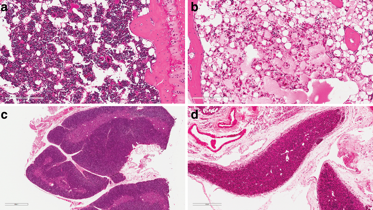
H&E-stained sections of hematolymphoid organs from mice of the busulfan time-course study.
There were 10 unscheduled deaths between week 9 and 17. The mortality in this group was due to busulfan hematotoxicity with the added occurrence of lymphoblastic lymphomas. The hematopoietic tissues in 9 out of 10 animals failed to be repopulated with the bone marrow and the thymus showing marked to severe decreased cellularity. In addition, 5 out of 10 animals had localized lymphomas. Lymphoma was observed in the spleen only in four animals and in the spleen and the thymus in the last animal. Two animals had focal lymphoid hyperplasia in the spleen (at week 9). No hyperplasia was observed after week 10 (Table 2).
There were three unscheduled deaths between week 18 and 36. The mortality in this group was related to the presence of generalized lymphoblastic lymphomas only, involving tissues such as spleen, thymus, bone marrow, lung, liver, kidney, and gastrointestinal tract (Table 2).
The study of the lens provided insights into the onset of lenticular degeneration following busulfan administration. It was not observed in the 18 unscheduled deaths from the week 0 to 8 group. Only 1 animal from the 10 unscheduled deaths from the week 9 and 17 group had a minimal lenticular degeneration and 3 out of 3 unscheduled deaths from the week 18 to 36 group had the change at a moderate or marked severity.
Busulfan: terminal sacrifice. Nonproliferative lesions
In the 52-week study, 5 out of 28 Th3/+ males mice had, at the end of the study, moderate lenticular degeneration. This finding was not observed in females in this study. Ovarian atrophy and seminiferous tubule atrophy were observed in virtually all females and males, respectively, at a marked to severe severity.
In the time-course study, most B6C3F1 mice had lenticular degeneration and severity appeared to increase with time. Animals sacrificed at week 18 had the change at a minimal or mild severity, animals sacrificed at week 24 had a mild to moderate severity, and animals sacrificed at week 36 had moderate or marked severity. In the hematopoietic organs, findings considered to be related to busulfan were observed in the spleen (mild to moderate extramedullary hemopoiesis), the thymus (increased incidence and severity of the cortical atrophy), and the bone marrow (minimal to mild decreased cellularity). The decreased cellularity observed in the bone marrow is believed to represent an incomplete repopulation of the tissues in animals not receiving murine HSPC transplantation. The minimal lymphoid infiltration frequently seen in various tissues from control animals (e.g., salivary glands and kidney) was not observed in busulfan-treated animals.
Busulfan: terminal sacrifice. Proliferative lesions
Th3/+ mice had relatively few proliferative findings, and all were part of the background lesions usually observed in aging animals (Table 3). Specifically, five localized pleomorphic lymphomas were observed in control animals (no conditioning regimen and no transplantation N = 30) and one in busulfan-treated animals (N = 28).
The time-course study using busulfan focused on the hematopoietic system evaluation of B6C3F1 mouse. Therefore, only the lymphoproliferative disorders are discussed. Only two lymphoblastic lymphomas were observed in the thymus of 2 out of 15 remaining treated animals sacrificed at the end of the study. None was observed in control animals at the end of the study (week 36). Table 3 summarizes the occurrence of lymphoproliferative changes in this study.
Immunostaining with anti-CD3 (T cells) and anti-CD45R (B cells) antibodies was performed on all hyperplasia and lymphomas from this study. All the lymphomas were clearly CD3 positive and CD45R negative (Fig. 1b, c, e, f). Hyperplasia appeared characterized by cells positive for CD3 staining and CD45R negative. However, the intensity of the staining was usually lower than in the normal T area of the spleen, potentially indicating immature lymphoid cells.
Discussion
In the context of pre-clinical studies to support the development of GTMP, animals undergoing transplantation of gene-modified HSPC receive a chemotherapy or radiotherapy treatment to create space for the graft. Conditioning treatment should be tailored to reach good levels of engraftment (and bone marrow repopulation), while minimizing toxicity, considering the different susceptibility of strain or disease model mice. Deep knowledge of the effects of conditioning regimens administered to disease models with specific biological background is required to provide a science-based rational for the study design (duration, number of animals, and gender) and for the interpretation of outcomes of the study. The selection of the conditioning regimen in the studies reviewed was mainly driven by their toxicity in the mouse models.
Both conditioning regimens, TBI and busulfan, induced in the reviewed studies expected degenerative lesions in highly proliferative tissues such as lens, reproductive organs, and bone marrow. However, no degenerative lesion (e.g., villous atrophy) was observed in the gastrointestinal tract. All these findings are described in the literature 24 –28 and are related to the mode of action of the conditioning regimens with direct effect on the DNA of proliferating cells. Busulfan, being an alkylating agent, induces DNA cross-linking, and ionizing energy (X-rays or gamma rays), used for TBI, induces DNA double-strand breaks. These findings are translatable to human and similar changes are described in the clinical setting 29 –31
The time-course study using busulfan has highlighted the tendency for the lenticular degeneration to increase in incidence and severity with time, whereas the reproductive organs tended to be affected at a marked severity earlier (as observed in the unscheduled deaths from all the studies). There was no clear difference in the incidence and severity of these three lesions between the two conditioning regimens. However, there is a slight tendency for TBI to induce higher severity of lenticular degeneration than busulfan, and a tendency for busulfan to induce higher severity of seminiferous tubular atrophy. More studies are needed to draw a definitive conclusion. Lenticular changes can potentially impact on visual acuity and affect behavioral assessment. These changes in reproductive organs were not considered to be reversible in the studies reviewed, and no strain difference was observed. However, it has been reported in the literature 27 that Swiss mice, but not Balb/C mice, treated with a single dose of 40 mg/kg of busulfan might recover from the seminiferous tubule atrophy after 90 days. This could depend on the number of germ cells affected by the conditioning regimen. Because the aim of the conditioning regimen in the studies reviewed was to sufficiently deplete the bone marrow before an HSPC graft, the doses used (25 or 40 mg/kg, 4 times) were likely too high to allow such reversibility.
Glomerulonephropathy represents a delayed manifestation of TBI only, is dose dependent, 28,32 and is responsible for late lethality, 33,34 particularly in some strains (e.g., MLD mice). Along with the delayed manifestations in kidneys, a late and lethal complication of thoracic irradiation was described in lungs as pleural effusions, which conceivably interfere with pulmonary expansiveness. 35 –37 The strain sensitivity to TBI should be considered when designing a study to avoid invalidating it due to high mortality and cofounding effects of sequelae (Supplementary Table S2).
In the studies reviewed, there was a relatively low incidence of proliferative lesions. TBI induced rare nonhemolymphoid tumors such as gastrointestinal adenomas or adenocarcinomas. The incidence of the adrenal cortical hyperplasia was particularly higher than what is usually observed and reported in the literature. Up to 70% of the irradiated animals presented the change, whereas the background incidence is usually below 10%. 38 This needs to be considered when evaluating the toxicity of the GTMP.
Lymphomas are probably the proliferative lesion that receives the most attention when dealing with ex vivo gene therapy using HSPC. They are classically seen in aging mice and are reported to be the most commonly observed spontaneous tumor in CD1 mice after 50 weeks on study 39 and in C57BL/6 mice at 12 months of age. 40 Incidence varies by strain, stock, sex, and age with incidences between 5% and 24% for CD-1 and B6C3F1 mice. Therefore, it is important to use various controls, including the specific experimental untreated controls and facility historical controls. 41 –43 In addition, it is reported that their development is accelerated and/or enhanced by irradiation or busulfan. 28,43 –45 However, relatively few lymphomas were observed in the studies reviewed. Hence, it is difficult to draw a definitive conclusion regarding the effect of the conditioning regimen in the context of HSPC transplantation. In the studies reviewed, we did not observe any major difference between control animals and irradiated animals; the dose used could explain, in part, the low incidence of lymphoma observed compared to the literature.
Similarly, in the study using busulfan as the conditioning regimen (Th3/+), there was no difference in the lymphoma incidence (Table 3). Conditioning before HSPC infusion in SCID-X1 mice was reported to protect mice from development of thymic lymphoma when a small number of progenitors were administered, 46 suggesting that these tumors might arise as a consequence of sustained thymocyte proliferation from a limited input of administered progenitors subjected to the stronger replicative stress and lack or limited replenishment from the bone marrow. It should be noted that lymphoblastic lymphomas were the common lymphoma observed in animals receiving the conditioning regimen, whereas pleomorphic lymphomas were mainly observed in control animals. Lymphoblastic lymphomas in conditioned animals tended to occur early during the study (from week 23) and were generalized (i.e., involving multiple tissues). Lymphomas in control animals were observed at terminal sacrifice (i.e., in animals older than 52 weeks of age) and were mainly pleomorphic, localized, and involved usually one tissue, usually the Peyer's patches or the mesenteric lymph node (Table 3). It is important to differentiate the type of lymphomas that develop in controls from those that appear in treated mice. The age of the mouse and the anatomical locations of the primary or multicentric lymphomas are important, as well as phenotype (T cell, B cell, etc.) and immunohistochemistry can identify different gene expression patterns for early lymphomas. For example, it is unusual for lymphomas to arise in the thymuses of young mice, except for specific mouse strains. However, if lymphomas are disseminated at the time of illness or death, it may be difficult to determine the origin of the lymphomas. 47
The time-course study using busulfan has helped to understand the progression of the lesion. Splenic lymphoid hyperplasia was observed in premature deaths, mainly before 8 weeks postadministration. The first lymphoblastic lymphoma (localized to the thymus and/or spleen only) was observed at week 10, and up to week 17, the lymphomas were still localized to these tissues. From week 17 postadministration, 3 lymphomas were observed (unscheduled deaths) and no hyperplasia. These lymphomas were generalized with involvement of the liver and kidney. Intraperitoneal injection of busulfan in an acetone/peanut oil solution can induce lymphomas. 48,49 Our work illustrates an early occurrence of lymphoid hyperplasia in the spleen and the early occurrence of the lymphoblastic lymphoma, and describes its progression with time (Table 2). Moreover, we showed that busulfan-induced lymphomas are of T cell origin. The pleomorphic lymphoma is the most common lymphoma in aged mice of most strains. On the contrary, lymphoblastic lymphoma, mostly of thymic origin, is not common in young mice. 40 Its incidence is described to be increased in both sexes by irradiation and to result in early mortality, 28,50 and we showed that this is also the case with busulfan. Characterization of the pattern of lymphoma, its distribution, the histotype, and time of onset with increased mortality provides evidence of relationship with the treatment.
The genome of inbred mice can contain endogenous retroviral elements, including murine leukemia virus (MuLV), 19 and this needs to be taken into account when evaluating lymphoproliferations. An endogenous MuLV that originated from the EMV 30 provirus has been indeed confirmed to be associated with the induction of acute myeloid leukemia in non obese diabetic scid gamma (NSG) mice 51 and T cell lymphomas arising in NSG mice can harbor murine retroviruses as demonstrated in some T lymphocytes by immunohistochemistry using an anti-p30 core protein. 52
The tumor incidences in the studies reviewed are reported from groups with limited number of animals, and therefore, no statistical analysis has been performed. Tumor incidence is influenced by different factors such as genetics, possible presence of a mouse retrovirus in the tissues, gender, environmental conditions, diet, and group size; therefore, our studies provide only a guide on expected incidence and type of tumors and underline the importance of concurrent controls to increase the robustness of the assessment.
Conclusions
When evaluating the pre-clinical safety of gene therapy medical products, understanding the spectrum of degenerative changes induced by the conditioning regimens is important. Except for the TBI-specific glomerulonephropathy, they were similar between TBI and busulfan. The incidence and severity of those changes can be influenced by the strain or model and may have an impact on the maximum feasible study duration. Proliferative changes can be induced by the conditioning agent, and lymphomas will still be of concern. It should be noted that, they tend to occur earlier and be of the lymphoblastic type when induced by the conditioning regimens.
We believe that this information can represent a critical weight of evidence when evaluating the effect of the HSPC transplantation on the proliferative changes. The data collected from these studies highlight the importance to collect historical pathology data of mice used in genetically engineered mouse experiments; it does not, however reduce the requirement for wild-type controls to be included in the experiments. The disease models are not defined strains and are produced in different laboratories, and variation in biology and pathology is expected. The characterization of emerging conditioning regimens and the effect of conditioning on the nonhematopoietic components of the bone marrow niche in different models of human diseases represent a further goal to improve the translatability of pre-clinical studies.
Footnotes
Acknowledgments
The authors would like to acknowledge Alessandro Aiuti, Alessandra Biffi, and Bernhard Gentner for their contribution in the design of studies, and Martina Rocchi and Valentina Mauro for tissue processing support. We are also grateful to Paola Albertini, Luca Monti, and Edoardo Stradella (QA Unit) for the assistance ensuring collected data meet the GLP standards of quality. Finally, we would like to thank Jan Klapwijk and Giovanni Pellegrini for pathology contribution of MLD and C57BL/6 studies, respectively.
Author Disclosure
No competing financial interests exist.
Funding Information
No funding was received for this article.
Supplementary Material
Supplementary Table S1
Supplementary Table S2
Supplementary Figure S1
Supplementary Figure S2
Supplementary Figure S3
References
Supplementary Material
Please find the following supplemental material available below.
For Open Access articles published under a Creative Commons License, all supplemental material carries the same license as the article it is associated with.
For non-Open Access articles published, all supplemental material carries a non-exclusive license, and permission requests for re-use of supplemental material or any part of supplemental material shall be sent directly to the copyright owner as specified in the copyright notice associated with the article.