
Abstract
Chronic granulomatous disease (CGD) is an inherited blood disorder of phagocytic cells that renders patients susceptible to infections and inflammation. A recent clinical trial of lentiviral gene therapy for the most frequent form of CGD, X-linked, has demonstrated stable correction over time, with no adverse events related to the gene therapy procedure. We have recently developed a parallel lentiviral vector for p47phox-deficient CGD (p47phoxCGD), the second most common form of this disease. Using this vector, we have observed biochemical correction of CGD in a mouse model of the disease. In preparation for clinical trial approval, we have performed standardized preclinical studies following Good Laboratory Practice (GLP) principles, to assess the safety of the gene therapy procedure. We report no evidence of adverse events, including mutagenesis and tumorigenesis, in human hematopoietic stem cells transduced with the lentiviral vector. Biodistribution studies of transduced human CD34+ cells indicate that the homing properties or engraftment ability of the stem cells is not negatively affected. CD34+ cells derived from a p47phoxCGD patient were subjected to an optimized transduction protocol and transplanted into immunocompromised mice. After the procedure, patient-derived neutrophils resumed their function, suggesting that gene correction was successful. These studies pave the way to a first-in-man clinical trial of lentiviral gene therapy for the treatment of p47phoxCGD.
Introduction
Chronic Granulomatous Disease (CGD) is a genetic disorder of the innate immune system caused by a defective nicotinamide adenine dinucleotide phosphate (NADPH) oxidase complex, the enzyme that drives pathogen clearance through production of reactive oxygen species and regulation of phagosomal pH and ionic content. 1,2 Around 1:200,000 individuals are born with CGD worldwide and suffer from life-threatening infections and inflammatory complications. There are X-linked and autosomal recessive forms of the disease depending on which subunit of the NADPH enzymatic complex is affected by the genetic mutation. The most common autosomal recessive form is p47phox deficiency that accounts for 25% of CGD cases in Western countries with a higher frequency in Eastern countries where the number of consanguineous marriages is elevated. 3,4 Although generally less severe than the X-linked form, p47phox-deficient CGD (p47phoxCGD) still causes significant morbidity and mortality mainly due to severe gastrointestinal disorders. 5 For years, hematopoietic stem cell transplantation 6 has been the only cure for individuals affected by CGD but the advent of ex vivo gene therapy has unveiled new therapeutic options. 7
We have previously reported on the efficacy of a lentiviral vector designed for the gene therapy of p47phoxCGD in a mouse model of p47phox−/− upon challenge with the Salmonella Typhimurium pathogen, a prominent cause of septicemia in CGD patients.
8
The lentiviral transfer plasmid used in the study, pCCLCHIM-p47phox, contains a myeloid internal promoter that drives expression preferentially in granulocytes mainly affected by CGD.
9
Similar lentiviral vectors, containing either the CYBB or the ITGB2 transgene are currently used in phase I/II clinical trials for the X-linked form of CGD (X-CGD) and for leukocyte adhesion deficiency type-1 (LAD-I), respectively (trial registry numbers NCT02234934/NCT01855685, NCT03812263/NCT03825783). Both trials have shown promising results in terms of efficacy
10,11
with no vector-related adverse events (more than 3 years of follow-up for some X-CGD patients). Comprehensive genotoxicity studies had been conducted, before the start of clinical trials, to confirm the safety profile of each vector.
12,13
Data from those can be used to inform the safety profile of the lentiviral vector that we propose for the gene therapy of p47phoxCGD (here referred as
The present work reports on safety studies for the
This work supports the implementation of a phase I/II lentiviral gene therapy trial for p47phoxCGD.
Results and Discussion
Clinical vector
The self-inactivating (SIN) lentiviral transfer plasmid pCCLCHIM-p47phox, depicted in Supplementary Fig. S1, is described in Schejtman et al. 8 The vector was produced by the manufacturing department of Indiana University Cell and Gene Therapy Manufacturing facility (IU-CGTM) according to good manufacturing practice (GMP) and its titer was determined on HT29 cells by quantitative polymerase chain reaction (qPCR) as described in Supplementary Data.
Aim and strategy
The main aim of this study was to test the safety profile of the
The second aim of this study was to test the clinical vector (p47CGD-18-2-VP-27, vector titre 6.3 × 10⁹ IG/mL) on patient's cells using the combination of poloxamer F10814 (LentiBOOST) and Protamine Sulfate as transduction enhancers, following protocols already established in our laboratory. 15
Summary of data
Genotoxicity
In vitro
The in vitro immortalization (IVIM) assay performed on murine Lineage-negative cells (Lin−) was used to assess the genotoxicity of the
Despite high VCN values, our candidate vector showed proliferation rates within the expected ranges of previously measured Mock controls (MA-Mock), as did the Mock and RSF91 controls (Supplementary Fig. S2). This argues against a general toxic effect of the vector supernatant or the transgene. Three days after a split, on day 11 of the assay, LV.p47 samples showed a slight but still significant reduction in proliferation compared with MA-Mock. However, the cultures recovered steadily after day 13. After the replating step by limiting dilution on day 15, the cells were cultured for 2 weeks before screening the wells for clonal outgrowth based on four categories: C1, C2, C3, and C4 (see Supplementary Materials and Methods for more details), C1 being the most obvious case of immortalized cultures. Table 1 shows the number of positive wells, either microscopically identified as C1/2 or by the 3-(4,5-dimethylthiazol-2-yl)-2,5-diphenyltetrazolium bromide (MTT)-assay in dubious cases.
Microscopic score and MTT-assay score of emerging clones in the in vitro immortalization assay
MTT, 3-(4,5-dimethylthiazol-2-yl)-2,5-diphenyltetrazolium bromide; MTT-RF, MTT-replating frequency; VCN, average vector copy number per cell.
Mock control plates contained wells of microscopic category C2 to C4. No C1 clones were detected for Mock.
The positive control RSF91 showed C1 wells, in eight out of nine plates. After the sensitive and more objective MTT-analysis, all nine positive control vector plates were above the quantification threshold and could be classified as proper clones. The incidence of plates with insertional mutants and the mean replating frequency (2.9 × 10⁻³) was statistically not different from the meta-data of RSF91 (Fig. 1). For the

IVIM assay determining the risk of insertional mutagenesis. RF of the control samples Mock or RSF91 and the test vector
In vivo
We performed an in vivo genotoxicity study using the p47phox−/− murine model. For this set of experiments, we transduced p47phox−/− lineage-negative cells with the
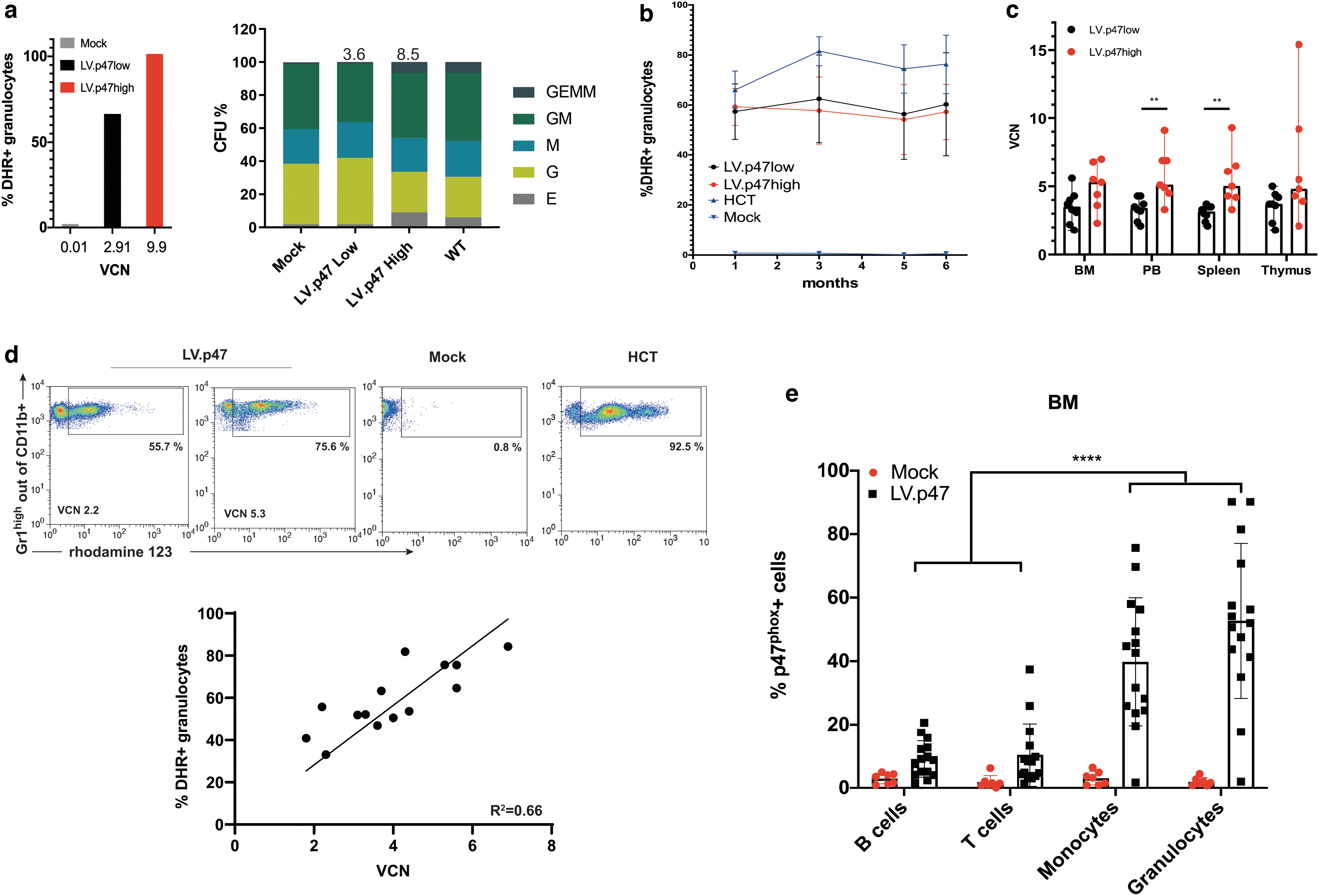
Biochemical correction of p47phox−/− mice by lentiviral gene therapy. p47phox−/− Lin− cells were transduced with the
Mice transplanted with untransduced p47phox−/− (Mock group; n = 8) or wild-type (HCT, hematopoietic cell transplantation group; n = 8) Lin− cells were used as negative and positive control, respectively. We also included a cohort of C57BL/6 (WT group; n = 6) and p47phox−/− (KO group; n = 4) animals to rule out any toxicity due to the gene therapy procedure itself.
Following transplantation, mice were monitored for up to 26 weeks with regular tail vein bleeds to check for the persistence of corrected cells over time (Fig. 2b and Supplementary Table S2). One mouse in the HCT group had to be sacrificed shortly after the gene therapy procedure due to engraftment failure. One mouse in the Mock group died during a tail vein bleed. One mouse in the LV. p47high group had to be sacrificed 4 months after transplantation following the development of a nasal granuloma, which has previously been shown to be a recurrent spontaneous infection in this mouse model.
18,19
Macroscopic examination of internal organs did not reveal any anomaly. Of note, this mouse (#10) had no gene therapy-corrected cells in the blood at any time point as shown by the lack of DHR positivity and VCN, suggesting that the graft was not successful (Supplementary Table S2). At the end of the experiment (6 months after transplantation), we analyzed the biochemical reconstitution of gene therapy-treated mice and the number of vector integrants. The analysis of VCN revealed that the majority of gene therapy-treated mice had between 3 and 6 vector copies in hematopoietic organs (Fig. 2c), confirming multiple integration events, a finding that underpins the validity of this genotoxicity study. We also found a good correlation (R
2
= 0.66) between vector copies and DHR positivity in the bone marrow of gene therapy-treated mice, with around 2 vector copies needed to achieve more than 30% functional neutrophils (Fig. 2c). Finally, we confirmed in this setting as previously shown
8
that the
In terms of general health, all animals increased body weight over time and similar growth trajectories were exhibited among all experimental groups (Fig. 3a). Flow cytometric analysis of the B, T, and myeloid compartment of peripheral blood, bone marrow, spleen, and thymus (Fig. 3b and Supplementary Fig. S3) showed no difference in hematopoiesis among the experimental groups. The spleen of one mouse belonging to the LV.p47low group (Supplementary Fig. S3) exhibited an elevated number of myeloid cells by flow cytometry. However, that sample contained >90% dead events, a confounding factor that could have impacted the technical validity of the data. Of note, the same mouse had normal lineage distributions in the other organs, normal blood parameters (Supplementary Fig. S4a–d), normal spleen weight (Supplementary Fig. S4e), and normal histology as discussed below. Hematological analysis showed that hemoglobin levels (HGB) and total count of erythrocytes (RBCs), white blood cells (WBCs), or circulating platelets (PLT) were similar between all groups.
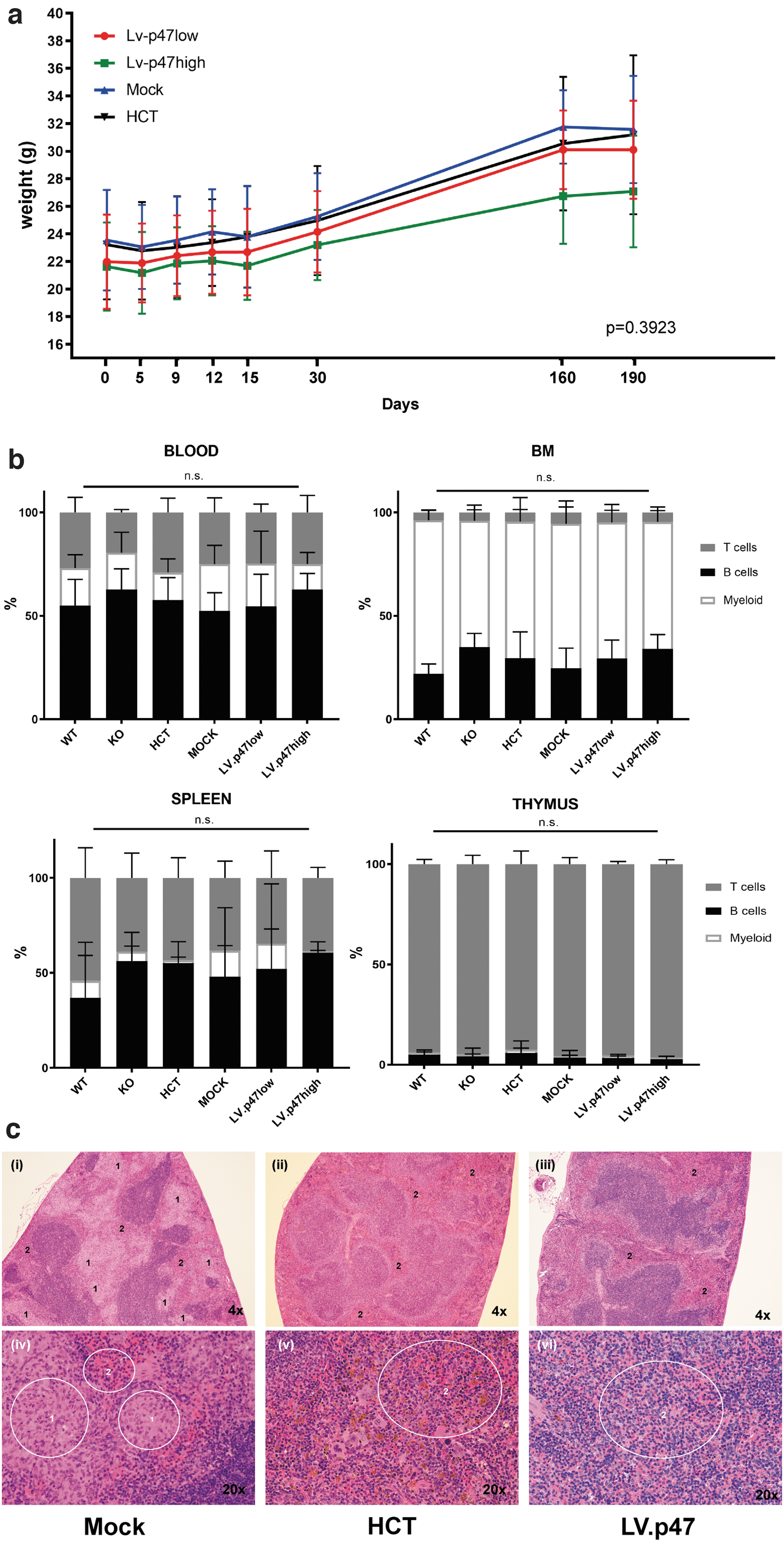
In vivo genotoxicity: body weight, hematological and histopathological analysis of transplanted mice.
Histopathological analysis of hematopoietic organs did not reveal any evidence of hematopoietic malignancy (Supplementary Data). The thymus had normal cellular density of lymphocytes in most animals and minimal-to-mild hypercellularity of the bone marrow was observed in all five experimental groups with no obvious difference between groups with regard to bone marrow histology. Splenic architecture and cellularity was normal in the majority of animals. Of note, four mice, two in the KO group (50%) and two in the Mock group (29%) showed macrophage infiltration either of the bone marrow or of the splenic red pulp. Infiltrating macrophages were often clustered together forming multifocal granuloma-like lesions, indicative of an inflammatory state. 20 Granuloma in the splenic red pulp of a representative mouse belonging to the Mock group (i, iv), characterized by macrophages with abundant pale cytoplasm compressing surrounding cells, is shown in Fig. 3c. In contrast, in the HCT (ii, v) and LV.p47 (iii, vi) groups the splenic red pulp showed an even dispersion of multiple cell types, with no mechanical tissue displacement, suggesting a rescue of the inflammatory phenotype by gene therapy.
From this set of experiments we can conclude that transduction with the
Biodistribution
To conclude the safety analysis of our lentiviral gene therapy strategy, we evaluated the effects of lentiviral transduction on the engraftment and repopulation ability of human CD34+ cells from mobilized blood, which is the most common starting material for clinical gene therapy. Indeed, transduction of hematopoietic stem cells in the context of gene therapy results in the expression of the therapeutic gene in progenitor cells and may alter their innate homing and engraftment properties or their ability to differentiate into myeloid and lymphoid lineages. Therefore, we performed a biodistribution study using human CD34+ cells that were transplanted into immunodeficient non-obese diabetic (NOD)-SCID Il2rg−/− (NSG) mice to evaluate any potential accumulation of gene-transduced cells in nonhematopoietic organs (Supplementary Data), as already described in Carriglio et al.
22
Following the protocol of the current gene therapy trial for X-CGD,
10
we transduced CD34+ cells with the
The hCD45 staining of bone marrow showed good levels of human cell engraftment in both UN and LV.p47 mice, with no significant difference among the groups (Table 2 and Supplementary Fig. S5b). Vector copies ranging between 1.0 and 2.8 were found in the bone marrow (BM) of experimental mice with no difference between sex (Table 2 and Supplementary Fig. S5c). In both experimental groups, we found a high percentage of CD19+ B cells and a smaller percentage of CD13+ myeloid cells as expected in NSG mice that predominantly sustain the development of B cells 23 (Supplementary Fig. S5d). The UN and LV.p47 groups had similar levels of human cell engraftment and lineage representation in peripheral blood, spleen, and thymus (data not shown) demonstrating once more that the vector does not have a negative impact on the engraftment capacity of gene therapy-treated cells. As expected, engraftment of human cells into the hematopoietic organs (blood, bone marrow, spleen, and thymus) of the recipient mice was much higher than in nonhematopoietic organs (Table 3). The average engraftment in the bone marrow was ∼50%, whereas engraftment of human cells in all nonhematopoietic organs was between 0.5% and 3.6% in liver, lung, muscle, and kidney and lower than 0.5% in brain, gonads, and heart, as assessed by ddPCR. We did not detect any difference in the distribution of human cells in nonhematopoietic tissues between the LV.p47 and UN groups. Importantly, vector copies were found only in the presence of the human albumin genome and in a similar range as in target organs, suggesting that the vector is present only in human cells and that vector-bearing cells do not accumulate in nontarget organs.
Human cell engraftment and average vector copy number per human cell in hematopoietic organs of NSG mice transplanted with untransduced (UN) or
Percentage of human cells detected by flow cytometry (hCD45+) and ddPCR (hAlb: hAlb/hAlb+mTitin) in the peripheral blood and lymphoid organs with respective average vector copy number per human cell; see Supplementary Materials and Methods for more information. Results are shown as mean ± SD calculated on numerical values >LOQ from animals showing successful engraftment (defined as ≥1% human CD45+ cells in BM at week 11). Statistical analysis: one-way ANOVA followed by Tukey's multicomparison, ns = not significant between UN and LV. p47 groups in all the organs.
Three samples showing an engraftment of 86% and 82% and 61% according to ddPCR were removed (outliers were detected by a Grubb's test considering a totality of 36 samples in PB).
ANOVA, analysis of variance; BM, bone marrow; ddPCR, droplet digital polymerase chain reaction; LOQ, limit of quantification; N/A, not available; NSG, immunodeficient non-obese diabetic (NOD)-SCID Il2rg−/−; PB, peripheral blood; SD, standard deviation.
Human cell engraftment and average vector copy number per human cell in nonhematopoietic organs of NSG mice transplanted with untransduced (UN) or
Percentage of human cells detected by ddPCR (hAlb: hAlbumin/hAlbumin+mTitin) in nontarget organs with respective VCN; see Supplementary Materials and Methods for more information. Results are given as mean ± SD calculated on numerical values >LOQ from animals showing successful engraftment (defined as ≥1% human CD45+ cells in BM at week 11). Statistical analysis: one-way ANOVA followed by Tukey's multicomparison. n.s. between UN and LV.p47 groups in all the organs.
Only one animal.
Clinical protocol for the gene therapy of human p47phoxCGD CD34+ cells
A bottleneck for any gene therapy clinical trial is the cost and availability of large quantities of “GMP-grade” lentiviral vectors. While we were performing preclinical studies, several reports highlighted the importance of transduction enhancers to reduce vector quantities required for transduction, minimizing the time for ex vivo manipulation of CD34+ cells.
15,24
We used GMP-grade clinical vector in combination with 1 mg/mL LentiBOOST and 4 μg/mL Protamine Sulfate to transduce CD34+ cells isolated from healthy donors. To combine experiments using different MOIs of the
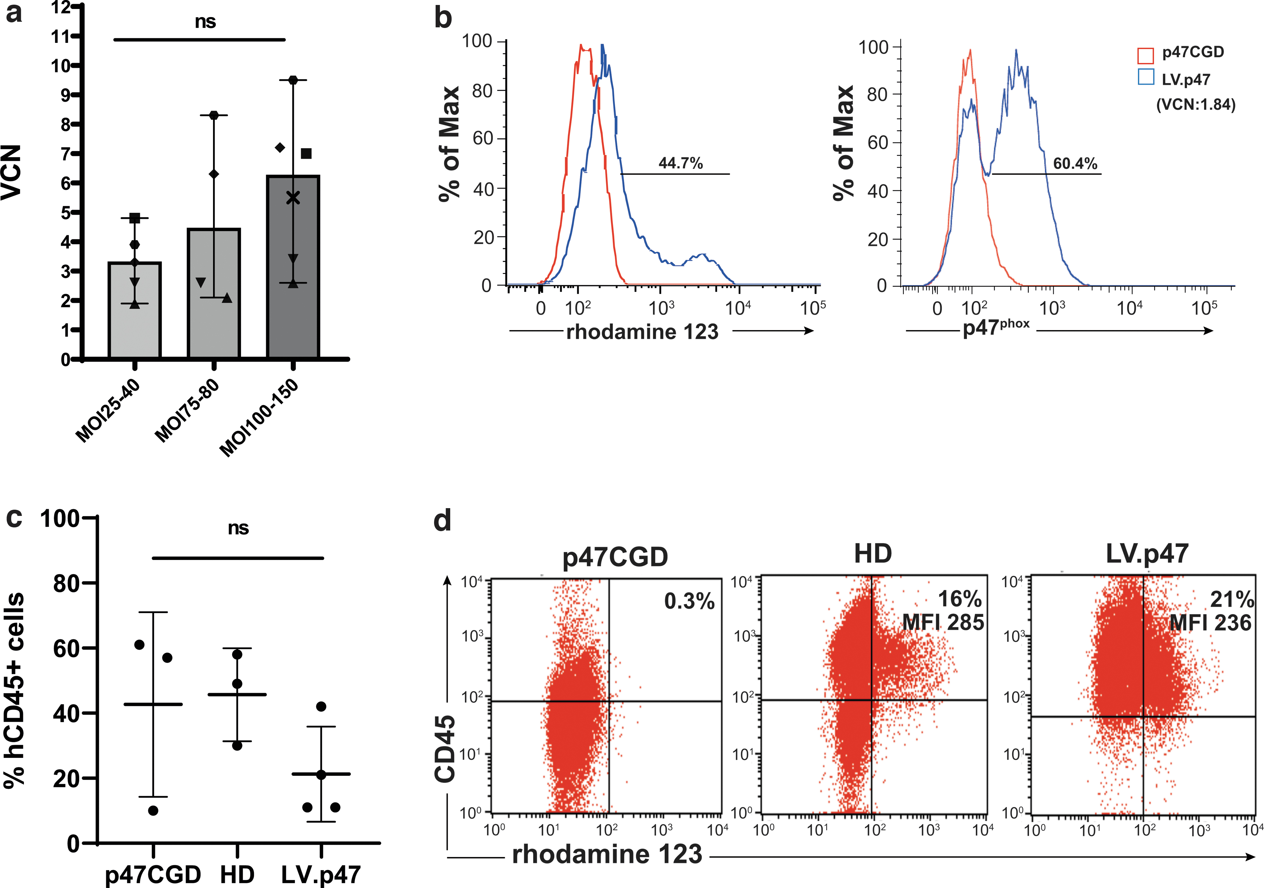
Vector performance on CD34+ cells from a healthy donor and from a p47phoxCGD patient.
Based on our observations in animal models,
8
VCN between 2 and 3 resulted in >30% functional neutrophils. Of note, transduction of CD34+ HSPCs using the clinical grade
When the high MOI protocol (MOI 130) was applied to p47phoxCGD CD34+ cells, we achieved a VCN of 1.84 with 60.4% of the cells positive for p47phox and 44.7% of the cells functionally corrected after 14 days of in vitro culture (Fig. 4b). The rather low vector copy numbers observed in the CGD samples could be partlially explained by the freezing and thawing of the vector (this was done to preserve vials of the clinical batch). Transduced cells (LV.p47) were able to engraft busulfan-conditioned NSG mice and an average of 5.8 ± 0.7 VCN was found in the bone marrow of gene therapy-treated animals, 16 weeks after transplantation. Due to the high variability in the percentage of hCD45+ cells found in the LV.p47 group (n = 4) and in mice transplanted with naive p47phoxCGD (p47CGD, n = 3) or healthy donor (HD, n = 3) cells, we could not draw any conclusion on the effects of lentiviral transduction on human cell engraftment in this experiment (Fig. 4c). CD34+ cells were isolated from pooled bone marrows of NSG mice from each group (LV.p47, p47CGD, HD) and cultured in differentiation media for 3 weeks to assess the percentage of functional neutrophils. We found not only a similar percentage of functional neutrophils between the LV.p47 and HD samples but also similar levels of NADPH oxidase activity as suggested by the values of mean fluoresce intensity in each DHR plot (Fig. 4d).
Conclusions
Early gene therapy trials for the X-linked form of CGD have highlighted the safety issues concerning the use of gamma retroviral vectors, which could cause insertional mutagensis and clonal expansion. 28,29
This study shows that the
In xenotranplantation experiments, we have also shown that the integration of the lentiviral vector in human CD34+ cells does not alter the engraftment or the differentiation ability of HSPCs nor the biodistribution of progeny hematopoietic cells. The clinical protocol, using transduction enhancers, resulted in a VCN higher than 2 in almost all the samples analyzed, regardless of the MOI used (from 25 to 150), and in good rescue of NADPH oxidase function when the vector was tested in p47phoxCGD cells.
In conclusion, in this study, we report that our lentiviral gene therapy protocol is ready for translation into the clinic.
Footnotes
Acknowledgments
The authors thank Drs. Martyn Dow and Richard Virgile from Propath for the histopatholgy data; Ms. Marta Zinicola and Dr. Catarina Cunha Santos for assistance in processing murine tissues.
Author Disclosure
The authors declare no conflict of interest. A.J.T. is on the Scientific Advisory Board of Orchard Therapeutics and Rocket Pharmaceuticals. H.L.M. is on the Scientific Advisory Board of Orchard Therapeutics and on the Safety Monitoring Board of Rocket Pharmaceuticals. H.B.G. is Chief Executive Officer for Orchard Therapeutics.
Funding Information
The work was supported by the UCL Technology Fund, the NIHR Biomedical Research Centre at Great Ormond Street Hospital for Children NHS Foundation Trust, the National Institute for Health Research (NIHR)-Blood and Transplant Research Unit, 2014-10074, University College London and by the intramural program of the National Institute of Allergy and Infectious Diseases, NIH under project nos. ZIA AI000644 and ZIA AI000989. This research was funded in part by the Wellcome Trust (217112/Z/19/Z). M.R. and
Supplementary Material
Supplementary Data
Supplementary Table S1
Supplementary Table S2
Supplementary Figure S1
Supplementary Figure S2
Supplementary Figure S3
Supplementary Figure S4
Supplementary Figure S5
References
Supplementary Material
Please find the following supplemental material available below.
For Open Access articles published under a Creative Commons License, all supplemental material carries the same license as the article it is associated with.
For non-Open Access articles published, all supplemental material carries a non-exclusive license, and permission requests for re-use of supplemental material or any part of supplemental material shall be sent directly to the copyright owner as specified in the copyright notice associated with the article.