
Abstract
In our previous studies, a novel gene therapy approach was developed based on a plasmid vector pSecTag2B in which recombinant HNP1 gene was regulated under a cytomegalovirus promoter to encode a mature human neutrophil peptide-1 (HNP1) form. We showed for the first time in various tumor models, including human cancer xenografts, that overexpression of HNP1 in the tumor milieu by intratumoral pSecTag-HNP1 (pHNP1) administration efficiently attenuated in vivo tumor progression, mediated host immune responses to tumors, and produced a synergistic effect when combined with chemotherapeutics. In this study, a preclinical safety investigation of HNP1 gene therapy was conducted in nonhuman primates. Eleven cynomolgus monkeys were divided into three groups of three to four animals each and received either repeated s.c. injections of pHNP1/cationic liposome complexes at a low (0.625 mg/kg) or a high (2.5 mg/kg) dose or glucose as control. Significant HNP1 in vivo accumulation was detected after consecutive administrations. All primates reached the end of the study with good body conditions. Injection site inflammation was the only obvious toxic reaction during observation period. In addition, elevation of monocyte/macrophage and neutrophil as well as decline of lymphocyte were detected in the peripheral blood of pHNP1-treated primates. These alterations were partially alleviated at the end of observation period. Besides, dose-related histopathological changes of the immune organs were observed at necropsy, including a minimal thymic lymphocyte decrease and a minimal-to-mild lymph node erythrocyte increase, but which cannot be excluded from HNP1-induced immune reactions. Together, these data support future clinical studies of pHNP1-based local gene delivery in tumor patients.
Introduction
Human neutrophil peptides (HNP1–4), also called human α-defensins, are a family of evolutionarily closely related small cationic antimicrobial peptides, which constitutively expressed by neutrophils and secreted at an increased level during infection. 1 Among them HNP1–3 showed a high degree of similarities in amino acid sequence and bioactivities. 2 HNP1–3 expression has been reported in many types of human tumors, 3 –7 correlated with cancer cell cytolysis and apoptosis, 8 –11 angiogenesis inhibition, 12 and reversion of tumor immune microenvironment. 13
Given the toxicities of HNP1 to viral gene delivery systems, 14 –17 a plasmid DNA delivery system was constructed previously, encoding a secretable form of mature HNP1. We showed for the first time in several cancer models, including human cancer xenograft, that HNP1 overexpression in the tumor milieu could efficiently attenuate the in vivo tumor progression, and at the same time, mediated a potent systemic antitumor effect. Intratumorally expressed HNP1 effectively induced tumor apoptosis, inhibited angiogenesis, and simultaneously triggered tumor-specific host immune responses. 18,19 Our further investigations found that when co-administrated with chemotherapeutics, HNP1 showed a synergistic treatment effect and markedly enhanced the intracellular drug accumulation. 20 Recently, plasmid HNP1 was also proved as an effective immunotherapeutic agent against Leishmania infection in mouse models. 21 Based on these findings, HNP1-based gene therapy exhibits promising potentials for clinical application.
In this study, the preclinical safety of locally administrated pHNP1/cationic liposome complexes were evaluated in nonhuman primates by repeated s.c. injections. The property, degree, dose-effect and time-effect relationships, and reversibility of potential pathological changes were comprehensively investigated.
Materials and Methods
For Materials and Methods please refer Supplementary Data.
Results and Discussions
Relevance for clinical trials
This study was conducted to support a phase I clinical trial of plasmid pSecTag-HNP1 (pHNP1)-based gene therapy for local tumor treatment. The delivery system encodes a secretable form of mature HNP1, and cationic liposome was used to enhance the transfection efficiency.
Objective and study design
This study was designed to evaluate the preclinical safety of pHNP1 therapy and aimed to investigate (1) the property, degree, dose-effect and time-effect relationships, and reversibility of possible toxic reactions, (2) to speculate the possible toxic target organs, (3) to characterize the toxicokinetics and relationship between exposure dose and toxicological results.
According to previous effectiveness studies in mice tumor models, 18 –20 the dosages of pHNP1 were designated as 0.625 and 2.5 mg/kg, which were one- and fourfold of the proposed clinical dose, respectively. A total of 11 cynomolgus monkeys were randomly assigned to the following groups: control, pHNP1 low dose (0.625 mg/kg), and high dose (2.5 mg/kg). The control group was consisted with two females and one male, whereas the other two groups comprised two females and two males. The inside of limbs were injected s.c. with pHNP1/liposome complexes every 3 days for a total of three times, followed by a 2-week observation. The dosage was adjusted according to the most recent weight measurement. To exclude the possible influence caused by local injection, primates in the control group were given the same volume of 5% glucose. The first day of administration was designated as day 1, and primates were sacrificed on day 23 for histological evaluation. Clinical observation was conducted every day. Body weights and food consumptions were measured weekly. Body temperature was detected at 2, 24, and 48 h before and after the first administration. II lead electrocardiogram and blood pressure data were collected at 2, 24, and 48 h before and after the first dose, and 2 h after the last dose. Clinical chemistry, hematology, coagulation parameters, and immune indices were evaluated on days 0, 5, 13, and 23.
Summary of data
Local toxicity at the injection site
There were no obvious toxicity reactions during the observation period except injection site redness and swelling. All primates reached the end of the study with good body conditions. Two days after the first administration, injection site inflammation occurred in all primates from the pHNP1 groups. On day 5, 2/4 primates in the low-dose group and 4/4 primates in the high-dose group developed skin ruptures of ∼5 mm in diameter (Fig. 1A), which were gradually alleviated from days 8 to 13. Besides, no abnormalities were observed in body weight or food consumption (Supplementary Table S1). There were no toxicological significant changes in body temperature or cardiovascular system (Supplementary Table S2 and S3).
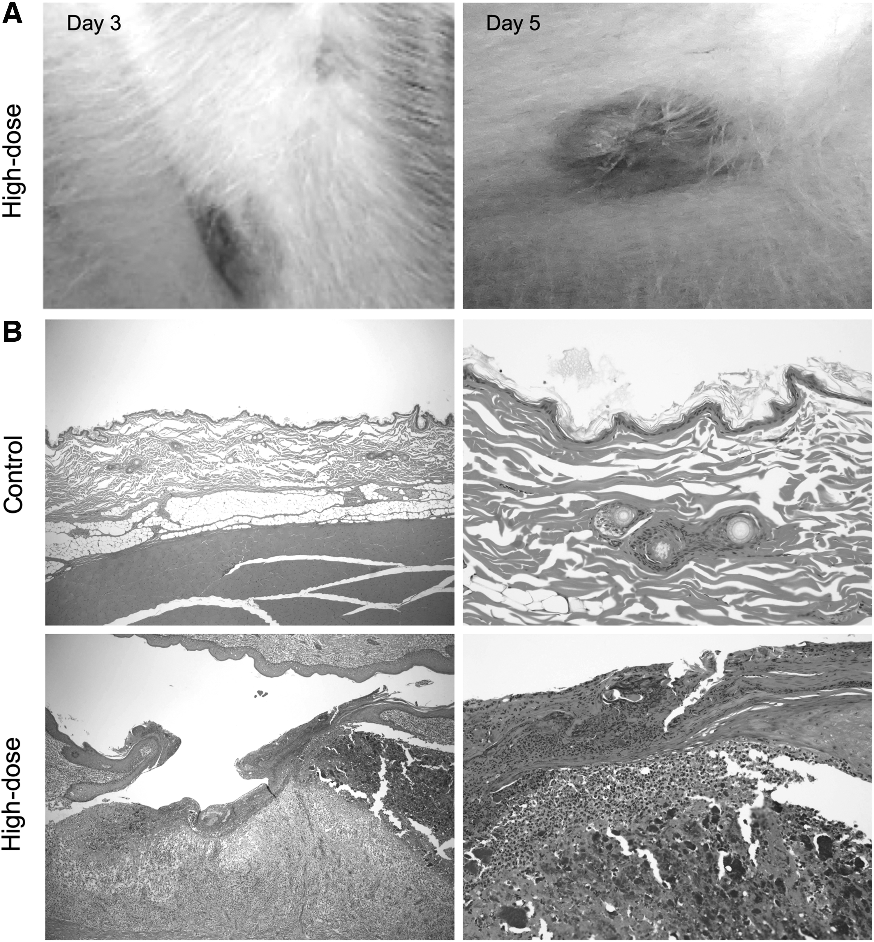
Local toxicities of pHNP1 administration. Nonhuman primates were received consecutive s.c. injections of pHNP1/liposome complexes.
In addition, thickening and crusting of the injection site skin were observed at necropsy. In the low-dose group skin thickening was found in 2/4 primates, whereas the others had scab formation at the administration site. The average area of thickened skin was 11 × 10 × 2 mm, and the scab diameter ranged from 2 to 7 mm. In the high-dose group, skin thickening occurred in 1/4 primates, and all primates scabbed at the injection site. The average area of thickened skin was 11 × 6 × 3 mm, and the scab diameter ranged from 1 to 7 mm. Furthermore, hematoxylin and eosin (H&E) staining showed that ulceration and foreign body granuloma developed at pHNP1 injection site (Fig. 1B). In the low-dose group, one primate had multiorgan granulomas that were different from those observed in other primates and thus considered as spontaneous lesions. The remaining three primates were noted with a moderate-to-severe degree of foreign body granuloma (3/3), and two of which had a mild-to-moderate degree of ulceration. In the high-dose group, moderate-to-severe foreign body granulomas were observed in all primates (4/4), as well as a minimal-to-mild degree of ulceration (Table 1).
Microscopic findings at necropsy on day 23 (14 days post the last dose)
Level: 1 = minimal, 2 = mild, 3 = moderate, 4 = severe, 5 = very severe.
Together, these findings indicated that pHNP1/liposome complexes had obvious toxicity to the administration site. The binding of HNP1–3 to cellular membrane has been reported to be nonspecific and can lead to membrane disruption and cellular content leakage. 22 One rational explanation for this local inflammation is that the regional aggregation of HNP1 gave rise to nonspecific binding and death of normal cells, which released a large number of cellular contents and caused subsequent infiltration of inflammatory cells into the administration site.
Immune system
HNP1–3 have been shown to regulate host innate and adaptive immunity by functioning as chemoattractant for immune cells such as monocytes and T lymphocytes. 23,24 In addition, bacterial-derived pDNA has been reported to have strong immunostimulatory and proinflammatory capacities because of the largely unmethylated DNA sequence. 25,26 To assess the influence of pHNP1 local delivery on the immune system, peripheral blood immune cells, and pathological changes of immune organs (thymus, axillary lymph node, and spleen) were analyzed. Flow cytometry detection showed that peripheral CD3−CD14+ monocyte/macrophage percentage began to increase in pHNP1-treated groups 4 days after the first dose (day 5), with a further increment 4 days after the last dose (day 13), especially in the high-dose group (p < 0.05), whereas CD20+ B cell percentage tended to decline from day 5. This trend was maintained to the end of the observation period (Table 2). Besides, no significant abnormalities were found in T lymphocytes or NK cells.
Immunological analysis in cynomolgus monkeys treated with pSecTag-human neutrophil peptide-1/liposome complexes
Results are given as means ± SD.
Indicate a statistically significant difference (p < 0.05) when compared with the control group.
SD, standard deviation.
Furthermore, hematological test revealed significant elevation of neutrophil as well as decline of lymphocyte after pHNP1 administration. On day 13, compared with the control group, the average peripheral neutrophil percentage in pHNP1-treated groups showed a 1.5-fold increase and the absolute neutrophil count remarkably raised. Meanwhile, the average peripheral lymphocyte percentage showed a onefold reduction, and the absolute lymphocyte count slightly decreased in the high-dose group. This trend was alleviated at the end of the observation period. Corresponding to flow cytometry results, significant monocyte percentage increase was also observed in the high-dose group on day 5, and this trend was maintained to the end of the observation period.
At necropsy, a dose-related thymic lymphocyte decrease as well as erythrocyte elevation in the lymph node were observed in pHNP1-treated primates by histological examination. H&E staining of thymus showed a minimal degree of lymphocyte decrease in 1/4 primates of the low-dose group and 3/4 primates of the high-dose group (Table 1 and Fig. 2A). Meanwhile, 1/4 primates in the low-dose group and 2/4 primates in the high-dose group showed minimal-to-mild antral erythrocyte increase in the axillary lymph node, respectively (Table 1 and Fig. 2B). No obvious histological abnormalities were noted in the spleen. No significant changes were observed in organ weight or organ indexes (Supplementary Table S5). Together, these finding indicated that local administration of pHNP1/liposome complexes caused dose-related changes in the immune system, but which cannot be excluded from HNP1-induced immune reactions.

Histopathologic findings of toxicities to the immune systems in nonhuman primates after pHNP1 administration.
Hematopoietic system
CpG motifs in bacterial-derived pDNA have been reported to contribute substantially to cationic lipid-pDNA complex induced adverse hematological changes, typified by profound leukopenia and thrombocytopenia. 27 To determine the influence of pHNP1 local administration on hematopoietic system, hematological test and bone marrow smear detection were conducted (Supplementary Table S6). In peripheral blood, no significant abnormalities were found in white blood cell count, eosinophil, basophil, red blood cell, hemoglobin, hematocrit, mean corpuscular volume, mean corpuscular hemoglobin, mean corpuscular hemoglobin concentration, platelet, reticulocyte, activated partial thromboplastin time, or prothrombin time (Supplementary Table S4). In contrast, hematological analysis of bone marrow smear showed significant reduction in granulocyte/erythroid (G/E) ratio as well as elevations of erythrocyte, rubricyte, and eosinophil percentages in pHNP1-treated primates, especially the high-dose group (p < 0.05). Compared with the control group, the average G/E ratio and erythroid percentage showed a 1.3-fold decrease and a 1.2-fold increase in pHNP1-treated groups, respectively. Meanwhile, in the high-dose group, increases in the percentages of eosinophil (2.4-fold) and rubricyte (1.6-fold) were observed (p < 0.05), and this trend was also observed in the low-dose group but to a lesser degree. However, the erythrocytosis is relatively mild and common, and thus not considered as significant histopathological alteration. Besides, no significant abnormalities were observed in granulocyte, myeloblast, promyelocyte, basophil, pronormoblast, prorubricyte, metarubricyte, lymphocyte, monocyte, megakaryocyte, or other cells (Supplementary Table S6). No hyperactive nor arrested cell proliferations of nucleated cells were noticed in the bone marrow. These findings suggested that local administration of pHNP1/liposome complexes had no significant influence on the hematopoietic systems.
Liver system
Overexpression of HNP-1 has been reported to induce hepatic fibrosis in nonalcoholic steatohepatitis by inducing hepatic stellate cell proliferation,
28
and enhance both hepatic fibrosis and hepatocyte apoptosis in alcoholic liver disease.
29
In addition, systemic administration of cationic lipid-pDNA complexes has been reported to induce serologic changes typified by elevated levels of serum transaminases, indicating potential liver injury.
30,31
In this study, serum chemistry detection showed that serum albumin and albumin/globulin (A/G) ratio trended to decrease in pHNP1-treated primates from days 5 to 23, whereas
Toxicokinetic analysis following repeated administration
For toxicokinetic analysis, serum HNP1 concentrations were detected at 0, 1, 8, 24, 48, and 72 h after the first and last doses by enzyme-linked immunosorbent assay. Before the initiation of treatment, HNP1 concentrations in all primates were below the lower limit of quantitation (LLOQ = 0.500 ng/mL), indicating no background systemic exposure. In the control group, HNP1 was undetectable throughout the experiment. At 1 h after the first injection, blood HNP1 was quantifiable in most pHNP1-treated primates. At each time point, the average serum HNP1 concentrations were approximate between the two groups, which was possibly affected by the DNA transfection efficiency and restriction of gene expression-related enzymes (Fig. 3A). After the last injection, the average serum HNP1 concentration in the high-dose group was marked higher than that in the low-dose group at different time points (Fig. 3B).
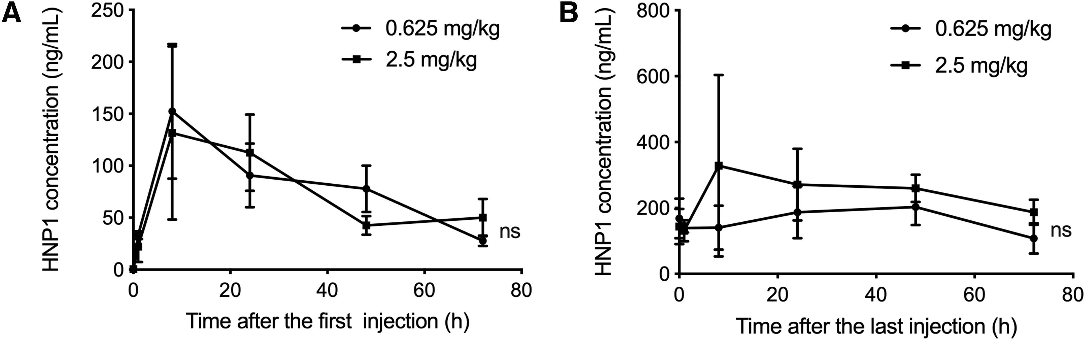
Serum HNP1 concentrations after pHNP1 injections. Blood samples were collected at 0, 1, 8, 24, 48, and 72 h after the first (day 1) and last doses (day 9).
Systemic exposure to HNP1 was measured by maximum concentration (Cmax) and area under the concentration-time curve (AUClast). After the first injection, the average maximum time (Tmax) to peak concentration were 18 and 16 h in the low- and high-dose groups, respectively. The average values of Cmax and AUClast were approximate between the two pHNP1 groups. When dosage increased four times, Cmax and AUClast showed 1.041- and 0.921-fold elevation each. After the last injection, the average Tmax values were 18 and 22 h in the low- and high-dose groups, respectively. Compared with that after the first dose, the average Cmax showed 1.432- and 2.555-fold increases in the low- and high-dose groups, respectively, whereas the average AUClast showed 2.061- and 3.365-fold increases, indicating substantially higher systemic exposure in the 2.5 mg/kg pHNP1 group. And the average AUClast was 18272.707 h × ng/mL in the high-dose group after the last administration. When dosage increased four times, Cmax and AUClast showed 1.859- and 1.503-fold elevations each. The increase rate of AUClast exhibited a lower growth rate compared with that of the pHNP1 dosage, indicating in vivo HNP1 accumulation after consecutive administrations (Table 3).
Systemic exposure to human neutrophil peptide-1 after subcutaneous injection of pSecTag-human neutrophil peptide-1/liposome complexes in cynomolgus monkeys
Tmax, maximum time; Cmax, maximum concentration; AUClast, area under concentration-time curve 0 − t.
Conclusions
In this study, we evaluated the preclinical safety of a plasmid vector-based HNP1 gene therapy by repeated s.c. administrations in cynomolgus monkeys. In vivo HNP1 accumulation was observed after consecutive injections. Systemic exposures to HNP1 were approximate between the high-dose group and the low-dose group after the first injection, and substantially raised after consecutive administrations, especially in the high-dose group. All primates reached the end of the study with good body conditions. Injection site inflammation was the only obvious toxic reaction during observation period. Furthermore, elevation of monocyte/macrophage and neutrophil as well as decline of lymphocyte were detected in the peripheral blood of pHNP1-treated primates. These alterations were partially alleviated at the end of observation period. Besides, histopathological alterations of the immune system (axillary drainage lymph node and thymus) were detected at necropsy. However, since HNP1 has been shown to function as an immune regulator in both innate and adaptive immunity, these changes cannot be excluded from HNP1-induced immune reactions.
In the previous study that HNP1 as immunotherapeutic agent against Leishmania infection, pHNP1-based gene therapy has shown even more effective treatment effect than folded HNP1, indicating the promising future of HNP1 gene therapy. 21 In this study, our data described potential dose-related administration site and immune system alterations after consecutive pHNP1 therapies. Considering these histopathological changes observed in preclinical safety evaluation, it is better and safer to choose local instead of systemic treatment of pHNP1 in clinical applications.
Footnotes
Author Disclosure
No competing financial interests exist.
Funding Information
This study was supported by National Major Scientific and Technological Special Project for “Significant New Drugs Development” (2013ZX09301003-005).
Supplementary Material
Supplementary Data
Supplementary Figure S1
Supplementary Table S1
Supplementary Table S2
Supplementary Table S3
Supplementary Table S4
Supplementary Table S5
Supplementary Table S6
Supplementary Table S7
References
Supplementary Material
Please find the following supplemental material available below.
For Open Access articles published under a Creative Commons License, all supplemental material carries the same license as the article it is associated with.
For non-Open Access articles published, all supplemental material carries a non-exclusive license, and permission requests for re-use of supplemental material or any part of supplemental material shall be sent directly to the copyright owner as specified in the copyright notice associated with the article.