
Abstract
Scaffold-guided viral gene therapy is a novel, powerful tool to enhance the processes of tissue repair in articular cartilage lesions by the delivery and overexpression of therapeutic genes in a noninvasive, controlled release manner based on a procedure that may protect the gene vehicles from undesirable host immune responses. In this study, we examined the potential of transferring a recombinant adeno-associated virus (rAAV) vector carrying a sequence for the highly chondroregenerative transforming growth factor beta (TGF-β), using poly(ɛ-caprolactone) (PCL) films functionalized by the grafting of poly(sodium styrene sulfonate) (pNaSS) in chondrogenically competent bone marrow aspirates as future targets for therapy in cartilage lesions. Effective overexpression of TGF-β in the aspirates by rAAV was achieved upon delivery using pNaSS-grafted and ungrafted PCL films for up to 21 days (the longest time point evaluated), with superior levels using the grafted films, compared with respective conditions without vector coating. The production of rAAV-mediated TGF-β by pNaSS-grafted and ungrafted PCL films significantly triggered the biological activities and chondrogenic processes in the samples (proteoglycan and type-II collagen deposition and cell proliferation), while containing premature mineralization and hypertrophy relative to the other conditions, with overall superior effects supported by the pNaSS-grafted films. These observations demonstrate the potential of PCL film-assisted rAAV TGF-β gene transfer as a convenient, off-the-shelf technique to enhance the reparative potential of the bone marrow in patients in future approaches for improved cartilage repair.
Introduction
Articular cartilage defects remain critical issues in orthopedics due to the inadequate ability of the articular cartilage for self-repair in adult individuals 1,2 due to the lack of tissue vascularization that may provide access to chondroregenerative cells such as mesenchymal stromal cells (MSCs). 3,4 Also, there is currently no clinical treatment capable of restoring a fully functional hyaline cartilage with mechanically adapted extracellular matrix (proteoglycans and type-II collagen) in sites of cartilage lesions, even using with procedures such as microfracture and isolated cell transplantation, leading instead to the production of fibrocartilage (type-I collagen) prone to osteoarthritis progression. 1,2,5
Administration of bone marrow aspirates that can deliver chondrogenically competent MSCs in a more natural microenvironment in cartilage defects using less invasive techniques has been further attempted as a means to improve the quality of the repair tissue in treated patients, 6 but again without affording the production of hyaline cartilage. 6
Scaffold-guided gene therapy emerged as a promising tool to enhance translational cartilage repair by supporting the controlled delivery of therapeutic gene vectors for prolonged, safe expression of gene-based products in cartilage defects. 7,8 While early work focused on the release of nonviral 9 –11 and lentiviral vectors, 12,13 recent studies evaluated the benefits of such an approach using recombinant adeno-associated virus (rAAV) vectors, 14 –19 a class of vector that is more effective than transient nonviral vectors (rAAV transgenes persist in their targets for months to years) 20 and safer than lentiviral vectors that may activate oncogenes upon genome integration (rAAV are maintained as transcriptionally active episomes). 20,21
Furthermore, scaffold-guided rAAV gene therapy has the potential to protect the rAAV capsid proteins from neutralization by host (humoral) immune responses, 22 which may prevent direct rAAV therapy in patients. 7 Specifically, rAAV vectors have been used for experimental cartilage research upon release from hydrogels (fibrin, alginate, self-assembling peptides, polypseudorotaxanes, and poloxamers/poloxamines) 14 –17 and from solid scaffolds [poly(ɛ-caprolactone)—PCL]. 18,19
Solid materials such as PCL display a number of features that are critical for cartilage repair strategies as they offer biocompatible, solid, and mechanically stable scaffolding systems to support cell viability, activation, and targeting with rAAV, 23 –26 especially when further grafted with poly(sodium styrene sulfonate) (pNaSS), a compound that upholds reparative biological responses in musculoskeletal cells. 18,19,24,26
In light of our previous findings showing the feasibility of using pNaSS-grafted PCL films as an efficient rAAV gene-controlled release system to safely modify chondroreparative bone marrow aspirates 18 and enhance their chondrogenic potential when using an rAAV vector coding for the cartilage-specific sex-determining region Y-type high-mobility group box 9 (SOX9) transcription factor, 19 our goal in this study was to further test the possibility of transferring other candidate rAAV vectors in such aspirates to improve the reparative outcomes.
We focused on applying the highly chondrogenic and pleiotropic transforming growth factor beta (TGF-β) 4 by pNaSS-grafted PCL films due to the ability of this agent to stimulate anabolic, chondrogenic, and proliferative events in bone marrow aspirates following direct (scaffold free) rAAV transduction 27 as an alternative to the use of SOX9 that had no impact on cell proliferation in these samples. 19
The data show the superiority of pNaSS-grafted PCL films to mediate the delivery and overexpression of the therapeutic rAAV-human TGF beta (hTGF-β) vector for up to 21 days in human marrow aspirates over other conditions (rAAV/ungrafted PCL films, lack of rAAV coating on pNaSS-grafted and ungrafted PCL films as this vector class does not change the chondrogenic potential of PCL-treated aspirates), 18,19 promoting higher deposition of major cartilage matrix components (proteoglycans and type-II collagen) and cell proliferation, while counteracting undesirable mineralization and hypertrophy. These results provide evidence of the value of using PCL-guided rAAV gene therapy in future applications, aiming at enhancing cartilage repair in patients.
Materials and Methods
Reagents
Reagents were from Sigma-Aldrich (Munich, Germany), including 4-styrenesulfonic acid sodium salt hydrate (NaSS) (cat. no. 434574), unless otherwise indicated. The anti-TGF-β (V) antibody was obtained at Santa Cruz Biotechnology (Heidelberg, Germany), the anti-type-II collagen (AF-5710) and anti-type-I collagen (AF-5610) antibodies at Acris (Hiddenhausen, Germany), and the anti-type-X collagen (COL-10) antibody at Sigma-Aldrich. The biotinylated secondary antibodies and ABC reagent were purchased at Vector Laboratories (Alexis Deutschland GmbH, Grünberg, Germany).
The AAVanced Concentration Reagent was obtained at System Bioscience (Heidelberg, Germany), the TGF-β Quantikine enzyme-linked immunosorbent assay (ELISA) from R&D Systems (Mannheim, Germany), and the Cell Proliferation Reagent WST-1 at Roche Applied Science (Mannheim, Germany).
Bone marrow aspirates
Bone marrow aspirates (∼15 mL) were obtained from the distal femurs of patients undergoing total knee arthroplasty (n = 12, age 74 ± 3 years as ultimate targets for therapy) with approval from the Ethics Committee of the Saarland Physicians Council (Ärztekammer des Saarlandes, reference number Ha06/08). All patients gave their informed consent before inclusion in the evaluation performed according to the Helsinki Declaration. Aspirates containing MSCs (0.5–1.2 × 10⁹ cells/mL) were placed in 96-well plates (150 μL aspirate/well, 6.1 × 10⁷ cells) at 37°C 18,19,27 until immediate application of the various films as described below.
Preparation of the PCL films
The PCL films were generated by spin coating as described. 24 Briefly, PCL (60% [w/v] in dichloromethane) was dropped for spinning on a glass slide (30 s at 1,500 rpm) using a SPIN150-v3 SPS. The films were air dried for 2 h, vacuum dried for 24 h, and cut in 4-mm disks for grafting with pNaSS (1.3 × 10⁻⁵ mol/g) by ozonation (10 min at 30°C), followed by incubation in degassed NaSS (15% [w/v] in distilled water) for graft polymerization (3 h, 45°C) (Fig. 1). The films were washed in distilled water, 0.15 M NaCl, and phosphate-buffered saline and rinsed for vacuum drying. Some films were left ungrafted as controls. Characterization of the systems (charge, mechanical properties, etc.) has been already reported by us in previous work. 24,26,28 –30
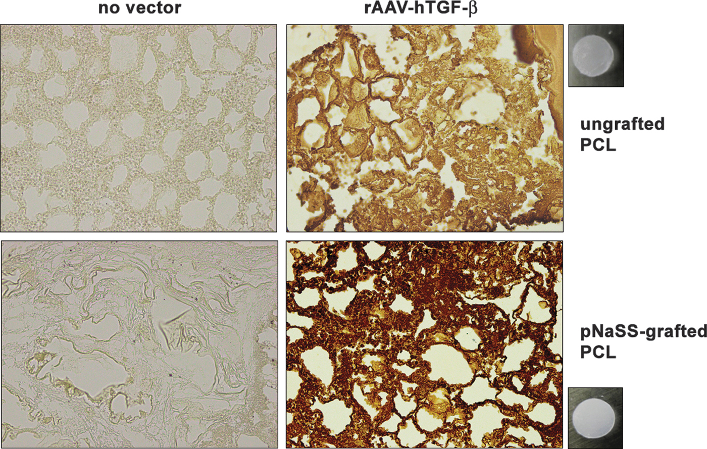
Detection of transgene (TGF-β) overexpression in human bone marrow aspirates transduced with rAAV TGF-β-coated PCL films. pNaSS-grafted and ungrafted PCL films (macroscopic photographs) were coated with rAAV-hTGF-β (40 μL, 8 × 10⁵ transgene copies) or left without vector coating before incubation with the aspirates in chondrogenic medium (150 μL, 6.1 × 10⁷ MSCs, i.e., MOI = 75). TGF-β expression was monitored by immunohistochemistry after 21 days of culture as described in the Materials and Methods (magnification × 20; all representative data). hTGF-β, human transforming growth factor beta; MOI, multiplicity of infection; MSCs, mesenchymal stromal cells; PCL, poly(ɛ-caprolactone); pNaSS, poly(sodium styrene sulfonate); rAAV, recombinant adeno-associated virus; TGF-β, transforming growth factor beta.
Generation of the rAAV vectors
The vectors were created using a parental AAV-2 genomic clone (pSSV9). 31,32 rAAV-hTGF-β carries a 1.2-kb hTGF-β cDNA sequence controlled by the cytomegalovirus immediate-early (CMV-IE) promoter. 27 Packaging of conventional vectors (not self-complementary) was performed by helper-free (two-plasmid) transfection in 293 cells using the packaging plasmid pXX2 and the adenovirus helper plasmid pXX6. 18,19 Vector purification was then performed using the AAVanced Concentration Reagent 18,19 and vector titration was managed by real-time PCR. 18,19,27 The method allowed to generate vector solutions at about 1010 transgene copies/mL (i.e., about 1/500 functional recombinant viral particles).
rAAV immobilization on PCL films
The rAAV vectors (40 μL, 8 × 10⁵ transgene copies) were incubated with 0.002% poly-
rAAV gene transfer
Aliquots of human bone marrow aspirates (150 μL/well in 96-well plates) containing MSCs (6.1 × 10⁷ cells) 18,19,27 were incubated with rAAV-coated PCL films (multiplicity of infefction [MOI] = 75) in the presence of fibrinogen/thrombin (17 mg/mL/5 U/mL) (Baxter, Volketswil, Switzerland). 18,19
The systems were placed either in defined chondrogenic differentiation medium (Dulbecco's modified Eagle's medium [DMEM] high glucose 4.5 g/L, 100 U/mL penicillin, 100 μg/mL streptomycin, 0.1 μM dexamethasone, 50 μg/mL ascorbic acid, 40 μg/mL proline, 110 μg/mL pyruvate, 6.25 μg/mL of insulin, 6.25 μg/mL transferrin, 6.25 μg/mL selenous acid, 1.25 μg/mL bovine serum albumin, 5.55 μg/mL linoleic acid, and 10 ng/mL TGF-β3) 18,19,27 or in osteogenic differentiation medium (StemPro Osteogenesis Differentiation kit with 100 U/mL penicillin and 100 μg/mL streptomycin) where indicated 18,19,27 (Life Technologies GmbH, Darmstadt, Germany) at 37°C under 5% CO2 for up to 21 days before the analyses.
Transgene expression
TGF-β expression was monitored by immunohistochemistry using a specific primary antibody, a biotinylated secondary antibody, and the ABC method with diaminobenzidine (DAB) as a chromogen for evaluation under light microscopy (Olympus BX45; Olympus, Hamburg, Germany). 27 TGF-β expression was also detected with a specific ELISA. 27 Measurements were performed on a GENios spectrophotometer/fluorometer (Tecan, Crailsheim, Germany).
Biological analyses
The systems were digested with papain and the proteoglycan contents were measured by binding to dimethylmethylene blue dye by normalization to the total cellular proteins (Pierce Thermo Scientific Protein Assay; Thermo Fisher Scientific, Schwerte, Germany). 18,19,27 Measurements were performed on a GENios spectrophotometer/fluorometer (Tecan). Cell viability was monitored with the Cell Proliferation Reagent WST-1 with OD450nm being proportional to the cell numbers 18,19,27 on a GENios spectrophotometer/fluorometer (Tecan).
Histology and immunohistochemistry
The systems were fixed in 4% formalin, dehydrated in graded alcohols, embedded in paraffin, and sectioned (3 μm). Sections were stained with hematoxylin and eosin (H&E) for cellularity, with toluidine blue for matrix proteoglycans, and with alizarin red for mineralization. 18,19,27 Immunohistochemistry was performed to monitor the type-II, type-I, and type-X collagen expression with specific primary antibodies, biotinylated secondary antibodies, and the ABC method with DAB. 18,19,27 Controls with lack of primary antibodies were tested to check for secondary immunoglobulins. Sections were examined under light microscopy (Olympus BX45).
Histomorphometric analyses
The % of TGF-β+ cells (TGF-β+ cells/total cell numbers on immunohistochemical sections), the intensities of toluidine blue and alizarin red staining (histological sections) and of type-II, type-I, and type-X collagen deposition (immunohistochemical sections), and the cell densities (cells/mm2) on H&E histological sections were assessed using four sections per condition with the SIS AnalySIS program (Olympus) and Adobe photoshop (Adobe Systems, Unterschleissheim, Germany). 18,19,27
Sections (toluidine blue, alizarin red, type-II/type-I/type-X collagen) were scored blind by two individuals for uniformity and density with a modified Bern grading score 18,19 as follows: 0 = no staining, 1 = heterogeneous and/or weak staining, 2 = homogeneous and/or moderate staining, 3 = homogeneous and/or intense staining, and 4 = very intense staining.
Real-time RT-PCR analysis
Total cellular RNA was extracted using the RNeasy Protect Mini Kit and an on-column RNase-free DNase treatment (Qiagen, Hilden, Germany). The RNA was eluted in 30 μL RNase-free water. Reverse transcription was next performed with 8 μL eluate using the 1st Strand cDNA Synthesis kit for RT-PCR (AMV) (Roche Applied Science). Real-time PCR amplification was performed on an Mx3000P QPCR system (Stratagene, Agilent Technologies, Waldbronn, Germany) with 3 μL cDNA product using the Brilliant SYBR Green QPCR Master Mix (Stratagene) 18,19,27 and with the following conditions: 10 min at 95°C, 55 amplification cycles (30-s denaturation, 95°C; 1-min annealing, 55°C; and 30-s extension, 72°C), denaturation 1 min at 95°C, and final incubation 30 s at 55°C.
The primers (Invitrogen GmbH) were SOX9 (chondrogenic marker; forward 5′-ACACACAGCTCACTCGACCTTG-3′; reverse 5′-GGGAATTCTGGTTGGTCCTCT-3′), type-II collagen (COL2A1; chondrogenic marker; forward 5′-GGACTTTTCTCCCCTCTCT-3′; reverse 5′-GACCCGAAGGTCTTACAGGA-3′), aggrecan (ACAN; chondrogenic marker; forward 5′-GAGATGGAGGGTGAGGTC-3′; reverse 5′-ACGCTGCCTCGGGCTTC-3′), type-I collagen (COL1A1; osteogenic marker; forward 5′-ACGTCCTGGTGAAGTTGGTC-3′; reverse 5′-ACCAGGGAAGCCTCTCTCTC-3′), type-X collagen (COL10A1; marker of hypertrophy; forward 5′-CCCTCTTGTTAGTGCCAACC-3′; reverse 5′-AGATTCCAGTCCTTGGGTCA-3′), and glyceraldehyde-3-phosphate dehydrogenase (GAPDH; housekeeping gene and internal control; forward, 5′-GAAGGTGAAGGTCGGAGTC-3′; reverse, 5′-GAAGATGGTGATGGGATTTC-3′) (all at a final concentration of 150 nM). 18,19,27
Control reactions included water and non-reverse-transcribed mRNA. The specificity of the generated products was confirmed by melting curve analysis and agarose gel electrophoresis. The threshold cycle (Ct) value for each gene was measured for each amplification using the MxPro QPCR software (Stratagene), with normalization of the value to GAPDH expression using the 2-ΔΔCt method. 18,19,27
Statistical analysis
Data are given as mean ± standard deviation of separate experiments. All conditions were performed in triplicate in three independent experiments per patient, using all patients in all experiments. Data were analyzed by two individuals blinded with respect to the different groups. The t test and the Mann–Whitney Rank Sum Test were employed where appropriate, with p-value <0.05 considered statistically significant.
Results
Effective TGF-β overexpression in human bone marrow aspirates mediated by the application of rAAV-hTGF-β/pNaSS-grafted PCL films
The candidate rAAV-hTGF-β vector was coated on pNaSS-grafted PCL films (TGF-β/pNaSS-grafted PCL) versus ungrafted films (TGF-β/ungrafted PCL) to evaluate capacity of the system to promote TGF-β expression over time in human bone marrow aspirates compared with other treatments (pNaSS-grafted and ungrafted PCL films lacking rAAV, i.e., no vector/pNaSS-grafted PCL and no vector/ungrafted PCL, respectively). 18,19
Efficient overexpression of TGF-β by rAAV gene delivery was noted in human bone marrow aspirates treated with the TGF-β/pNaSS-grafted and TGF-β/ungrafted PCL films for 21 days in chondrogenic medium as evidenced by significantly higher levels of TGF-β immunodetection versus samples receiving films without vector coating (up to 4-fold difference of % TGF-β+ cells, p ≤ 0.001) (Fig. 1 and Table 1). Notably, the % of TGF-β+ cells was higher when providing TGF-β/pNaSS-grafted films relative to TGF-β/ungrafted films (1.3-fold difference, p ≤ 0.001) (Fig. 1 and Table 1).
Histomorphometric and biological analyses in human bone marrow aspirates transduced with the recombinant adeno-associated virus-coated poly(ɛ-caprolactone) films
TGF-β is in % TGF-β+ cells/total cell numbers on immunohistochemically stained sections. Sections for toluidine blue staining, type-II/type-I/type-X collagen immunostaining, and alizarin red staining were scored using a modified Bern grading score, 18,19 with 0 = no staining, 1 = heterogeneous and/or weak staining, 2 = homogeneous and/or moderate staining, 3 = homogeneous and/or intense staining, and 4 = very intense staining. The proteoglycan contents are in ng/μg total proteins, the cell densities on H&E-stained sections in cells/mm2, and the results of the WST-1 assay as OD450nm. Values are provided as mean ± SD. Statistically significant relative to ano vector/ungrafted PCL, bno vector/pNaSS-grafted PCL, and cTGF-β/ungrafted PCL.
H&E, hematoxylin and eosin; PCL, poly(ɛ-caprolactone); pNaSS, poly(sodium styrene sulfonate); SD, standard deviation; TGF-β, transforming growth factor beta.
These results were corroborated by the findings of a TGF-β ELISA revealing prolonged, significantly higher levels of growth factor production when applying films coated with rAAV-hTGF-β relative to the other treatments (up to 12.4-fold difference, p ≤ 0.001) and a superior effect when using pNaSS-grafted PCL films (up to 2.3-fold difference relative to ungrafted films, p ≤ 0.001) (Table 2). Of further note, increases in TGF-β expression were significantly measured over time when using films coated with rAAV-hTGF-β (up to 2.3-fold difference between days 14 and 21, p ≤ 0.001) (Table 2).
Production of transforming growth factor beta in human bone marrow aspirates transduced with the recombinant adeno-associated virus-coated poly(ɛ-caprolactone) films
Values are expressed as mean ± SD in pg/mL/24 h. Statistically significant relative to ano vector/ungrafted PCL, bno vector/pNaSS-grafted PCL, cTGF-β/ungrafted PCL, and dearlier time point.
Overexpression of TGF-β by TGF-β/pNaSS-grafted PCL films enhances the deposition of proteoglycans and type-II collagen and the levels of cell viability in human bone marrow aspirates
The rAAV-hTGF-β candidate vector was next provided to human bone marrow aspirates by coating on pNaSS-grafted versus ungrafted PCL films to evaluate the potential of the systems to stimulate over time the biological activities and the chondrogenic events in these samples over time compared with other conditions.
Application of rAAV-hTGF-β using the pNaSS-grafted and ungrafted PCL films promoted significantly stronger proteoglycan deposition in the aspirates maintained for 21 days in chondrogenic medium, as seen by a more important toluidine blue staining relative to the other treatments (up to 3.2-fold difference, p ≤ 0.001) and with a superior effect when using pNaSS-grafted PCL films (1.2-fold difference relative to ungrafted films, p = 0.033) (Fig. 2 and Table 1).
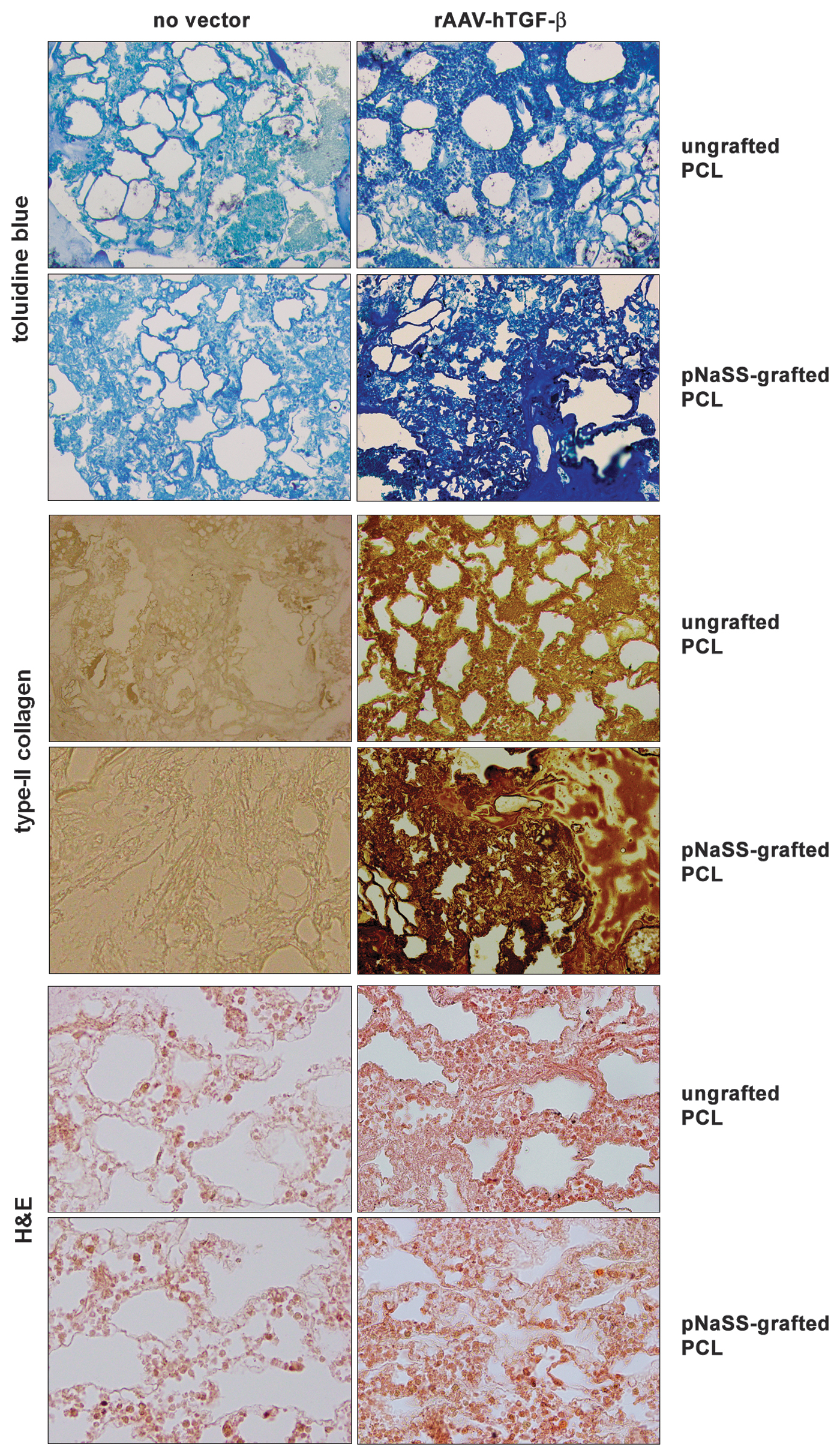
Analysis of chondroreparative activities in human bone marrow aspirates transduced with rAAV TGF-β-coated PCL films. pNaSS-grafted and ungrafted PCL films were coated with rAAV-hTGF-β or left without vector coating before incubation with the aspirates in chondrogenic medium as described in Figure 1. Matrix proteoglycans (toluidine blue staining), type-II collagen (immunohistochemistry), and cellularity (H&E staining) were assessed after 21 days of culture as described in the Materials and Methods section (magnification × 20, except for H&E at magnification × 40; all representative data). H&E, hematoxylin and eosin.
These findings were substantiated by an estimation of the proteoglycan contents in the samples, with higher levels achieved with TGF-β/pNaSS-grafted and TGF-β/ungrafted PCL films relative to the other treatments (up to 1.5-fold difference, p ≤ 0.007) and with a superior effect following pNaSS grafting (1.2-fold difference relative to ungrafted films, p ≤ 0.001) (Table 1).
Similar findings were noted when monitoring type-II collagen deposition, with stronger type-II collagen immunostaining using TGF-β/pNaSS-grafted or TGF-β/ungrafted PCL films relative to the other treatments (up to 4.5-fold difference, p ≤ 0.001) and with a superior effect when applying pNaSS-grafted PCL films (1.3-fold difference relative to ungrafted films, p = 0.018) (Fig. 2 and Table 1).
Furthermore, gene transfer of rAAV-hTGF-β using the pNaSS-grafted and ungrafted PCL films significantly increased the cell proliferation indices in the aspirates relative to the other treatments (cell densities: up to 1.7-fold difference, p ≤ 0.001; WST-1 assay: up to 2.3-fold difference, p ≤ 0.001) and with a superior effect when using pNaSS-grafted PCL films (cell densities: 1.2-fold difference relative to ungrafted films, p ≤ 0.001; WST-1 assay: 1.9-fold difference relative to ungrafted films, p ≤ 0.001) (Fig. 2 and Table 1).
Overexpression of TGF-β by TGF-β/pNaSS-grafted PCL films reduces mineralization and type-X collagen deposition in human bone marrow aspirates
The rAAV-hTGF-β candidate vector was then administered to human bone marrow aspirates by coating on pNaSS-grafted versus ungrafted PCL films to determine the potential of the systems to limit premature osteogenic events and hypertrophy in these samples over time compared with other conditions.
Application of rAAV-hTGF-β using the pNaSS-grafted and ungrafted PCL films significantly decreased matrix mineralization in the aspirates maintained for 21 days in osteogenic medium as seen by a reduced alizarin red staining relative to the other treatments (up to 4.7-fold difference, p ≤ 0.001) and with a superior effect when using pNaSS-grafted PCL films (2.3-fold difference relative to ungrafted films, p = 0.025) (Fig. 3 and Table 1).
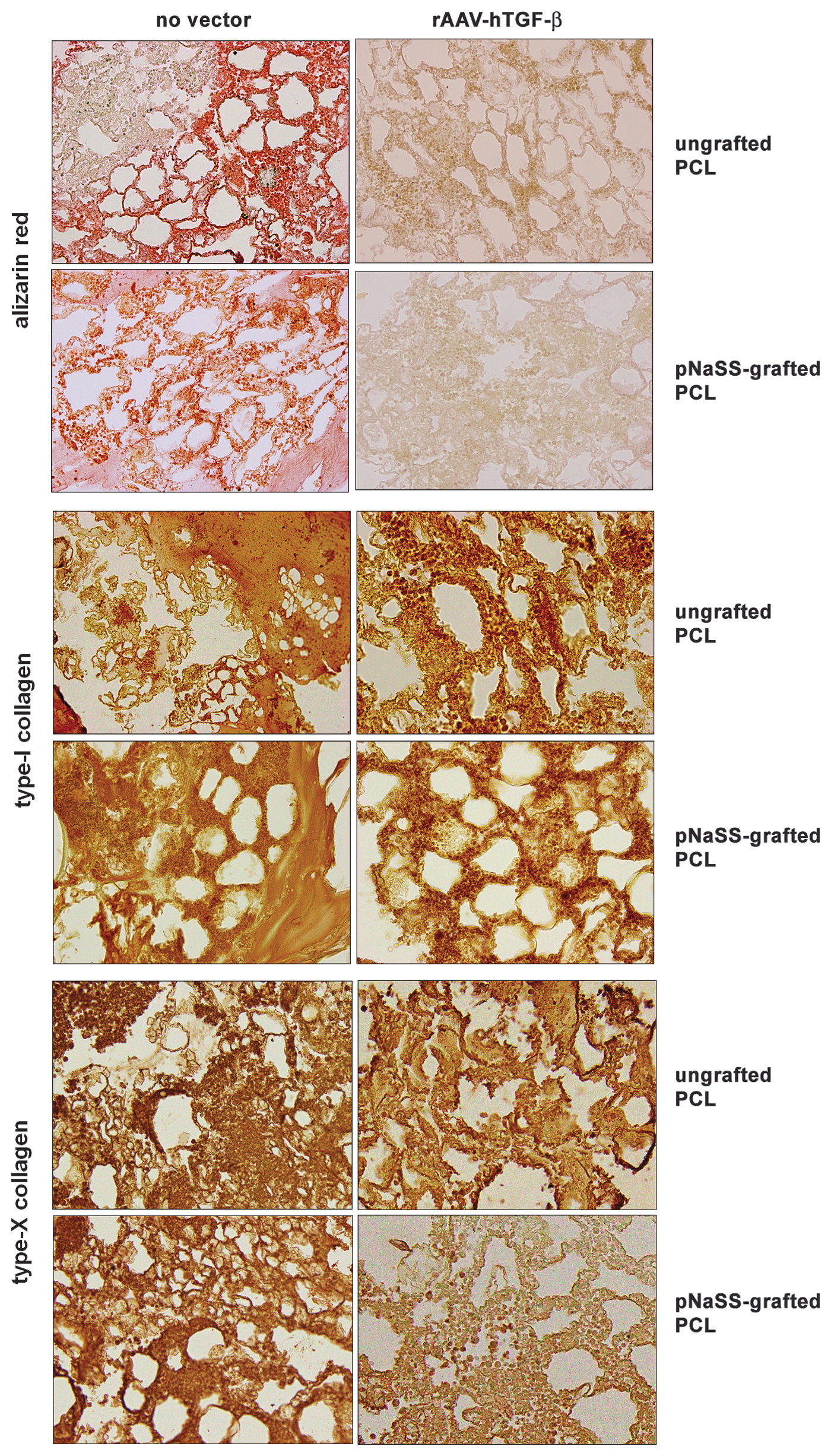
Analysis of mineralization and type-I and type-X collagen deposition in human bone marrow aspirates transduced with rAAV TGF-β-coated PCL films. pNaSS-grafted and ungrafted PCL films were coated with rAAV-hTGF-β or left without vector coating before incubation with the aspirates in osteogenic medium as described in Figures 1 and 2. Mineralization (alizarin red staining) and type-I and type-X collagen (immunohistochemistry) were assessed after 21 days of culture as described in the Materials and Methods section (magnification × 20; all representative data).
In addition, while there was no significant effect of rAAV-hTGF-β on osteogenic type-I collagen deposition in the aspirates relative to the other treatments, regardless of the type of film applied (p ≥ 0.173) (Fig. 3 and Table 1), administration of the therapeutic candidate by the pNaSS-grafted films significantly decreased hypertrophic type-X collagen deposition relative to the use of ungrafted PCL films and to the other treatments (up to 2.4-fold difference, p ≤ 0.001) (Fig. 3 and Table 1).
Overexpression of TGF-β by TGF-β/pNaSS-grafted PCL films stimulates the chondrogenic expression profiles and limits those for hypertrophy in human bone marrow aspirates
Overall, such findings were confirmed by real-time RT-PCR analyses of gene expression profiles in the aspirates after 21 days of culture with the candidate rAAV-hTGF-β vector provided with pNaSS-grafted and ungrafted PCL films versus the other treatments.
In a chondrogenic environment, application of rAAV-hTGF-β using the pNaSS-grafted and ungrafted PCL films significantly increased the levels of chondrogenic SOX9, COL2A1, and ACAN expression in the aspirates relative to each respective control treatment (2-, 2.4-, and 1.5-fold difference in the TGF-β/ungrafted compared with the ungrafted PCL films, respectively; 12.8-, 5.1-, and 2.4-fold difference in the TGF-β/pNaSS-grafted compared with the pNaSS-grafted PCL films, respectively; p ≤ 0.001) (Fig. 4A, B) and with a superior effect when using pNaSS-grafted PCL films (14.5-, 4.6-, and 2.2-fold difference in the TGF-β/pNaSS-grafted compared with the TGF-β/ungrafted PCL films, respectively; p ≤ 0.001) (Fig. 4C).
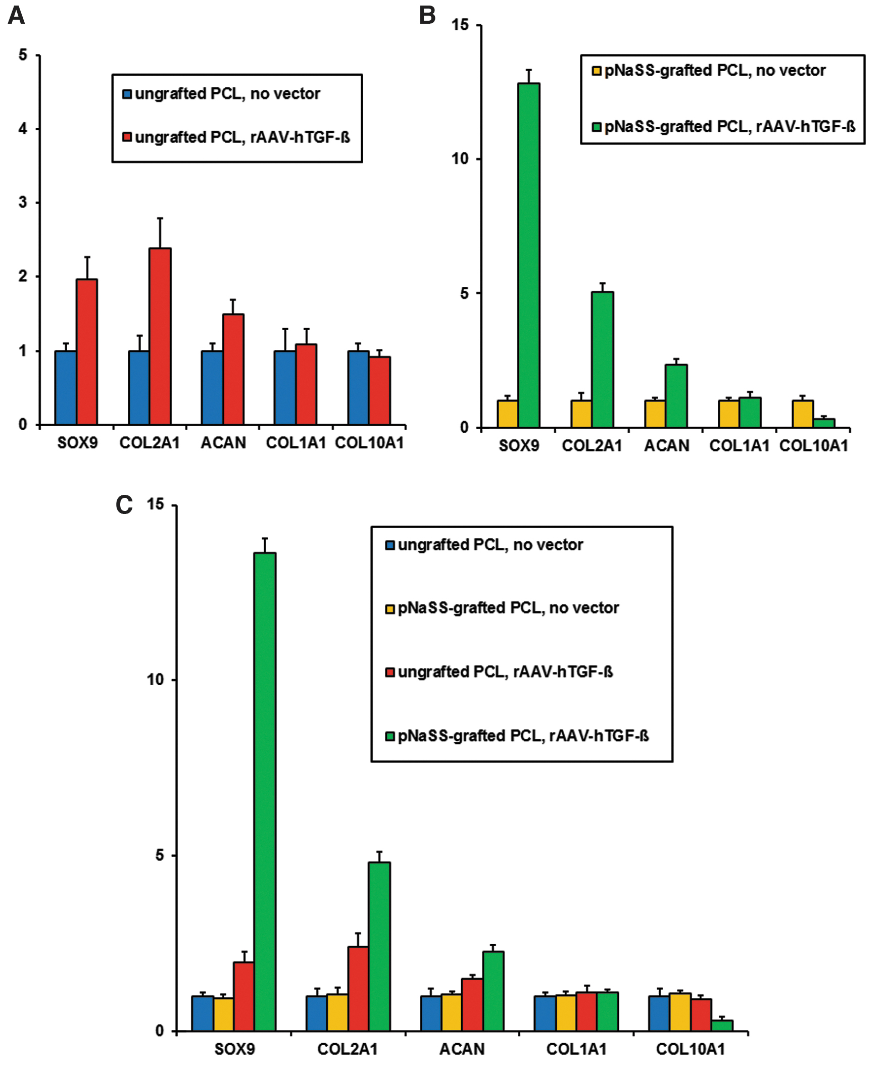
Analysis of the gene expression profiles in human bone marrow aspirates transduced with rAAV TGF-β-coated PCL films. pNaSS-grafted and ungrafted PCL films were coated with rAAV-hTGF-β or left without vector coating before incubation with the aspirates as described in Figures 1–3. The SOX9, COL2A1, ACAN, COL1A1, and COL10A1 expression profiles were evaluated by real-time RT-PCR versus GAPDH after 21 days of culture (SOX9, COL2A1, and ACAN: chondrogenic medium; COL1A1 and COL10A1: osteogenic medium) as described in the Materials and Methods section
In an osteogenic environment, while there was no significant effect of rAAV-hTGF-β on osteogenic COL1A1 expression in the aspirates relative to the other treatments, regardless of the type of film applied (p ≥ 0.165) (Fig. 4A–C), application of rAAV-hTGF-β using the pNaSS-grafted PCL films significantly decreased the levels of hypertrophic COL10A1 expression in the aspirates relative to the use of ungrafted PCL films and to the other treatments (up to 3.5-fold difference; p ≤ 0.001) (Fig. 4A–C).
Discussion
Biomaterial-guided gene transfer is a promising approach to support the persistent and safe improvement of cartilage repair processes 7,8 as an off-the-shelf tool to control the delivery of candidate vectors in sites of injury, like when using clinically fitted rAAV vehicles, 14 –19 offering more stable protocols than scaffold-free gene therapy 7,8,27 and less complex strategies than those based on the implantation of genetically modified samples. 33 –36
Specifically, in this study, we examined the feasibility of delivering a highly chondrogenic TGF-β 4 sequence by rAAV in human bone marrow aspirates 27 using solid PCL films functionalized with a bioactive pNaSS molecule, 18,24,26 as a noninvasive strategy to further stimulate the chondroregenerative processes in these samples relative to the outcomes achieved when applying a candidate rAAV SOX9 construct. 19
These data first show that TGF-β can be successfully overexpressed by rAAV in human bone marrow aspirates following vector delivery using PCL films for up to 21 days (the longest time point evaluated), especially using films grafted with pNaSS (92.6% transduction efficiencies) relative to the other conditions, probably due to the effective controlled release of rAAV from such films evidenced in our earlier work using reporter vectors. 18
These findings also extend our previous observations in similar samples treated with film-free rAAV-hTGF-β gene delivery 27 and confirm similar observations using an rAAV SOX9 vector. 19 Remarkably, the levels of TGF-β production achieved using PCL films, especially those grafted with pNaSS, increased over the period of evaluation, in contrast to findings where rAAV-hTGF-β was applied to bone marrow aspirates in a free form, showing significant, but decreasing TGF-β synthesis over time (from 113 to 16 pg/mg total proteins/24 h between days 14 and 21), 27 again possibly due to the effective controlled release of rAAV from such films. 18
The findings next indicate that effective TGF-β overexpression through application of rAAV vectors coated on PCL films led to higher levels of matrix proteoglycans and type-II collagen and of cell proliferation after 21 days in the bone marrow aspirates, particularly when using films grafted with pNaSS, compared with the other conditions, concordant with the activities of the growth factor 4 and with our previous work using similar samples transduced with film-free rAAV-hTGF-β. 27 The increases in matrix compound deposition were probably due to the enhanced expression of SOX9 in the corresponding samples as noted by real-time RT-PCR analysis, in good agreement with the properties of this transcription factor. 37
Interestingly, while the levels of matrix components achieved using PCL-guided TGF-β overexpression in this study were in the range of those reported when using film-free rAAV TGF-β administration (53–60 ng/mg total proteins), 27 the indices of cell proliferation were higher than those reached when using film-free rAAV TGF-β gene transfer (5,201 ± 76 and 6,475 ± 89 cells/mm2 with ungrafted and pNaSS-grafted films, respectively, versus 119 ± 27 cells/mm2 at similar vector doses, i.e., an up to 54.4-fold difference). 27 Such stimulating effects of PCL-guided rAAV TGF-β delivery on cell proliferation demonstrate the potential additional benefits of using this candidate gene for reparative purposes relative to the transfer of an rAAV SOX9 construct using similar films that promoted higher matrix deposition (70–83 vs. 48–53 ng/mg total proteins in this study), but had no proliferative effect in bone marrow aspirates. 19
Equally important, PCL-guided administration of rAAV-hTGF-β was capable of reducing premature matrix mineralization and hypertrophy (type-X collagen expression) in the aspirates for 21 days, especially when applying the films grafted with pNaSS, compared with the other conditions, as noted when employing film-free rAAV-hTGF-β gene delivery 27 or when transferring an rAAV SOX9 construct with the films, 19 possibly due to the increased expression of anti-hypertrophic SOX9 38 in the TGF-β-treated samples.
Interestingly, there was no effect of rAAV-hTGF-β on type-I collagen expression regardless of the type of film employed, in contrast to findings using film-free rAAV-hTGF-β gene transfer where expression of this marker was significantly decreased. 27 Nevertheless, in this previous study, free rAAV-hTGF-β was applied at a lower MOI (10 vs. 75 in this stduy), suggesting that high rAAV-hTGF-β doses may have a limited impact on type-I collagen in such a system, in contrast to the administration of an rAAV SOX9 construct through similar films at the same MOI (75), which was capable of significantly reducing the levels of this osteogenic marker, 19 in agreement with the activities of the transcription factor. 39
Conclusion
This work reveals the ability of PCL films to deliver a therapeutic rAAV TGF-β vector in human bone marrow aspirates as an effective off-the-shelf compound for chondroreparative purposes, especially upon functionalization by pNaSS grafting. The possible chondroreparative benefits and immune protection of the PCL-guided rAAV TGF-β gene transfer are currently being evaluated over free gene vector delivery in orthotopic models of cartilage defects in vivo, 9,10,33 –36,40 where anti-AAV immune responses may occur. 22
The therapeutic benefits of this system may further be improved by combined delivery of additional rAAV constructs, like for instance, an rAAV SOX9 vector that may further stimulate matrix deposition and reduce osteogenic and terminal differentiation in the aspirates. 19 This might be performed by simultaneous or independent coating of various rAAV on the films since co-transduction of such vectors can be successfully established without viral interference. 41,42
Overall, such a scaffold-guided gene therapy approach to enhance the chondrogenic potential of noninvasively prepared human bone marrow aspirates has a broad potential in translational applications that aim at reinforcing the processes of healing by implantation of the modified aspirates in cartilage defects. Alternatively, therapeutic rAAV-coated scaffolds may also directly be applied to sites of cartilage damage as a means to stimulate the chondrogenic activities of local bone marrow cells that repopulate the defects. Taken together, this study provides evidence supporting the concept of applying rAAV using solid scaffolds in protocols aiming at improved cartilage repair.
Footnotes
Acknowledgments
The authors would like to thank R.J. Samulski (The Gene Therapy Center, University of North Carolina, Chapel Hill, NC), X. Xiao (The Gene Therapy Center, University of Pittsburgh, Pittsburgh, PA), and E.F. Terwilliger (Division of Experimental Medicine, Harvard Institutes of Medicine and Beth Israel Deaconess Medical Center, Boston, MA) for providing the genomic AAV-2 plasmid clones and the 293 cell line.
Authors' Contributions
J.K.V.: methodology, software, validation, formal analysis, investigation, writing-original draft preparation, writing-review and editing, and visualization; X.C.: methodology, software, validation, formal analysis, investigation, writing-original draft preparation, writing-review and editing, and visualization; W.M., A.R.-R., G.S., S.S.-M., C.F.-D., A.L., and H.M.: methodology, validation, formal analysis, investigation, and visualization; V.M.: conceptualization, methodology, validation, formal analysis, investigation, resources, data curation, writing-original draft preparation, writing-review and editing, visualization, and funding acquisition; M.C.: conceptualization, methodology, software, validation, formal analysis, investigation, resources, data curation, writing-original draft preparation, writing-review and editing, visualization, supervision, and funding acquisition. All authors have read and agreed to the published version of the article.
Author Disclosure
No competing financial interests exist.
Funding Information
This research was supported by the Deutsche Forschungsgemeinschaft (DFG VE 1099/1-1) and by University Paris 13, Sorbonne Paris Cité. We acknowledge support by the Saarland University within the funding program Open Access Publishing.