
Abstract
Implantation of genetically modified chondrogenically competent human bone marrow-derived mesenchymal stromal cells (hMSCs) is an attractive strategy to improve cartilage repair. The goal of this study was to examine the potential benefits of transferring a sequence coding for the bone morphogenetic protein 3 (BMP-3) that modulates bone and cartilage formation, using recombinant adeno-associated virus (rAAV) vectors on the chondroreparative activities of hMSCs. Undifferentiated and chondrogenically induced primary human MSCs were treated with an rAAV-hBMP-3 construct to evaluate its effects on the proliferative, metabolic, and chondrogenic activities of the cells compared with control (reporter rAAV-lacZ vector) condition. Effective BMP-3 expression was noted both in undifferentiated and chondrogenically differentiated cells in the presence of rAAV-hBMP-3 relative to rAAV-lacZ, stimulating cell proliferation and extracellular matrix (proteoglycans, type-II collagen) deposition together with higher levels of chondrogenic sex-determining region Y-type high-mobility group box 9 (SOX9) expression. rAAV-hBMP-3 also advantageously decreased terminal differentiation, hypertrophy, and osteogenesis (type-I/-X collagen and alkaline phosphatase expression), with reduced levels of osteoblast-related runt-related transcription factor 2 (RUNX-2) transcription factor and β-catenin (osteodifferentiation mediator) and enhanced parathyroid hormone-related protein expression (inhibitor of hypertrophic maturation, calcification, and bone formation). This study shows the advantage of modifying hMSCs with rAAV-hBMP-3 to trigger adapted chondroreparative activities as a source of improved cells for transplantation protocols in cartilage defects.
INTRODUCTION
The adult hyaline articular cartilage, the tissue responsible for smooth gliding and weight-bearing functions in the articulations with a unique cell population (articular chondrocytes), has a restricted ability for self-healing in the absence of vascularization that may be a source of reparative cells in response to injury. 1 –3 As a consequence, articular cartilage defects resulting from trauma may persist and further progress toward irreversible osteoarthritis, even following therapeutic interventions like bone marrow-stimulating techniques (microfracture, abrasion arthroplasty, pridie drilling) or cell implantation procedures that mostly promote the formation of a fibrocartilaginous repair tissue of inadequate quality (type-I collagen vs. hyaline type-II collagen and proteoglycans) and poor mechanical integrity. 1,2,4,5
Administration of progenitor cells such as mesenchymal stromal cells (MSCs) has strong potential to improve the natural reparative processes in the injured cartilage since these cells can reliably differentiate in the chondrocyte lineage 6 –9 and have been safely used in patients with articular cartilage lesions. 10 Yet, their adapted use for cartilage repair is challenging as only a low percentage of MSCs can effectively undergo differentiation processes to generate a proper repair tissue while they may commit toward premature, undesirable terminal differentiation, hypertrophy, and osteogenesis. 11,12
Gene therapy is an attractive, potent tool to address such limitations by stably enhancing the intrinsic chondrogenic activities of MSCs in approaches aiming at improving cartilage repair. 13 –15 A clinically workable gene transfer strategy may be performed with recombinant adeno-associated virus (rAAV) gene vectors that are best suited in translational protocols in vivo 16 –18 and can target MSCs at very high and durable efficiencies (up to 100% for at least 21 days), due to their long maintenance (months to years) as stable, actively expressed episomes in their targets, 19 –21 without modifying their commitment toward chondrogenesis. 22 –27
While various gene candidates have been applied to human bone marrow-derived mesenchymal stromal cells (hMSCs) to achieve this goal, including the transforming growth factor beta (TGF-β), 23,27 insulin-like growth factor I (IGF-I), 26 basic fibroblast growth factor (FGF-2), 24 and the cartilage-specific sex-determining region Y-type high-mobility group box 9 (SOX9) transcription factor, 25 none has definitely promoted adequate chondrogenic differentiation profiles, showing the critical need to identify new, more effective chondrotherapeutic sequences.
The bone morphogenetic protein 3 (BMP-3, osteogenin), a member of the TGF-β superfamily, may constitute an interesting candidate to regulate MSC chondrogenesis as it has been described as an antagonist of osteogenic BMPs, negatively regulating bone formation/density and directing de novo cartilage formation. 28 –30 Interestingly, there is no evidence thus far showing the potential benefits of overexpressing BMP-3 through rAAV-mediated gene transfer in human MSCs as a tool to enhance chondrogenesis in these cells in vitro.
The goal of the present study was therefore to test the ability of rAAV to deliver a BMP-3 gene sequence to primary adult human MSCs as a possible, clinically adapted tool to regulate the proliferative, metabolic, and chondrodifferentiation activities of such cells for future implantation strategies in cartilage defects in translational applications in vivo.
MATERIALS AND METHODS
Reagents
Reagents were purchased at Sigma (Munich, Germany), except for the recombinant FGF-2 (rFGF-2) and recombinant TGF-β (rTGF-β) (R&D Systems, Wiesbaden-Nordenstadt, Germany), the AAVanced Concentration Reagent (System Bioscience, Heidelberg, Germany), the dimethylmethylene blue dye (Serva, Heidelberg, Germany), the BMP-3 ELISA (human BMP-3 ELISA, NBP2-69993; Novus Biologicals, Wiesbaden, Germany), and the Cell Proliferation Reagent WST-1 (Roche Applied Science, Mannheim, Germany). The anti-BMP-3 (C-20), anti-SOX9 (C-20), anti-CD34 (C-18), anti-CD71 (C-20), and anti-CD105 (T-20) antibodies were purchased at Santa Cruz Biotechnology (Heidelberg, Germany), the anti-type-I (AF-5610) and anti-type-II collagen (AF-5710) antibodies at Acris (Hiddenhausen, Germany), the anti-type-X collagen (COL-10) antibody at Sigma, and biotinylated secondary antibodies with the ABC reagent at Vector Laboratories (Alexis Deutschland GmbH, Grünberg, Germany).
Cell culture
Human bone marrow aspirates (∼15 mL) were collected from the distal femurs of patients undergoing total knee arthroplasty (n = 8), with approval from the Ethics Committee of the Saarland Physicians Council (Ärztekammer des Saarlandes; Reference No. Ha06/08) and with informed consent before inclusion in the study, all in accordance with the Helsinki Declaration. Human bone marrow-derived mesenchymal stromal cells (hMSCs) were isolated and expanded in culture using standard protocols. 8,24 –27,31,32
Briefly, the aspirates were washed in Dulbecco's modified Eagle's medium (DMEM) and centrifuged to resuspend the pellet in Red Blood Cell Lysing Buffer/DMEM (1:1). The resulting fraction was washed, pelleted, and resuspended in growth medium (DMEM, 10% fetal bovine serum, penicillin/streptomycin—100 U/mL/100 μL/mL—pen/strep). Cells were plated at 2 × 10⁵ cells/cm2 (T-75 flasks) at 37°C, 5% CO2 in a humidified atmosphere, with medium change containing rFGF-2 (1 ng/mL) after 48 h and every 2–3 days. The cells were replated for the experiments at appropriate densities (passage <2) and tested by flow cytometry for expression of stem cell surface markers (CD71+, CD105+, CD34−). 8,24 –27,31,32
Plasmids and rAAV vectors
The constructs were produced using the parental AAV-2 genomic clone, pSSV9. 33,34 rAAV-lacZ carries the Escherichia coli β-galactosidase (lacZ) reporter gene and rAAV-hBMP-3 a 1.416 bp human BMP-3 sequence (pCMV6-Entry [PS100001]) (Origene, Herford, Germany) instead of lacZ (the fragment was confirmed by sequencing), both under the control of the cytomegalovirus immediate–early (CMV-IE) promoter. 24 –27 rAAVs were packaged as conventional (not self-complementary) vectors in the 293 cell line, an adenovirus-transformed human embryonic kidney cell line, using a helper-free, two-plasmid transfection system based on the packaging plasmid pXX2 and the adenovirus helper plasmid pXX6. 27 The rAAV vector preparations were purified using the AAVanced Concentration Reagent according to the manufacturer's recommendations and titered by real-time PCR, 24 –27 averaging 1010 transgene copies/mL (ratio virus particles to functional vectors = 500/1). 24 –27
rAAV-mediated gene transfer
Monolayer cultures of undifferentiated hMSCs (4 × 10⁴ cells) were treated with rAAV (40 μL vector, i.e., MOI = 20) and maintained in growth medium for 21 days at 37°C, 5% CO2 in a humidified atmosphere with medium change every 2–3 days, starting after 5 days. 24 –27 For chondrogenic differentiation studies, hMSC aggregate cultures (2 × 10⁵ cells) were prepared in defined chondrogenic medium composed of DMEM high glucose (4.5 g/L), pen-strep, ITS+ Premix (insulin 6.25 μg/mL, transferrin 6.25 μg/mL, selenous acid 6.25 μg/mL, linoleic acid 5.35 μg/mL, bovine serum albumin 1.25 μg/mL), pyruvate (1 mM), ascorbate 2-phosphate (37.5 μg/mL), dexamethasone (10–7 M), and rTGF-β (10 ng/mL) at 37°C, 5% CO2 in a humidified atmosphere with medium change every 2–3 days. 24 –27
Free-floating masses with cells formed within 24 h and were treated with rAAV (40 μL vector, i.e., MOI = 4) 1 day after aggregate formation. Aggregates were then maintained in defined chondrogenic medium for 21 days at 37°C, 5% CO2 in a humidified atmosphere with medium change every 2–3 days. 24 –27 A control condition lacking the application of rAAV was not tested here since it was already described in our previous work, showing no difference in terms of hMSC chondrogenic potency relative to the use of the current control reporter rAAV (lacZ) vector. 24,25
BMP-3 production
To monitor BMP-3 production, the samples were washed twice and placed for 24 h in serum-free medium. The culture supernatants were then collected at the denoted time points and centrifuged to remove debris. BMP-3 secretion was tested by quantitative ELISA using a GENios spectrophotometer/fluorometer (Tecan, Crailsheim, Germany). BMP-3 production was also analyzed by immunocyto-/histochemical analyses with a specific primary antibody. 24 –27
Histology, immunocyto-/histochemistry
Monolayer and aggregate cultures were collected and fixed in 10% buffered formalin. Aggregate cultures were dehydrated in graded alcohols, embedded in paraffin, and sectioned (5 μm). Samples were processed for transgene (BMP-3) expression by immunocyto-/histochemical analyses using a specific primary antibody, a biotinylated secondary antibody, and the ABC method with diaminobenzidine (DAB) as the chromogen. 24 –27 Expression of SOX9, type-I, type-II, and type-X collagen was detected on sections of chondrogenically induced aggregate cultures by immunohistochemistry using specific antibodies, biotinylated secondary antibodies, and the ABC method with DAB as the chromogen. 24 –27
All samples were processed with omission of the primary antibody to control for secondary immunoglobulins. Sections of chondrogenically induced aggregate cultures were also stained with hematoxylin/eosin (H&E) (cellularity), Toluidine Blue (matrix proteoglycans), and Alizarin Red (matrix mineralization) according to routine protocols. 24 –27 All samples were visualized under light microscopy (Olympus BX 45, Hamburg, Germany).
Histomorphometry
The percentage of BMP-3-expressing cells (i.e., transduction efficiencies as cells positively stained for BMP-3 expression to the total number of cells), 26,27 the aggregate diameters, the cell densities (cells/mm2 on H&E-stained sections from chondrogenically induced aggregate cultures), the extent of Toluidine Blue and Alizarin Red staining and of SOX9, type-I, type-II, and type-X collagen immunostaining (positively stained surface to the total surface evaluated) were all measured at three standardized sites or on ten serial histological and immunohistochemical sections for each parameter, test, and replicate condition using SIS AnalySIS (Olympus), Adobe Photoshop (Adobe Systems, Unterschleissheim, Germany), and Scion Image (Scion Corporation, Frederick, MD, USA). 24 –27
Biochemical assays
Samples were collected and treated with papain digestion (aggregate cultures). 24 –27 Cell proliferation was assessed with the Cell Proliferation Reagent WST-1 with optical densities being proportional to the cell numbers. 24 –27 The DNA contents were determined with a fluorimetric assay using Hoechst 33258 and the proteoglycan contents were measured by binding to dimethylmethylene blue dye, with normalization to total cellular proteins using a protein assay (Pierce Thermo Scientific; Fisher Scientific, Schwerte, Germany). 24 –27 The measurements were performed on a GENios spectrophotometer/fluorometer (Tecan).
Total RNA extraction and real-time polymerase chain reaction analyses
Total cellular RNA was prepared from the cultures with the RNeasy Protect Mini Kit with an on-column RNase-free DNase treatment (Qiagen, Hilden, Germany). 24 –27 The RNA was eluted in 30 μL RNase-free water and reverse transcription was performed using 8 μL of eluate and the First Strand cDNA Synthesis Kit for real-time-PCR (AMV) (Roche Applied Science). An aliquot (2 μL) of the cDNA product was amplified by real-time PCR with the Brilliant® SYBR® Green QPCR Master Mix (Stratagene, Agilent Technologies, Waldbronn, Germany) 24 –27 using a Mx3000P® QPCR operator system (Stratagene) and the following conditions: (95°C, 10 min), 40 cycle amplification (denaturation: 95°C, 30 s; annealing: 55°C, 1 min; extension: 72°C, 30 s), denaturation (95°C, 1 min), and terminal incubation (55°C, 30 s).
The following primers were employed (Invitrogen GmbH): SOX9 (chondrogenic marker) (forward 5′-ACACACAGCTCACTCGACCTTG-3′; reverse 5′-GGGAATTCTGGTTGGTCCTCT-3′), type-II collagen (COL2A1) (chondrogenic marker) (forward 5′-GGACTTTTCTCCCCTCTCT-3′; reverse 5′-GACCCGAAGGTCTTACAGGA-3′), type-I collagen (COL1A1) (osteogenic marker) (forward 5′-ACGTCCTGGTGAAGTTGGTC-3′; reverse 5′-ACCAGGGAAGCCTCTCTCTC-3′), type-X collagen (COL10A1) (marker of hypertrophy) (forward 5′-CCCTCTTGTTAGTGCCAACC-3′; reverse 5′-AGATTCCAGTCCTTGGGTCA-3′), alkaline phosphatase (ALP) (osteogenic marker) (forward 5′-TGGAGCTTCAGAAGCTCAACACCA-3′; reverse 5′-ATCTCGTTGTCTGAGTACCAGTCC-3′), runt-related transcription factor 2 (RUNX2) (osteogenic marker) (forward 5′-GCAGTTCCCAAGCATTTCAT-3′; reverse 5′-CACTCTGGCTTTGGGAAGAG-3′), β-catenin (mediator of the Wnt signaling pathway for osteoblast lineage differentiation) (forward 5′-CAAGTGGGTGGTATAGAGG-3′; reverse 5′-GCGGGACAAAGGGCAAGA-3′), parathyroid hormone-related protein (PTHrP) (anti-hypertrophic factor) (forward 5′-CGACGACACACGCACTTGAAAC-3′; reverse 5′-CGACGCTCCACTGCTGAACC-3′), and glyceraldehyde-3-phosphate dehydrogenase (GAPDH) (housekeeping gene and internal control) (forward 5′-GAAGGTGAAGGTCGGAGTC-3′; reverse 5′-GAAGATGGTGATGGGATTTC-3′) (all 150 nM final concentration). 24 –27
Control conditions included reactions with water and non-reverse-transcribed mRNA. The products' specificity was confirmed by melting curve analysis and agarose gel electrophoresis. Threshold cycle (Ct) values for each gene of interest were tested for each amplified sample using the MxPro QPCR software (Stratagene). Values were normalized to GAPDH expression with the 2-ΔΔCt method. 24 –27
Statistical analysis
Data are expressed as mean ± standard deviation of independent experiments. Each condition was performed in triplicate in three independent experiments for all donors. Data were obtained by two individuals blinded with respect to the treatment groups. The t-test and the Mann–Whitney Rank-Sum Test were employed where appropriate. p-Values <0.05 were considered statistically significant.
RESULTS
Effective, durable rAAV-mediated BMP-3 overexpression in undifferentiated monolayer cultures of human MSCs
The candidate rAAV-hBMP-3 vector was first applied to hMSCs in undifferentiated monolayer cultures to test the ability of rAAV to promote the overexpression of the growth factor over time in these cells at an undifferentiated stage compared with control rAAV-lacZ vector treatment.
Strong immunoreactivity to BMP-3 was specifically detected in cells treated for 21 days with rAAV-hBMP-3 relative to rAAV-lacZ treatment, with transduction efficiencies of 94.8% ± 1.5% versus 3.4% ± 1.1% (27.9-fold difference, p ≤ 0.001) (Fig. 1). This result was corroborated by an estimation of BMP-3 production by ELISA in the BMP-3-treated cells versus control cells (11-fold difference, p ≤ 0.001) (Table 1).

Immunodetection of BMP-3 in undifferentiated monolayer and chondrogenically differentiated aggregate cultures of hMSCs. Cells were transduced with rAAV-lacZ or rAAV-hBMP-3 (40 μL each vector) in monolayer or aggregate cultures as described in the Materials and Methods section and processed on day 21 to monitor transgene expression by analyzing the immunoreactivity to BMP-3 (monolayer culture: magnification × 20; aggregate culture: magnification × 100; representative data). rAAV, recombinant adeno-associated virus.
Analyses in undifferentiated hMSC monolayer cultures (day 21)
Values are given as mean (SD).
Statistically significant relative to rAAV-lacZ.
Effects of rAAV-mediated BMP-3 overexpression upon the proliferation and metabolic activities of undifferentiated hMSCs
Undifferentiated hMSCs in monolayer cultures were next treated with the candidate rAAV-hBMP-3 vector to determine the effects of BMP-3 overexpression over time on the proliferation levels and metabolic activities of these cells at an undifferentiated stage compared with rAAV-lacZ treatment.
Administration of rAAV-hBMP-3 significantly enhanced the proliferation rates (WST-1 assay), DNA contents, and proteoglycan contents of the cells relative to rAAV-lacZ treatment for 21 days (WST-1 assay: 1.3-fold difference, p = 0.001; DNA contents: 1.2-fold difference, p = 0.038; proteoglycan contents: 1.5-fold difference, p = 0.001; normalized proteoglycan contents: 1.2-fold difference, p = 0.043) (Table 1).
Effective, durable rAAV-mediated BMP-3 overexpression in chondrogenically induced human MSCs
The candidate rAAV-hBMP-3 vector was then applied to chondrogenically induced hMSCs in aggregate cultures to test the ability of rAAV to promote the overexpression of the growth factor over time in a three-dimensional environment allowing for chondrogenic differentiation compared with rAAV-lacZ treatment.
Consistent with the results in monolayer cultures, durable BMP-3 expression was mostly seen in aggregates treated for 21 days with rAAV-hBMP-3 relative to rAAV-lacZ treatment, with transduction efficiencies of 97.2% ± 1.9% versus 9.2% ± 1.9% (10.6-fold difference, p ≤ 0.001) (Fig. 1). This result was again corroborated by an estimation of BMP-3 production by ELISA in the BMP-3-treated aggregates versus control aggregates (3.9-fold difference, p ≤ 0.001) (Table 2).
Analyses in chondrogenically induced hMSC aggregate cultures (day 21)
Values are given as mean (SD).
Statistically significant relative to rAAV-lacZ.
Effects of rAAV-mediated BMP-3 overexpression upon the proliferative, metabolic, and chondrogenic activities of chondrogenically induced hMSCs
Chondrogenically induced hMSCs in aggregate cultures were next treated with the candidate rAAV-hBMP-3 vector to investigate the effects of BMP-3 overexpression on the hMSC proliferative, biosynthetic, and differentiative activities over time under chondrogenic induction compared with rAAV-lacZ treatment.
Administration of rAAV-hBMP-3 significantly increased the diameters and cell densities of the cell aggregates (H&E staining) after 21 days relative to rAAV-lacZ treatment (diameters: 1.9-fold difference, p ≤ 0.001; cell densities: 1.5-fold difference, p ≤ 0.001) (Fig. 2 and Table 2). Delivery of rAAV-hBMP-3 also significantly enhanced the proliferation rates (WST-1 assay) and the DNA contents of the cell aggregates relative to rAAV-lacZ treatment for at least 21 days (WST-1 assay: 1.4-fold difference, p ≤ 0.001; DNA contents: 1.8-fold difference, p ≤ 0.001) (Table 2).

Proliferative, metabolic, and chondrogenic activities in chondrogenically differentiated aggregate cultures of hMSCs. Aggregate cultures were transduced with rAAV-lacZ or rAAV-hBMP-3 (40 μL each vector) as described in the Materials and Methods section and processed on day 21 for histological analyses (H&E, Toluidine Blue, and Alizarin Red staining) and for immunohistochemical analyses (SOX9 and type-II/-I/-X collagen deposition) as described in the Materials and Methods section (magnification × 40; inserts: magnification × 4; all representative data). H&E, hematoxylin/eosin.
Application of rAAV-hBMP-3 significantly increased the chondrodifferentiative and metabolic activities of hMSCs in the cell aggregates after 21 days relative to rAAV-lacZ treatment (Toluidine Blue staining: 1.9-fold difference with stronger matrix deposition around the cells when applying rAAV-hBMP-3, p ≤ 0.001; SOX9 expression: 2.3-fold difference, p ≤ 0.001; type-II collagen deposition: 1.4-fold difference, p ≤ 0.001) (Fig. 2 and Table 2).
Delivery of rAAV-hBMP-3 also significantly enhanced the proteoglycan contents of the cell aggregates after 21 days relative to rAAV-lacZ treatment (proteoglycan contents: 1.9-fold difference, p ≤ 0.001; normalized proteoglycan contents: 1.2-fold difference, p = 0.006) (Table 2). In contrast, administration of the BMP-3 vector significantly decreased matrix mineralization (Alizarin Red staining) and type-I and type-X collagen deposition in the cell aggregates after 21 days relative to rAAV-lacZ treatment (Alizarin Red staining: 3.8-fold difference, p ≤ 0.001; type-I collagen deposition: 4.2-fold difference, p ≤ 0.001; type-X collagen deposition: 2.9-fold difference, p ≤ 0.001) (Fig. 2 and Table 2).
The results of the biochemical, histological, and immunohistochemical evaluations were corroborated by findings of a real-time RT-PCR analysis (Fig. 3). Chondrogenic hMSC differentiation was seen after 21 days in all the aggregates, as noted by the detection of SOX9 and type-II collagen expression, but with significantly higher levels achieved when applying rAAV-hBMP-3 (11.6- and 1.8-fold higher SOX9 and COL2A1 expression levels with rAAV-hBMP-3 vs. rAAV-lacZ; p ≤ 0.001).
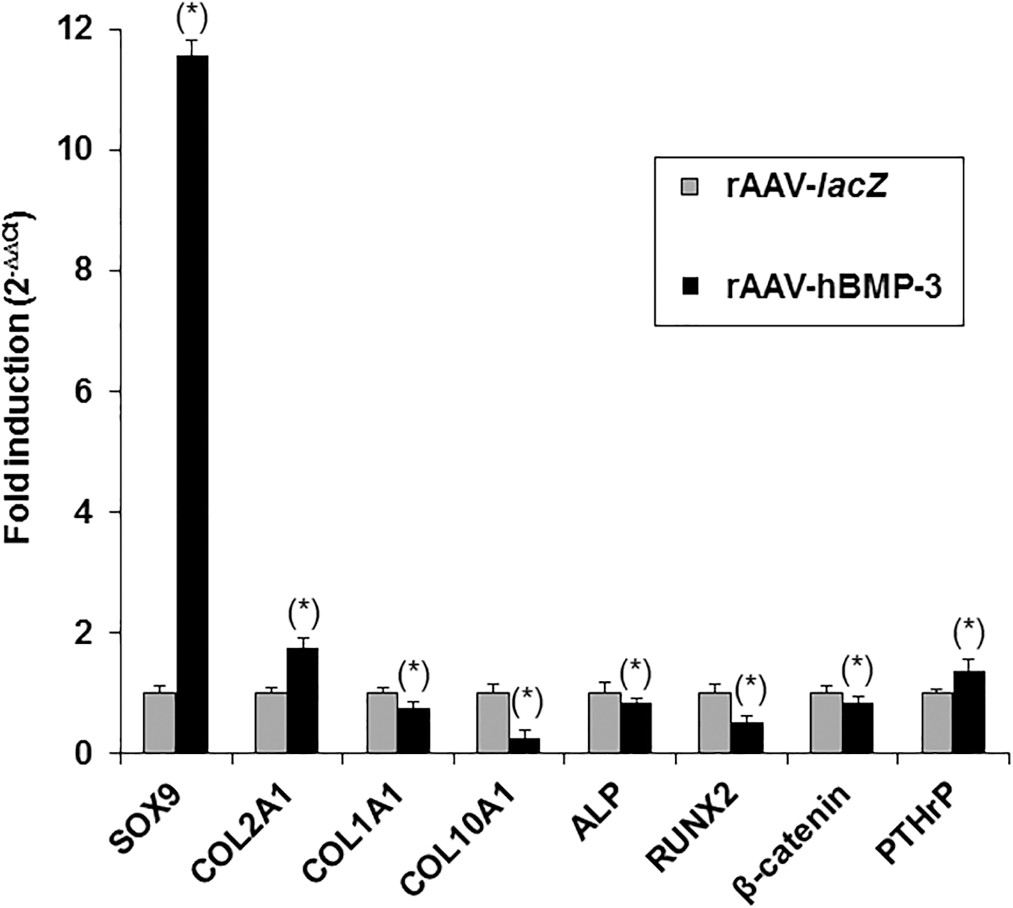
Gene expression profiles in chondrogenically differentiated aggregate cultures of hMSCs. Aggregate cultures were transduced with rAAV-lacZ or rAAV-hBMP-3 as described in Fig. 2 and processed on day 21 for gene expression analysis by real-time RT-PCR amplification following total cellular RNA extraction and cDNA synthesis as described in the Materials and Methods section. The genes analyzed were SOX9, COL2A1, COL1A1, COL10A1, ALP, RUNX2, β-catenin, and PTHrP, with GAPDH serving as a housekeeping gene and internal control. Ct values were obtained for each target and for GAPDH (control for normalization) and fold inductions (relative to rAAV-lacZ-treated aggregates) were measured using the 2−ΔΔCt method. *Statistically significant relative to rAAV-lacZ. GAPDH, glyceraldehyde-3-phosphate dehydrogenase; hMSC, human bone marrow-derived mesenchymal stromal cell; RT-PCR, real-time PCR.
Such a real-time RT-PCR analysis further revealed a decreased expression of type-I and -X collagen when applying rAAV-hBMP-3 (1.3- and 3.8-fold lower COL1A1 and COL10A1 expression levels with rAAV-hBMP-3 vs. rAAV-lacZ; p ≤ 0.001), concomitant with a decrease in ALP expression (1.2-fold lower ALP expression levels with rAAV-hBMP-3 vs. rAAV-lacZ; p ≤ 0.001). Interestingly, these results were associated with contrasting effects of rAAV-hBMP-3 on RUNX2 and β-catenin (1.9- and 1.2-fold lower expression levels, respectively, with rAAV-hBMP-3 vs. rAAV-lacZ; p ≤ 0.001) and with enhancing effects of rAAV-hBMP-3 on PTHrP (1.4-fold higher expression levels with rAAV-hBMP-3 vs. rAAV-lacZ; p ≤ 0.001).
DISCUSSION
Implantation of genetically triggered bone marrow-derived MSCs may allow to improve the natural processes of tissue repair in sites of articular cartilage injury by ameliorating their chondroreparative potential and reducing their premature terminal differentiation, hypertrophy, and osteogenesis. 11,13 –15 Among various genetic triggers, BMP-3 may be a strong candidate to stimulate hMSC chondrogenesis in light of its ability to suppress bone formation/density as an antagonist of osteogenic BMPs and to promote de novo cartilage formation. 28 –30 In this study, we examined, for the first time to our best knowledge, whether BMP-3 overexpression mediated by gene transfer using the clinically competent rAAV vectors can advantageously modulate chondrogenic events in primary human MSCs as a potential source of regenerative cells for transplantation.
The data first show that hMSCs are effectively and durably amenable to rAAV-mediated BMP-3 overexpression both in undifferentiated and chondrogenically induced culture conditions at very high efficiencies and over prolonged periods of time (up to 97% for at least 21 days, the longest time point evaluated, relative to control rAAV-lacZ treatment), in good agreement with findings using rAAV 22 –28 and probably due to the persistence of these vectors in their targets. 19 –21 The levels of BMP-3 production achieved here in hMSCs using rAAV-hBMP-3 were sustained and significantly higher than those in the control (rAAV-lacZ-treated) cells (between 4- and 11-fold difference after 21 days, a difference probably due to the presence of extracellular matrix in aggregate cultures that serves as a reservoir of BMP-335), in contrast with the use of short-lived recombinant BMP-3 (rBMP-3) 36 –38 or of transient nonviral 39 and retroviral gene transfer of BMP-3. 30
We next demonstrate that efficient and sustained BMP-3 overexpression through rAAV effectively and durably stimulated the proliferative, metabolic, and chondrogenic activities in undifferentiated and chondrogenically induced hMSCs relative to control rAAV-lacZ treatment (up to 1.8- and 1.9-fold difference in the proliferation indices and in the production of matrix proteoglycans and type-II collagen after 21 days), in agreement with previous work using rBMP-3. 36 –38 Enhanced deposition of proteoglycans and type-II collagen in hMSCs upon rAAV BMP-3 administration was probably due to the increased levels of SOX9 achieved in these cells, a transcription factor known for its stimulating effects on the expression of such major cartilage matrix components. 25,40
Equally important, BMP-3 overexpression reduced terminal differentiation, hypertrophy, and osteogenesis (type-I and type-X collagen, ALP, matrix mineralization) in the cells, concordant with the activities of this growth factor 30,36 and probably due to the increased expression of SOX9 and PTHrP (two factors with negative effects on osteogenesis and bone formation) 25,41 –43 and decreased RUNX2 and β-catenin expression (an osteoblast-related transcription factor and a mediator of the Wnt signaling pathway for osteodifferentiation, respectively). 44 –46
This is the first evidence, to the best of our knowledge, showing that BMP-3 overexpression through rAAV can trigger chondrogenesis and delay terminal differentiation, hypertrophy, and osteogenesis in hMSCs. It remains to be seen whether elevated BMP-3 levels too will not be detrimental as they may affect cartilage repair and formation 47,48 and cause thinner cortical bone formation, 49 although such effects were specifically reported during limb development, 47 at newborn stage, 49 or when using high amounts of rBMP-3 (100 ng) 48 compared with those achieved here with rAAV (0.39–0.44 ng/mL/24 h, i.e., a 227- to 256-fold difference).
It will be important to determine whether administration of MSCs modified by rAAV BMP-3 supports the competent healing of cartilage defects in vivo 20,21 without premature terminal differentiation and hypertrophy, especially in an inflammatory environment. 50 To this goal, the amounts of cells needed for implantation will have to be carefully considered, with the possible use of a supportive matrix, 16 of additional stimuli through rAAV (TGF-β, IGF-I, FGF-2, SOX9), 23 –27,51 and of regulatable (or lineage-specific) promoter elements to strictly control BMP-3 production.
Alternatively, bone marrow aspirates or concentrates may be employed instead of isolated MSCs as a more convenient, minimally invasive transplantation approach for cartilage repair. 52,53 Besides, it might be interesting to test in parallel whether a direct, cell-free application of the current rAAV-hBMP-3 construct at an equivalent vector dose will generate similar cartilage healing processes in vivo as a means to provide a more convenient therapeutic approach relative to the administration of genetically modified MSCs. 20,21 It will also allow to specify the advantages (and any superiority) of using BMP-3 through rAAV over other candidate genes, such as FGF-2 20 and sox9, 21 regarding a possible improvement of chondrogenesis with reduced hypertrophy and osteogenesis in vivo, like in a large model of cartilage defect for instance. 20,21
Yet, before initiating such extensive evaluations in clinically relevant (orthotopic) animal models, thorough analyses will first need to be strictly performed in animal MSCs in vitro to confirm the effects of BMP-3 reported in human cells in this study. Overall, the current work reveals the benefits of rAAV BMP-3 gene transfer in hMSCs as a future strategy for transplantation in sites of cartilage injury in affected patients.
Footnotes
AUTHORs' CONTRIBUTIONS
J.K.V.: methodology, software, validation, formal analysis, investigation, writing-original draft preparation, writing-review and editing, and visualization; G.S., S.S.-M., P.O., and H.M.: methodology, validation, formal analysis, investigation, and visualization; and M.C.: conceptualization, methodology, software, validation, formal analysis, investigation, resources, data curation, writing-original draft preparation, writing-review and editing, visualization, supervision, and funding acquisition. All authors have read and agreed to the published version of the article.
ACKNOWLEDGMENTS
The authors would like to thank Mary B. Goldring for helpful discussions and R.J. Samulski (The Gene Therapy Center, University of North Carolina, Chapel Hill, NC, USA), X. Xiao (The Gene Therapy Center, University of Pittsburgh, Pittsburgh, PA, USA), and E.F. Terwilliger (Division of Experimental Medicine, Harvard Institutes of Medicine and Beth Israel Deaconess Medical Center, Boston, MA, USA) for providing genomic AAV-2 plasmid clones and the 293 cell line.
AUTHOR DISCLOSURE
No competing financial interests exist.
FUNDING INFORMATION
This research was supported by a grant from the World Arthrosis Foundation (P264-A15-Madry-EP2-cucc8-allgemein-pr–136k-2013-15) (M.C. and H.M.).