
Abstract
The treatment of malignant tumors has always been one of the challenges that have plagued researchers and clinicians. The ideal status in cancer treatment is to eliminate tumor cells while avoiding damage to normal tissues. Different approaches have been investigated to achieve such a goal, and suicide gene therapy has emerged as a novel mode of cancer treatment. This approach involves the delivery of genes encoding enzymes that activate non-toxic prodrugs into cytotoxic metabolites that cause the death of transfected cancer cells. Despite promising results obtained both in vitro and in vivo, this innovative approach has long been stalled in the clinic due to the lack of a suitable delivery system to introduce the suicide gene into cancer cells. Ultrasound-targeted microbubble destruction (UTMD) represents a valuable non-viral vector system for site-specific and noninvasive gene therapy. Ultrasound promotes intracellular uptake of therapeutic agents by increasing vascular and cell membrane permeability, especially in the presence of microbubbles. In this scenario, the true potential of suicide genes can be translated into clinically valuable treatments for patients. This review provides background information on suicide gene therapy and UTMD technology, summarizes the current state of knowledge about UTMD-mediated suicide gene delivery in cancer treatment, and presents an outlook on its future development.
INTRODUCTION
According to the GLOBOCAN cancer statistics produced by the International Agency for Research on Cancer (IARC), cancer is one of the leading causes of death worldwide, accounting for nearly 10.0 million deaths in 2020. 1 Nearly one in five deaths is due to cancer, and the global cancer burden is expected to rise by about 47% over the next two decades worldwide. 2,3 Tumorigenesis is thought to be the result of abnormal cell proliferation caused by genetic mutations in normal cells. Due to the metastasis, recurrence, heterogeneity of cancer cells, and their resistance to chemotherapy and radiotherapy, many conventional treatment methods become ineffective against many malignant tumors. Cancer therapeutic options have advanced a lot as research into the mechanism of tumorigenesis has progressed. 4
One of the potential approaches is gene therapy, which is rapidly opening new frontiers for translation in the cancer arena. Significant advances have been reported in numerous cancer gene therapy approaches, including immune gene therapy, antisense gene therapy, oncolytic virus therapy, and suicide gene therapy. 5 Suicide gene therapy has attracted special attention by virtue of its unique mechanism of action. It allows the introduction of suicide genes into tumor cells, resulting in the exclusive expression of an enzyme capable of converting a non-toxic prodrug into a lethal drug and inhibiting tumor growth. 6 Achieving the specified purpose requires vehicles that encapsulate the gene and deliver it, particularly to cancer cells and the cancer local environment of the tumor.
The lack of safe, efficient, and noninvasive gene delivery systems remains a major obstacle impeding the clinical translation of cancer suicide gene therapy. 7 An optimal transfer system for gene therapy should meet the following criteria: (1) improved transfection efficiency; (2) reduced off-target transfection; and (3) lowered immune response. Many efforts have been made in this area, and a wide range of viral and non-viral vectors have been developed.
Viral vectors are the most commonly used gene delivery strategies due to their high transduction efficiency. However, they have been linked to a number of issues, including inflammatory and immune responses, insertional mutagenesis, limited loading capacity, and difficulty in production. Non-viral gene delivery systems include chemical vehicles (e.g., naked DNA, liposomes, polyplexes, or nanoparticles, etc.) and physical strategies (e.g., electroporation, gene gun, hydrodynamic injection or jet injection, etc.). These methods are of interest because they can bypass many of the side effects associated with viral vectors. However, their progress stalled due to the low transfection efficiency of chemical vehicles and the intrusiveness of physical strategies. 8
Ultrasound-based modalities offer new opportunities, as ultrasound can be used not only for clinical diagnostic imaging but also for therapeutic purposes. Ultrasound imaging is usually performed in combination with contrast agents, which mainly refers to microbubbles (MBs). They are known for use as echo-enhancers and have proven to be excellent gene delivery devices as well. 9 Ultrasound can be easily focused on the target tissue or organ; therefore, gene delivery and expression should be restricted to the insonified zone. Consequently, it may be possible to develop an efficient and safe tissue- or organ-specific delivery method through MB targeting and focused ultrasound. As a result, ultrasound-targeted microbubble destruction (UTMD) technology came into being, it has attracted attention as a theranostic tool due to its advantages of convenience, repetitive applicability, noninvasiveness, and cost-effectiveness. The UTMD technology, therefore, holds considerable promise as an effective gene transfer method to overcome the limitations of current delivery strategies.
The aim of the present review is to discuss about the following: (1) Current status of suicide gene therapy in cancer; (2) Progress and applications of MB carriers and UTMD technology; (3) Investigation of previous studies about UTMD in the role of a vehicle for suicide genes.
CURRENT STATUS OF SUICIDE GENE THERAPY IN CANCER
Gene-directed enzyme prodrug therapy (GDEPT) is a type of suicide gene therapy, which has gained significant attraction for cancer therapy as a promising alternative to conventional treatments. This strategy uses genetic engineering techniques to transfer suicide genes (prodrug-converting enzyme genes) into tumor cells for expression. The products convert non-toxic or low-toxic prodrugs into highly toxic metabolites that interfere with cell synthesis as synthetic nucleocapsid substitutes, thereby causing tumor cell death. 6
There are numerous types of suicide gene/prodrug systems, and the single suicide gene/prodrug system, represented by the herpes simplex virus-thymidine kinase/ganciclovir (HSV-TK/GCV) system and the Escherichia coli cytosine deaminase/5-fluorocytosine (CD/5-FC) system, has been most extensively studied in experimental models and clinical trials. 10 –13 In addition, other suicide genes include E. coli uracil phosphoribosyl transferase (UPRT), purine nucleoside phosphorylase (PNP), nitroreductase (NTR), and inducing caspase 9 (iCasp9). 14
As research continued, it was discovered that when two or more suicide genes are combined, or when suicide genes are combined with other genes into a new fusion gene, their different mechanisms of action can complement each other, thus greatly improving the efficiency of tumor killing. 15,16 Due to the limitation of transfer capacity of vectors, most of them are double suicide gene systems at present. The most commonly used double suicide gene systems are CD/TK and CD/UPRT. 17,18 Furthermore, a triple-suicide gene GDEPT approach using TK, CD, and UPRT has been reported. Simultaneous expression of these genes significantly enhanced the cytotoxicity and radiosensitivity of the prodrug in vitro and in vivo, exhibiting superior antitumor effects compared to the double-suicide gene approach. 19
The mechanism of action of the suicide gene has been advanced and updated in extensive exploration, which is currently thought to eliminate tumor cells in three main ways. (1) Direct killing, in which a suicide gene is transferred to tumor cells via a vector, encodes an enzyme that selectively converts a low or non-toxic prodrug into a toxic metabolite, leading to apoptosis and necrosis of tumor cells. 20 –22 Each suicide gene/prodrug system works on essentially the same principles and produces broadly similar intracellular effects, differing only in the substrates and toxic products of their respective actions. (2) The bystander effect, in which untransfected cells are eliminated along with transfected cells, is the main advantage of suicide gene therapy. 23
In this way, a large range of tumor cells can be killed with a low dose of suicide genes, compensating to some extent for the low efficiency of gene transfection. Moreover, the bystander effect of double suicide genes is more powerful than that of a single suicide gene. 24 For example, the HSV-TK/GCV system relies primarily on gap junctional intercellular communication (GJIC) to exert bystander effects, 25,26 which means that its anticancer activity is greatly limited as GJIC is considerably compromised in many tumor tissues. In contrast, the toxic metabolite of the CD/5-FC system, 5-fluorouracil (5-FU), readily diffuses into cells due to its small size and neutral charge. Therefore, the CD/5-FC system has the unique advantage of remote bystander effect, which can make up for the deficiency of the HSV-TK/GCV system and achieve a wider range of killing effects. 27,28
In addition, the use of UPRT in conjunction with CD sensitizes cancer cells to a lower dose of 5-FC, 29,30 and it leads to a greater bystander effect than CD-expression alone. 31,32 Furthermore, many scholars have worked to improve the bystander effect using substances such as all-trans retinoic acid 33 and connexins, 34,35 so as to enhance the killing ability of suicide genes on tumor cells. (3) Cell signaling pathways, 36 based on evidence that expression-related products of suicide gene systems are associated with MAPK 37 –39 and RAS 40 signaling pathways, and that activation or inhibition of key elements of these signaling pathways can induce apoptosis or necrosis in tumor cells.
By virtue of its outstanding anticancer efficiency, the GDEPT strategy has been studied for various types of cancer, from basic research to clinical applications. Preclinical studies include leukemia, 41 –43 hepatoma, 44,45 glioma, 46 –48 bladder cancer, 49,50 breast cancer, 51,52 and others in various animal models. Promising preclinical findings have driven the development of clinical trials, some of which have entered Phase II/III clinical trials. 53 –55 In terms of improving transcriptional targeting regulation mechanisms, a number of tumor-specific promoters have been identified to enhance the specificity and regulatability of the GDEPT strategy to drive the expression of suicide genes for specific killing effects only in targeted tumor cells, but not in normal cells.
A variety of cancer/tissue-specific promoters have been developed, including human telomerase reverse transcriptase (hTERT) promoter, 56 carcinoembryonic antigen (CEA) promoter, 57 human α-fetoprotein (AFP) promoter, 58 and epithelial cell adhesion molecule (EpCAM) promoter. 59 With tumor-specific receptors or tissue-specific promoters as the site of action, which can, to a certain extent, avoid therapeutic failure due to insufficient tumor specificity and achieve more precise gene therapy. In addition, attempts have been made to use radiation induction to enhance the exogenous regulation mechanism of transgene expression, by combining suicide genes with radiotherapy, 60 and cytokines, 61 which can induce stronger antitumor immunity and further enhance tumor-killing potency.
Currently, the initial enthusiasm for the GDEPT strategy in the clinic has faded mostly due to the lack of a suitable delivery system (vector), yet only a few numbers of vectors are deemed safe and efficient to elicit a significant therapeutic response. To date, most vectors used for suicide gene therapy have been viral vectors (e.g., retrovirus, lentivirus, adenovirus, and adeno-associated virus), whose main limitations are the potential for significant immune response, high costs of production, and low specificity. Suicide genes have also found a good platform for better performance by riding on mesenchymal stem cells (MSCs). 62 However, MSCs are also known to confer pro-oncogenic effects, rendering them into double-sword weapons against neoplastic diseases.
In addition, a wide range of non-viral vectors (e.g., liposomes, polymers, and nanoparticles) characterized by higher gene-loading capacity and lower immune response are being investigated as safer gene transporters for suicide gene therapy. Nevertheless, non-viral vectors generally have the problem of a low transfection rate. If combined with acoustic pulse, ultrasound, and other technologies, their transfection efficiency and targeting can be significantly improved, which becomes an important direction for the future development of non-viral vectors.
PROGRESS AND APPLICATIONS OF MB CARRIERS AND UTMD TECHNOLOGY
MB carrier system
In recent years, MB ultrasound contrast agents (UCAs) have emerged as a new type of novel gene carrier that can mediate targeted release and transfection of therapeutic genes under a certain dose of ultrasonic irradiation. 63 MBs consist of a gas-filled core (e.g., perfluorocarbon, sulfur hexafluoride, or nitrogen) and a stabilizing shell membrane composed of phospholipids, proteins, or synthetic polymer materials, measuring ∼1–10 μm. 64 As non-viral expression vectors, MBs have unique advantages such as high safety profile, low preparation cost, easy and rapid metabolism, and active targeting of tumor receptors, bringing a lot of excitement with promising applicability in gene delivery.
However, the micro-size dimension restricts them from vasculature extravasation, resulting in reduced accumulation at the targeted site. Thus, possible extravasation of small-sized UCAs in tumor tissue has led to the development of nanometer-sized MBs, called nanobubbles (NBs), capable of reaching beyond the endothelial barrier in tumors. 65 Similar to MBs, NBs are composed of different core and shell materials and may be coupled with specific tissue ligands for targeted tissue delivery. NBs have a superior ability to efficiently diffuse within the tumor tissue based on the enhanced permeability and retention (EPR) effect, 66 and have better biocompatibility, stability, and ease of modification compared with MBs. 67 In the virtue of the prominent properties described above, nanocarriers inevitably become a research hotspot and a new generation of gene carriers. However, there is now a consensus that the EPR effect is very limited in humans due to heterogeneity, 68 such that the ability of NBs to passively target to tumor tissue is compromised.
To facilitate the translation of NBs to the clinic, and to allow for personalized and improved anticancer therapies, active targeting is a desirable approach by attaching modifiers, such as target molecules, specific antibodies, or ligands, to the surface of NBs. Targeted NBs can specifically bind to overexpressed receptors in tumors, thereby limiting local delivery of therapeutic gene loading carriers to tumors, improving efficacy and avoiding off-target side effects. 69,70 Active targeting thus enables highly selective accumulation of therapeutic agents in target tissues and minimizes deposition at undesired sites compared to passive targeting. Various targeted NBs have been developed and applied in preclinical studies. 71 –74
For example, Wang et al prepared a novel NB UCA targeted to the prostate-specific membrane antigen (PSMA-targeted NBs) and evaluated its effectiveness in ultrasound imaging of prostate cancer (PCa) in mice tumor models. The results demonstrated that PSMA-targeted NBs had increased extravasation and retention in PSMA-expressing orthotopic mouse tumors, leading to improved image-based detection and diagnosis of PCa. 72 It is believed that these advanced MB/NB carriers will serve as enablers and visualization guides for ultrasound medicine, providing a more accurate way of gene delivery for the treatment of various diseases.
UTMD technology
The combination of ultrasound and various functional MB/NB carriers forms the UTMD technology, a new non-viral gene transport mode. Although ultrasound has been shown to increase cell membrane permeability on its own, 75 –77 there is a significant additive effect of using MBs/NBs given their dual role in the action of UTMD. On the one hand, gene-loaded MBs/NBs themselves can be used as good UCAs for imaging at the tumor site; on the other hand, MBs/NBs serve as potentiators of ultrasound-mediated gene delivery, and they are broken down one by one upon ultrasound exposure, thus releasing genes to the tumor site. 78,79 Therefore, it is possible to visualize systemically administered MBs/NBs transit through tumor to allow for more precise gene delivery guided by imaging.
In addition, the cavitation effect generated in the process of UTMD weakens the membrane barrier effect, 80 which in turn enhances the intracellular transport of therapeutic genes, resulting in a significant increase in the therapeutic effect. Cavitation increases vascular permeability mainly by inducing the sonoporation effect. 81 MBs/NBs oscillate and rupture under the action of acoustic waves of different intensities, producing acoustic phenomena such as microflows, microjets, and shock waves, which then produce reversible or irreversible pores in the cell membrane. 82,83 This process is known as sonoporation, which is considered the main mechanism of UTMD-mediated gene delivery. Furthermore, sonoporation can be influenced by multiparameter such as acoustic pressure, MB and DNA dosage, and treatment time. 84,85 The optimization of these parameters allows an efficient and prolonged gene expression.
UTMD technology overcomes the immunogenicity associated with viral vectors and has a high spatial and temporal resolution, enabling real-time gene monitoring and capture, which provides unparalleled advantages for gene therapy to treat various cancers.
An increasing number of studies have demonstrated that UTMD technology not only improves transfection efficiency by several orders of magnitude, 86 –88 but also enables significant targeting specificity. 89 –91 For example, Wu et al synthesized small interfering RNA (siRNA)-loaded cationic NBs conjugated with A10-3.2 aptamers (siFoxM1-Apt-CNBs). Fluorescence imaging and flow cytometry evidenced that the siFoxM1-Apt-CNBs could specifically bind to PSMA-positive cells. The results showed that UTMD-mediated siFoxM1-Apt-CNBs significantly increased the transfection efficiency and killing effectiveness in vitro and led to significant inhibition of tumor growth and prolonged the survival time of xenograft tumor-bearing mice. 89 With its less invasive approach and highly specific gene delivery system, UTMD shows an attractive prospect as adjuvant therapy for malignant tumors in the future.
UTMD AS A VEHICLE FOR SUICIDE GENE THERAPY
The GDEPT strategy has been a widely explored approach with great potential to facilitate cancer treatment. A major obstacle to the clinical success of this therapeutic approach is the limitation of targeted delivery of therapeutic genes to the tumor and the achievement of adequate amounts of therapeutic gene expression through cancer cells. UTMD emerges as a promising new therapeutic option in precisely controlling the spatiotemporal delivery of therapeutic genes to tumors. Suicide genes are either adsorbed on the surface of MBs/NBs or wrapped inside them to avoid hydrolysis by enzymes in vivo, while specific antibodies or ligands are usually modified on the surface of MBs/NBs for targeted delivery (Fig. 1). Features of the UTMD-mediated suicide gene delivery system include low cytotoxicity/immunogenicity, high transfection efficiency, tissue specificity, and cost-effectiveness.
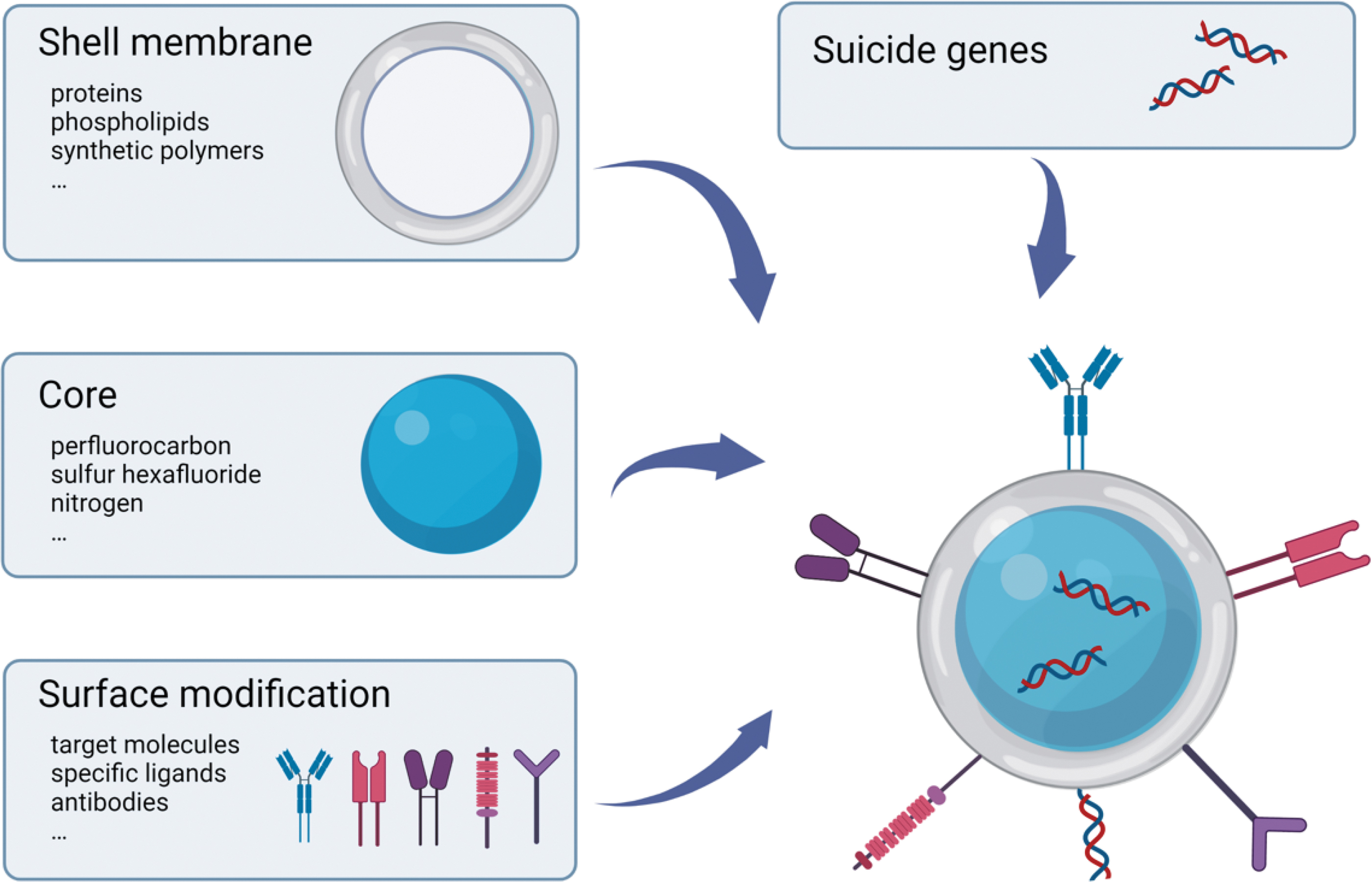
The composition of targeted MB/NB loaded with suicide genes. MB, microbubble; NB, nanobubble. Created with
As illustrated in Fig. 2, UTMD-mediated suicide gene therapy in the in vivo setting is typically performed first by either systemic or local administration of MBs/NBs along with the suicide genes. MBs/NBs can be accumulated to target organs or tissues through specific binding between antigen antibodies or ligand receptors. Low-frequency ultrasound is then selectively applied at the site of lesion to actuate sonoporation and the destruction of MBs/NBs may initiate a burst release of encapsulated suicide genes within this region. Finally, the enzymes generated by the expression of suicide genes in tumor cells are capable of converting the corresponding prodrugs into lethal drugs, thus leading to cell death.

UTMD-mediated suicide gene delivery in mice.
The first study using UTMD for suicide genes delivery was published by Aoi et al. 92 Low-intensity pulsed ultrasound and NBs were used to transduce the HSV-TK gene both in vitro and in vivo, leading to gene transfer. The results showed that UTMD-mediated HSV-TK gene transfer resulted in reduced cell survival and a dramatic reduction in tumor size compared to controls. These demonstrated the potential of UTMD as a new physical gene delivery method for cancer gene therapy. After these first encouraging experiments, a series of research has been performed in this field. Until now many studies have been practiced in cell and animal experiments using UTMD in cancer suicide gene therapy and have yielded promising therapeutic results and significant breakthroughs. A brief report of studies within this research area is embedded in Table 1. These studies demonstrate that UTMD can be applied universally for diverse organs, tumors, and with varied suicide genes.
Summary of ultrasound-targeted microbubble destruction-mediated suicide gene therapy in cancer
Annotation: ultrasound parameters refer to: frequency (MHz), intensity (W/cm2), duty ratio (%), and time (min/s).
CD/5-FC, cytosine deaminase/5-fluorocytosine; CW, continuous wave; HCC, hepatocellular carcinoma; HSV-TK/GCV, herpes simplex virus-thymidine kinase/ganciclovir; HUVECs, human umbilical vein endothelial cells; MI, mechanical index; NTR, nitroreductase; PNP, purine nucleoside phosphorylase; PRF, pulse repetition frequency; TNBC, triple-negative breast cancer; UPRT, uracil phosphoribosyl transferase.
Looking to previous studies we found that the most common transduced suicide gene is HSV-TK. Areas of experimental studies applying the UTMD-mediated HSV-TK/GCV system include hepatic carcinoma, 93 –96 glioma, 97 –99 ovarian cancer, 100 and squamous cell carcinoma. 101
It is worth mentioning that intracranial tumors are difficult to treat owing to the presence of the blood-brain barrier (BBB). UTMD has been shown to enhance the permeability of BBB 102,103 and transfect genes into the central nervous system, 104,105 allowing for the treatment of intracranial tumors. 98 Importantly, ultrasound beams can be focused on the brain tumor site, thus enabling local delivery of genes to the brain tumor and avoiding damage to normal brain tissues. Chang et al conducted an experiment on the treatment of glioma with the HSV-TK suicide gene mediated by UTMD. The results showed that the rat model of HSV-TK-expressing glioma cells exhibited a significant reduction in tumor volume after UTMD-induced BBB disruption. 97 Similar results were obtained by Yang et al 98 and Pan et al. 99 These indicated that UTMD is a viable therapeutic tool for noninvasive, effective, and targeted gene delivery to brain tumors.
In addition, UTMD-mediated HSV-TK/GCV system can also be used as a new adjuvant therapy after high-intensity focused ultrasound (HIFU) treatment, which is based on the principle that ultrasound can penetrate soft tissues and focus high-energy ultrasound on diseased tissues to kill tumor cells by virtue of the transient mechanical and thermal effects. 106 However, there are problems such as residual tumor tissue caused by incomplete ablation in the treatment of liver cancer. Zhou et al explored the antitumor effect of HIFU combined with HSV-TK gene-loaded ultrasound-targeted MBs on VX2 rabbit liver tumors. After HIFU ablation, rabbits in different groups received MBs wrapped around HSV-TK or HSV-TK solution via marginal ear veins and/or local ultrasonic irradiation to the tumor. Compared to other groups, HIFU combined with MBs wrapped HSV-TK suicide gene significantly inhibited tumor growth in vivo and prolonged the survival time of animals. 107 Therefore, UTMD-mediated HSV-TK/GCV system combined with HIFU has a synergistic effect on the treatment of cancer.
The CD/5-FC system is the second most commonly used suicide gene system after the HSV-TK/GCV system, and it is usually used in combination with the HSV-TK/GCV system to form a double suicide gene system. Areas of experimental studies applying the UTMD-mediated CD/TK dual suicide genes system include hepatic carcinoma, 108 cervical cancer, 109 bladder cancer, 110 –112 and breast cancer. 113 The application of dual suicide genes not only enhances the therapeutic effect but also reduces the dose of the respective prodrug, therefore weakening the toxic side effects of the system.
In addition to the above-mentioned double suicide genes, some scholars have studied the UTMD-mediated CD/UPRT and TK/NTR double suicide genes for the treatment of breast cancer accordingly. Paris et al used the UTMD technique to transfect CD/UPRT into decidua-derived mesenchymal stem cells (DMSCs), and the experimental results showed that DMSCs transfected with suicide genes were capable of inducing cell death in cocultured NMU cancer cells. 114 Devulapally et al found that UTMD-mediated TK/NTR fusion gene not only showed effective cytotoxicity in triple-negative breast cancer (TNBC) cells, but also displayed a high expression level of TK/NTR protein and a significant reduction in tumor volume when treated with GCV/CB1954 prodrugs in TNBC xenograft in vivo. 115 These suggested that the UTMD-mediated TK/NTR gene could be a potential clinical treatment option for patients with TNBC.
Furthermore, to obtain better therapeutic effects from limited levels of gene expression, it is desirable to have multiple therapeutic genes targeting multiple cellular mechanisms simultaneously. As such, suicide genes can form fusion genes with other genes that mediate tumor apoptosis and play a synergistic effect on tumor inhibition under the action of UTMD technology. Kumar et al synthesized a triple therapeutic gene, TK-p53-NTR, and showed that TK, NTR suicide genes, and the P53 tumor suppressor gene exhibited synergistic anticancer effects both in vivo and ex vivo. Combined with microRNAs (miRNAs), tumor cells were effectively sensitized to TK-p53-NTR-mediated prodrugs (GCV and CB1954) and enhanced apoptosis. In addition, UTMD technology significantly increased the intra-tumor delivery of miRNA and TK-p53-NTR-loaded NBs in vivo, allowing for more effective anticancer treatment than conventional therapies. 116
Current diagnostic imaging methods, such as ultrasound imaging, magnetic resonance imaging, photoacoustic imaging, and fluorescence imaging, all have their own limitations. In terms of imaging, MBs/NBs can be functionalized to enable them to be visualized with other imaging techniques to facilitate multi-modality imaging. 117 As such, the exploration of ultrasound-based multimodal imaging-guided synergistic therapy came into being. The developments of MBs/NBs as multifunctional contrast agents have combined ultrasound imaging with other imaging methods realizing comprehensive diagnosis and image-guided therapy. Thus, the combination of UTMD technology with other advanced therapeutic techniques allows suicide genes to be efficacious in a more refined manner. 118
Wang et al designed a dual-modality gold NB with ultrasound and near-infrared fluorescence to synergistically potentiate CD/TK transfection by photothermal therapy with UTMD. The results showed that the gold NBs provided good ultrasound imaging in vivo and ex vivo while exhibiting excellent tumor suppression, making them promising to be further explored as a combined platform for ultrasound imaging, photothermal therapy, and gene therapy. 119 It was demonstrated that ultrasound combined with NIR fluorescence imaging-mediated dual-fusion suicide gene is valuable for providing more comprehensive diagnostic information and guiding more accurate and effective synergistic cancer therapy. The combination of multiple therapies can complement each other, thus having more advantageous merits over monotherapy in terms of improving therapeutic efficiency.
PROBLEMS AND PROSPECTS
The future success of the GDEPT strategy relies heavily on the development of efficient in vivo compatible gene delivery vectors and noninvasive gene delivery modalities. The suicide gene delivery system mediated by UTMD has filled the gap in the field of malignant tumor treatment to some extent. UTMD-mediated suicide gene transfection is a noninvasive targeted gene transfer method with great potential for clinical application. Numerous preclinical studies have not only proved the feasibility of suicide gene delivery, but also demonstrated quantifiable therapeutic effects. However, most of the studies in this field are still stuck in cell and animal experiments, large animal studies, or a human proof-of-concept are still missing and might face some challenging hurdles. Only further phase I and phase II clinical trials will determine whether these exciting experimental observations can be translated into novel cancer therapies. How to bridge the gap from basic research to clinical application is the major problem to be solved urgently. A slow development in this area may be related to the following problems.
The first issue to point out is that UTMD-mediated suicide gene transfection revealed a drawback inferior to viral vectors in the course of the study. Aoi et al found that the expression of suicide genes in vivo decreased dramatically 48 h after transfection, indicating that the nature of UTMD-mediated gene transfection is transient. 92 This kinetics is in sharp contrast with adenovirus-mediated gene transfer, which usually lead to a maximal level of expression 48 h after transduction and can last several days at least. 120,121 This transient expression is likely to be the result of rapid plasmid DNA degradation. The transient nature of UTMD-mediated suicide gene transfection implies that the suicide gene must be delivered repeatedly, and that the prodrug must be given shortly after transduction.
In the experiments of Zhou et al, the method of multiple dosing of HSV-TK gene was applied to overcome the shortcoming that exogenous genes cannot constantly express in transient transfection. The method of multiple dosing of suicide gene also shows a great help for the treatment of tumor. 94 Therefore, it is crucial to improve the stability and safety of MBs so as to protect suicide genes from degradation and ensure the safety of multiple dosing. Perhaps there is also the option of loading prodrugs and suicide genes together into MBs/NBs as a single agent for topical delivery to further enhance therapeutic efficacy while minimizing toxicity. 115
Besides, the manner of MBs/NBs administration has a significant effect on the experimental results. Currently, the MBs/NBs are largely injected via the tail vein, which often produces unsatisfactory efficacy. This may be attributed to the small amount of suicide genes and MBs/NBs. Intratumoral injection also has the disadvantage of an uneven distribution. These may be the causes of the varying results obtained between in vitro and in vivo studies. 93 By direct arterial cannulation of the tumor blood supply, more MBs/NBs may be delivered to the tumor. Although direct cannulation is more invasive, higher levels of transduction may make this approach attractive for tumors with easily identifiable and accessible arterial blood supplies. 101
In addition to the problems mentioned above, there is another thorny issue with UTMD-mediated suicide gene therapy, which is the inconsistency of ultrasound parameters in the study (see Table 1 for details). At present, there is no unified standard ultrasound parameters in different basic experiments, including ultrasound irradiation time, sound intensity, pulse, duty cycle, and others. This leads to the need to screen the suitable ultrasound parameters before conducting each experiment, which wastes time and cost to some extent. Existing research on the optimization of ultrasound parameters is far from sufficient, 85,122 so there is a need to continuously explore the optimal conditions for gene transfection and make them consistent in the future, so as to maximize the efficiency of gene transfection while minimizing the damage to target tissues and target organs. A multidisciplinary approach holds the most promise to address the complexity of developing UTMD-mediated suicide gene delivery systems, there needs to be close collaboration between chemists, ultrasound engineers, and biologists to move this strategy to fruition.
Multimodality imaging with complementary advantages is the trend of UTMD technology applied to cancer treatment, which is expected to realize the integration of diagnosis and treatment of malignant tumors. It is believed that with the joint efforts of scholars at home and abroad, UTMD-mediated suicide gene therapy for malignant tumors will eventually transit from experimental research to clinical application and serve human health in a more comprehensive and practical way in the future.
Footnotes
ACKNOWLEDGMENTS
The authors are thankful to Department of Pharmacy, Shenyang Pharmaceutical University—Prof. Pingtian Ding's team, for their sincere support and cooperation throughout the study.
AUTHORs' CONTRIBUTIONS
T.W.: Conceptualization, writing—original draft, writing—review and editing, visualization. C.H.: Formal analysis. Y.Y. and Z.D.: Visualization. Z.L.: Conceptualization, supervision, project administration, funding acquisition. All authors have read and agreed to the published version of the article.
AUTHOR DISCLOSURE
Authors declare no conflict of interests.
FUNDING INFORMATION
This research was funded by The National Natural Science Foundation of China (grant number: 81801712).