
Abstract
Zinc finger FYVE-type containing 19 (ZFYVE19) deficiency, caused by biallelic ZFYVE19 complete loss-of-function variants, is a recently identified chronic hepatobiliary disorder characterized by obvious portal-tract fibrosis, increased numbers of bile ducts with malformations, and abnormal levels of serum markers of hepatobiliary injury. As liver-targeted adeno-associated virus (AAV) gene therapy has been used successfully in hepatobiliary diseases, liver-targeted gene therapy has been explored in a mouse model of this disorder. Three ZFYVE19 AAV vectors (AAV-hZFYVE19, AAV-hZFYVE19-m, and AAV-hZFYVE19-co) were constructed and injected into Zfyve19−/− mice, which were treated with alpha-naphthyl isothiocyanate, a hepatobiliary toxin. Hematoxylin/eosin, immunohistochemical staining, immunofluorescence staining, Sirius Red staining, real-time quantitative PCR, and Western blotting of liver tissue, along with serum hepatobiliary injury marker analyses, were performed to evaluate the effects of gene therapy. AAV-hZFYVE19 decreased serum hepatobiliary injury markers, portal-tract inflammation, ductal hyperplasia, and portal-tract fibrosis in the Zfyve19−/− model mice most substantially at a relatively low dose (1 × 10¹¹ vg/kg), whereas AAV-hZFYVE19 at a higher dose gradually lost the abovementioned benefits and even caused deterioration at the highest dose of 5 × 10¹² vg/kg. These observations verified the pathogenicity of ZFYVE19 deficiency and suggested that the ZFYVE19 gene needs to function well at an optimal level of expression; both too low and too high a ZFYVE19 expression may be harmful.
LAY SUMMARY
In this study, we conducted adeno-associated virus-based liver-targeted gene therapy with three types of human zinc finger FYVE-type containing 19 (ZFYVE19) sequences in an alpha-naphthyl isothiocyanate-treated Zfyve19−/− mouse model to evaluate the efficacy of the gene therapy, which had demonstrated the most effectiveness in preventing the formation of portal fibrosis by AAV-hZFYVE19 at the dose of 1 × 10¹¹ vg/kg. Too high an expression of ZFYVE19 may also cause biliary damage. This indicates that the optimal expression of the targeted gene should be considered in gene therapy.
INTRODUCTION
Z
Because the functions of ZFYVE19 and its related pathogenic mechanisms remain unclear, we generated Zfyve19−/− mice. Zfyve19−/− mice showed no significant differences from wild-type (WT) littermates in terms of general growth and life span in either sex until 40 and 80 weeks of age, and liver histopathology as well as blood biomarkers for hepatobiliary injury in males aged 8 weeks (Yang J, et al. Manuscript in preparation). Alpha-naphthyl isothiocyanate (ANIT) is a biliary toxin often used to produce mouse models of cholestasis or liver fibrosis. 4,5 ANIT undergoes glutathione conjugation within hepatocytes and is transported into the bile. The accumulation of ANIT in the bile then causes initial selective damage to bile duct epithelial cells. 6
When gavaged with ANIT three times (0, 7, and 14 days) at a dose that was tolerated well by male WT mice at the age of 6–8 weeks, Zfyve19−/− mice showed typical biochemistry and histopathologic features mimicking those of ZFYVE19-deficient patients (Animal Model Patent: No. 202210904552. X, China National Intellectual Property Administration (CNIPA).
ZFYVE19 deficiency disease, caused by complete loss-of-function ZFYVE19 variants, might be curable if gene therapy could introduce a normal gene before the damage. Adeno-associated virus (AAV) vectors have been used in gene therapies for various genetic diseases. They are considered nonpathogenic, relatively safe, and infecting a wide range of cells. 7 –9 Theoretically, ZFYVE19 can be expressed by AAV vectors integrating the human (h)ZFYVE19 gene to cure various clinical manifestations in ZFYVE19-deficient individuals. AAV843, a newly engineered vector with strong liver-targeting ability, has already been used in clinical trials for hemophilia therapy and offers great prospects for liver-related gene therapy. 10
This study was performed in the Zfyve19−/− mouse model using AAV843 vectors loaded with different hZFYVE19 sequences at different dosages. The optimal vector was preliminarily selected based on changes in body weight, liver weight/body weight ratio, and liver histopathological findings. The optimal vector and dosage were then determined based on transgenic hZFYVE19 expression in the liver, degree of liver fibrosis, and values of serum biomarkers of hepatobiliary injury (ALT, alkaline phosphatase [ALP], total bile acid [TBA], total bilirubin [TBIL]).
MATERIALS AND METHODS
Construction of AAV vector genomes
Three ZFYVE19-related sequences (hZFYVE19, hZFYVE19-m, and hZFYVE19-co) were designed and synthesized. The hZFYVE19 sequence (1,416 bp) was the same as the coding region of the full-length original hZFYVE19 gene transcript variant 1 (NM_001077268.2). The hZFYVE19-co sequence was referred to above the full-length hZFYVE19 sequence with codons optimized, which adopted human-preferred codons without changing the amino acids and avoiding the CpG sequence as far as possible to improve the efficiency of expression in human cells (shown in the patent with No. 202310044275.4, CNIPA). The hZFYVE19-m sequence was referred to as a 1,191 bp sequence in which the coding region of the first 75 amino acids of full-length ZFYVE19 was cut off from the original hZFYVE19 sequence. 1
The AAV843 vector, of which capsid has strong liver targeting, 10 was prepackaged with three plasmids, including the important terminal repeat (ITR)-plasmid, the capsid-plasmid, and the helper-plasmid with a conventional AAV packaging process. First, three ZFYVE19-related sequences were inserted into AAV-ITR transgenic skeleton plasmids with a strong nonspecificity promoter cytomegalovirus enhancer/chicken-β-actin promoter 11,12 using enzyme digestion and ligation, as shown in Fig. 1A. Then, three AAV843 plasmids were transfected with polyethylenimine into HEK 293 cells for culturing and packaging. After purification using iodixanol gradient ultracentrifugation, the viruses were further assessed and titrated using quantitative techniques, such as qPCR and silver staining. 13 –15

Vector construction and human ZFYVE19 expression in Huh7 cells.
Animals and animal manipulation
The mouse model used in this study was as described (Animal Model Patent: No. 202210904552. X, CNIPA. Briefly, Zfyve19−/− mice were established by deleting the exon 3–6 of Zfyve19 (NM_028054.3) using the CRISPR/Cas9 system in C57BL/6N mice. Successful deletion was confirmed at the DNA, mRNA, and protein levels. Zfyve19−/− mice did not show differences from WT mice; however, ZFYVE19 disease manifestations were consistently induced in male Zfyve19−/− mice, aged 6–8 weeks old, by three times (0, 7, and 14 days) gavage of ANIT (60 mg/kg).
All the experiments were performed using age-matched littermates of Zfyve19−/− and WT mice (Animal Model Patent: No. 202210904552. X, CNIPA). Mice aged 4–6 weeks were divided into 16 groups (n = 4–6). The experimental scheme is presented in Supplementary Fig. S1. Group 1 consisted of WT mice, and groups 2–16 consisted of Zfyve19−/− mice. Mice in group 1 were given 0.1 mL of phosphate-buffered saline (PBS), and mice in group 2 received 5 × 10¹³ vg/kg of AAV-Control vector via tail vein injection. Mice in groups 3–6 were given the AAV-hZFYVE19 vector via tail vein injection at 1 × 10¹¹, 5 × 10¹¹, 1 × 10¹², and 5 × 10¹² vg/kg, respectively.
Mice in groups 7–11 were given the AAV-hZFYVE19-m vector via tail vein injection at 1 × 10¹¹, 1 × 10¹², 5 × 10¹², 1 × 10¹³, and 5 × 10¹³ vg/kg, respectively. Mice in groups 12–16 were given the AAV-hZFYVE19-co vector via tail vein injection at 1 × 10¹¹, 5 × 10¹¹, 1 × 10¹², 5 × 10¹², and 1 × 10¹³ vg/kg, respectively. All 16 groups were intragastrically administered ANIT (60 mg/kg) 2, 3, and 4 weeks after tail vein injection.
The body weights of the mice were determined on days 0, 7, 14, 21, 28, and 30 days after intravenous injection. The liver was collected for histological analysis, and blood was collected to analyze serum markers of hepatobiliary injury from euthanized mice 48 h after last ANIT administration. Liver weight/body weight ratios were calculated after sample collection.
All animal protocols were approved by the Committee of the Care and Use of Laboratory Animals in Children's Hospital of Fudan University (Permit No. [2022] 103/104), according to the Institutional Animal Care and Use Committee guidelines.
Statistical analyses
All data are presented as mean ± standard deviation. Statistical analyses and mapping were performed using SPSS 20 or GraphPad Prism 6.0 software. Continuous data were tested for normality and analyzed using unpaired Student's t-tests or one-way ANOVA, as appropriate. Statistical significance is displayed as ns (not significant), *p < 0.05, **p < 0.01, ***p < 0.001, or ****p < 0.0001 unless otherwise specified.
More details are provided as the Supplementary Materials and Methods section.
RESULTS
In vitro expression of hZFYVE19 AAV vectors
Three ZFYVE19-related AAV vectors were constructed, and target protein expression was demonstrated in Huh7 cells (Fig. 1B, C). A substantially stronger expression of 60- and 50-kDa proteins from AAV-hZFYVE19 and AAV-hZFYVE19-co vectors, compared with control cells, indicated the successful extrinsic expression (Fig. 1B). However, less 50-kDa protein was expressed from the AAV-hZFYVE19-co vector than from the AAV-hZFYVE19 vector. The hZFYVE19-m vector was designed to express only the 50-kDa protein. A much stronger expression of the 50-kDa protein, but not the 60-kDa protein, was detected compared with that in control Huh7 cells (Fig. 1C), indicating the successful extrinsic expression of the hZFYVE19-m vector too.
We also used these three vectors to infect hZFYVE19-KO RPE1 (RPE1-KO) cells and demonstrated the successful expression of the target proteins (Supplementary Fig. S2). Although no obvious difference of the 50-kDa protein expression was observed in RPE1-KO cells infected with either the AAV-hZFYVE19 vector or the AAV-hZFYVE19-co vector, cells infected with the hZFYVE19-m vector showed the strongest 50-kDa protein expression. The 60-kDa protein expression was obviously stronger in RPE1-KO cells infected with the AAV-hZFYVE19-co vector than in those infected with the AAV-hZFYVE19 vector. Anyway, these results demonstrated the success of the vector construction and warranted further use in animal studies.
Effects of AAV vectors on mouse general condition
The initial body weights of the mice did not differ significantly among groups (Supplementary Fig. S3A). After injecting different AAV vectors at different doses, the body weight, measured weekly and just before sacrifice, did not differ significantly from that after PBS injection. The liver/body weight ratios also showed no significant differences among groups (Supplementary Fig. S3B).
Low-dose AAV-hZFYVE19 vector showed obvious therapeutic effects in the Zfyve19−/− mouse model
ANIT-treated Zfyve19−/− mice (AAV-Control group) reliably replicated the characteristic features of ZFYVE19-deficient disease, such as increased inflammatory cell infiltration, ductal hyperplasia, severe portal fibrosis, and elevated levels of serum ALT, ALP, and TBA (Animal Model Patent: No. 202210904552. X, CNIPA). To identify which of the three vectors (AAV-hZFYVE19, AAV-hZFYVE19-m, and AAV-hZFYVE19-co) achieved the best outcome, mouse livers were subjected to staining with hematoxylin/eosin (H&E), immunohistochemical staining for CD45 (marking leukocytes 16 ), and immunofluorescence staining for CK19 (marking cholangiocytes 17 ) to assess histopathological changes. H&E staining showed that inflammatory cell infiltration and ductal hyperplasia were lower in some groups treated with AAV-hZFYVE19 and AAV-hZFYVE19-m vectors but not in groups treated with the AAV-hZFYVE19-co vector (Fig. 2).

The representative liver histopathology (H&E staining) of mice receiving different vectors. ANIT-treated Zfyve19−/− mice receiving AAV-hZFYVE19 vector at 1 × 10¹¹ or 5 × 10¹¹ vg/kg, and AAV-hZFYVE19-m vector at 5 × 10¹² or 1 × 10¹³ vg/kg showed obvious improvement in portal-tract fibrosis, inflammation, and ductal hyperplasia. WT mice receiving PBS
The number of CD45-positive inflammatory cells was significantly decreased in mice treated with the AAV-hZFYVE19 vector at doses of 1 × 10¹¹ and 5 × 10¹¹ vg/kg, as well as in mice treated with the AAV-hZFYVE19-m vector at doses of 5 × 10¹² and 1 × 10¹³ vg/kg (Fig. 3A, B). The CK19-positive area was also significantly decreased in mice treated with the AAV-hZFYVE19 vector at doses of 1 × 10¹¹ and 5 × 10¹¹ vg/kg, as well as in mice treated with the AAV-hZFYVE19-m vector at doses of 5 × 10¹² and 1 × 10¹³ vg/kg (Fig. 4A, B). In particular, the group treated with the AAV-hZFYVE19 vector at a dose of 1 × 10¹¹ vg/kg showed nearly no differences from the WT mice group on H&E staining and no significant difference in terms of CD45+ cell number and CK19+ area (Fig. 3B and 4B).
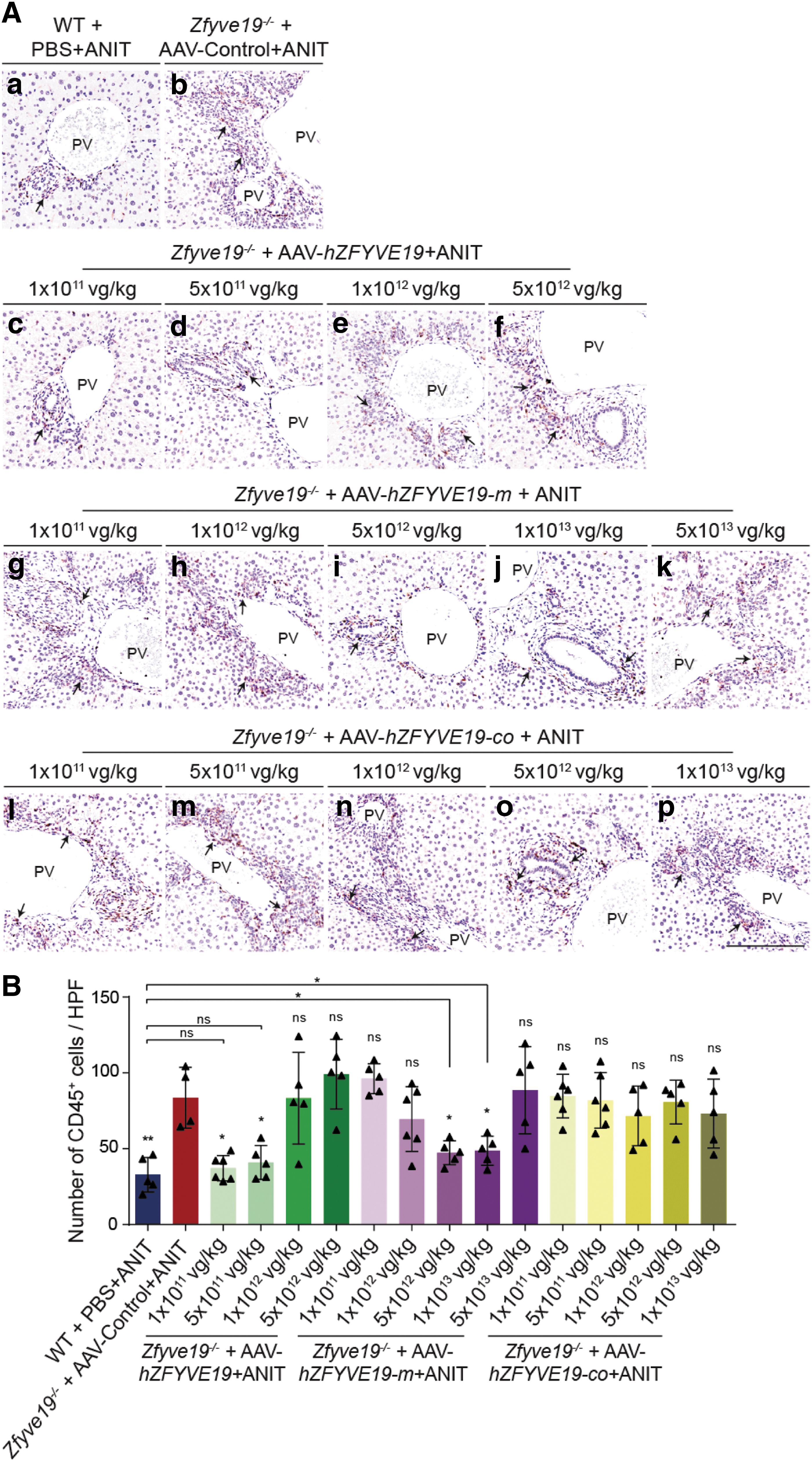
Immunostaining of CD45 on the livers of mice receiving different vectors.
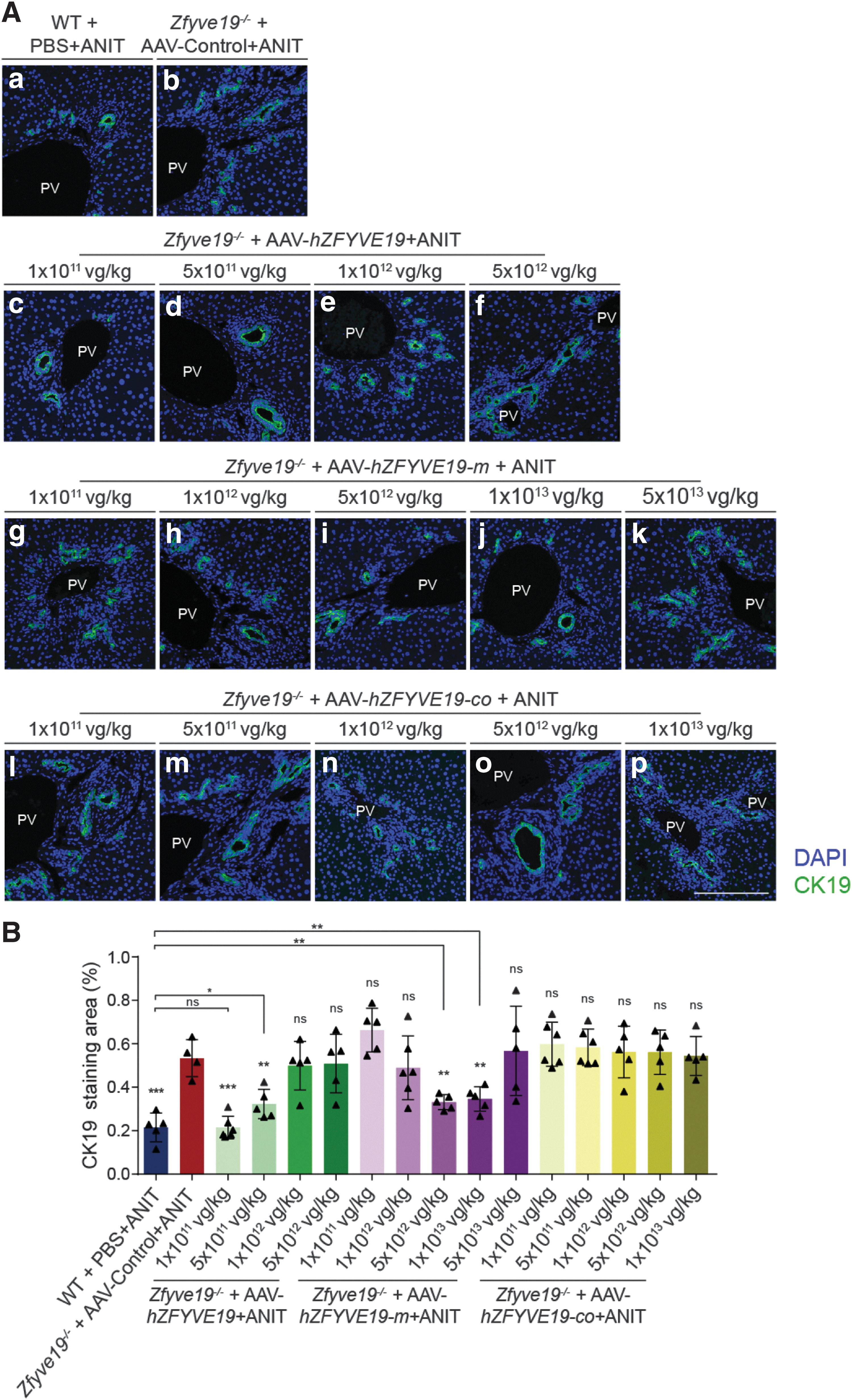
Immunofluorescence of CK19 on the livers of mice receiving different vectors.
Interestingly, higher doses of the AAV-hZFYVE19 vector gradually lost therapeutic benefits and even caused deterioration at the highest dose of 5 × 10¹² vg/kg. The same trend was observed in groups treated with the AAV-hZFYVE19-m vector (Figs. 3 and 4).
AAV-hZFYVE19 vector prevented the portal-tract fibrosis in the Zfyve19−/− mouse model
Liver fibrosis was further quantified using Sirius Red staining. Zfyve19−/− mice in the AAV-Control group showed a significantly larger area of Sirius Red uptake in the portal tract than WT mice (Fig. 5A, B). However, the areas of Sirius Red uptake in the portal tract were significantly decreased in the 1 × 10¹¹ and 5 × 10¹¹ vg/kg AAV-hZFYVE19 vector groups compared with the AAV-Control group (Fig. 5A, B), while the area of Sirius Red uptake varied greatly among individuals in the groups receiving the AAV-hZFYVE19 vector at either 1 × 10¹² or 5 × 10¹² vg/kg, not achieving a statistically significant difference in comparison with the group receiving the AAV-Control vector (Fig. 5A, B).

Sirius Red staining on the livers of mice receiving different vectors.
For AAV-hZFYVE19-m vector groups, portal-tract fibrosis was also significantly decreased at doses of 5 × 10¹² and 1 × 10¹³ vg/kg, but not at 5 × 10¹³ vg/kg (Fig. 5A, B). Mice in groups receiving AAV-hZFYVE19-co showed no effect on portal fibrosis.
AAV-hZFYVE19 vector corrected serum liver marker alterations in the Zfyve19−/− mouse model
Zfyve19−/− mouse model receiving the AAV-Control vector showed significantly increased serum ALT, ALP, and TBA levels compared with WT mice (Fig. 6A, C). However, mice in the groups receiving the AAV-hZFYVE19 vector at a dose of 1 × 10¹¹ vg/kg and the AAV-hZFYVE19-m vector at a dose of 1 × 10¹³ vg/kg showed significantly lower serum ALT levels (marker for hepatocyte injury) compared with the group receiving the AAV-Control vector (Fig. 6A). Serum ALP levels (marker for biliary injury) and TBA levels (marker for cholestasis) were significantly lower in the AAV-hZFYVE19 (1 × 10¹¹ vg/kg), AAV-hZFYVE19 (5 × 10¹¹ vg/kg), AAV-hZFYVE19-m (5 × 10¹² vg/kg), and AAV-hZFYVE19-m (1 × 10¹³ vg/kg) groups than in the group receiving AAV-Control vectors (Fig. 6B, C). No significant differences were observed in the TBIL levels among the groups (Fig. 6D).

Serum levels of hepatobiliary injury markers from mice receiving different vectors at different doses.
Dose-dependent expression of AAV-hZFYVE19 vector
Based on the above results, the AAV-hZFYVE19 vector at a lower dose seemed to have the best therapeutic effects, and so, the expression of the AAV-hZFYVE19 vector at different doses was further assessed. RT-qPCR analysis showed that hZFYVE19 mRNA (Supplementary Fig. S4A) was expressed in a dose-dependent manner in Zfyve19−/− mouse livers after tail vein injection of the AAV-hZFYVE19 vector at various doses compared with WT mice receiving PBS injection or Zfyve19−/− mice receiving the AAV-Control vector. Western blotting (WB) of hZFYVE19 showed that the recombinant protein (43-kDa and 60-kDa, Supplementary Fig. S4B, C) was dose dependently expressed in Zfyve19−/− mouse livers after injection of the AAV-hZFYVE19 vector at various doses compared with no expression in the livers of Zfyve19−/− mice receiving the AAV-Control vector. WB also showed dose-dependent expression of ZFYVE19 in Huh7 in vitro using the AAV-hZFYV19 vector with varying multiplicity of infection values (Supplementary Fig. S5).
DISCUSSION
Biallelic loss-of-function ZFYVE19 variants are associated with a chronic liver disease characterized by portal-tract fibrosis, increased numbers of malformed bile ducts, and abnormal values for serum biomarkers of hepatobiliary injury. 1 No cure other than liver transplantation exists, 1 to explore novel treatments is thus urgent. The correction of ZFYVE19 disease phenotypes in the Zfyve19−/− mouse model by certain vectors encoding hZFYVE19 at optimized doses not only further confirmed the causal relationship between ZFYVE19 variants and the disease, but also demonstrated the potential of gene therapeutics in the treatment of this disease.
Three ZFYVE19-related AAV vectors (AAV-hZFYVE19, AAV-hZFYVE19-m, and AAV-hZFYVE19-co) were tested in this study. Both AAV-hZFYVE19 and AAV-hZFYVE19-m prevented or reduced the development of liver histopathological abnormalities and the abnormal elevation of serum biomarkers of hepatobiliary injury (ALT, ALP, and TBA) at low doses, but the codon-optimized vector (AAV-hZFYVE19-co) showed no therapeutical effect at any doses. Although codon optimization is generally used to enhance the expression of targeted genes in gene therapy, it may not succeed every time. 18 However, we only created one optimized form of the protein and a different codon optimization strategy for the ZFYVE19 gene could perform better.
The therapeutically effective doses of the AAV-hZFYVE19-m vector were 5 × 10¹² and 1 × 10¹³ vg/kg, much higher than those of the AAV-hZFYVE19 vector (1 × 10¹¹ and 5 × 10¹¹ vg/kg). However, even higher doses of the AAV-hZFYVE19-m vector could not reach the same efficacy as the lower doses of the AAV-hZFYVE19 vector. The AAV-hZFYVE19 vector produces both 60- and 50-kDa proteins, whereas the AAV-hZFYVE19-m vector only produces 50-kDa protein, but at a higher yield (Fig. 1 and Supplementary Fig. S2). It has been suggested that the 50-kDa form, which starts translation from the second initiation codon of the ZFYVE19 transcript, 1 may play a major role in human beings. This study suggests that the 60-kDa protein, or the sequence between the traditional and alternative start codon, has important functions in unknown mechanisms and warrants further exploration.
It was also noted that the AAV-hZFYVE19 vector at lower doses (1 × 10¹¹ and 5 × 10¹¹ vg/kg) showed better efficacy than at higher doses, with a dose of 1 × 10¹¹ vg/kg being the best. At a dose of 5 × 10¹² vg/kg, some individuals showed even worse histopathological and blood hepatobiliary indices than those in the control group receiving the AAV-Control vector. This suggests that ZFYVE19 requires an optimal expression level to achieve the best outcome in gene therapy. A similar phenomenon has been observed in gene therapy experiments on Rett syndrome (RTT) and MeCP2-related disorders. 19,20 Gene replacement is considered the most amenable strategy to cure RTT and MeCP2-related disorders, but overexpression of MeCP2 results in severe neurological defects and liver damage in injected animals. 21,22
ZFYVE19 was known to delay the cytokinesis by forming a complex with VPS4 and CHMP4C on the midbody ring during cytokinesis to ensure complete separation of DNA. 23 Depletion of ZFYVE19 enabled cleavage furrow regression, multinucleation, and abscission failure in HeLa cells, while overexpression of ZFYVE19 induced a significant delay in abscission, as judged by a marked increase in cytokinetic profiles in vitro. 23 Therefore, both ZFYVE19 deficiency and overexpression may elicit pathogenic outcomes. It could be a possible explanation why the AAV-hZFYVE19 at a higher dose gradually lost the therapeutic benefits, and even caused deterioration in some individuals receiving the highest dose of 5 × 10¹² vg/kg. Our results reported here can provide a basis for future clinical gene therapy research on the prevention of the progression of the ZFYVE19 deficiency disease. However, the long-term hZFYVE19 expression, long-term effectiveness, and toxicology require further investigation.
CONCLUSION
The presented data demonstrate that bile duct hyperplasia, portal-tract fibrosis, and elevations in serum biomarkers of hepatobiliary injury (ALT, ALP, and TBA) that characterize the mouse Zfyve19−/− model of ZFYVE19 disease can be prevented or reduced by the introduction of hZFYVE19 via AAV. These data not only verify the pathogenic role of ZFYVE19 deficiency in one form of human chronic liver disease but may also suggest that ZFYVE19 needs to function well at an optimal level of expression; both too low and too high expressions may be harmful.
Footnotes
ACKNOWLEDGMENTS
The authors thank all the members of the collaborating laboratories for the thoughtful discussions.
DATA AVAILABILITY STATEMENT
The data that support the findings of this study are available on reasonable request from the corresponding authors.
ETHICS APPROVAL STATEMENT
All animal-related protocols were approved by the Committee of the Care and Use of Laboratory Animals in Children's Hospital of Fudan University (Permit No. [2022] 103/104), according to the Institutional Animal Care and Use Committee guidelines.
AUTHORs' CONTRIBUTIONS
J.-S.W., X.X., and X.W.: Conception, design, drafting, and revision of the work. Y.Z. and D.T.: Conception, experiment, analysis, visualization, and writing—original draft. J.Y. and L.W.: Analysis and interpretation of the data. All authors have read the final version of the article and approved its final submission.
AUTHOR DISCLOSURE
No competing financial interests exist.
FUNDING INFORMATION
The work was supported by the National Key Research and Development Program of China (Grant No. 2021YFC2700800) and the Shanghai Municipal Science and Technology Major Project (19140901600).
SUPPLEMENTARY MATERIAL
Supplementary Data
Supplementary Figure S1
Supplementary Figure S2
Supplementary Figure S3
Supplementary Figure S4
Supplementary Figure S5
References
Supplementary Material
Please find the following supplemental material available below.
For Open Access articles published under a Creative Commons License, all supplemental material carries the same license as the article it is associated with.
For non-Open Access articles published, all supplemental material carries a non-exclusive license, and permission requests for re-use of supplemental material or any part of supplemental material shall be sent directly to the copyright owner as specified in the copyright notice associated with the article.