




Abstract
MC3 and MC5 are both colorectal cancer-specific MAbs previously prepared in our laboratory that can detect colorectal cancer with high sensitivity and specificity. Thus far the distribution of MC3-Ag and MC5-Ag in colorectal cancer remains largely unknown. In the present study, we have firstly found that the expression of MC3-Ag and MC5-Ag was higher in moderate-differentiated and poor-differentiated colorectal cancer tissue than that in well-differentiated colorectal cancer tissue by immunohistochemistry. The expression of MC5-Ag in colorectal cancer tissue without metastasis was found to be significantly less than that in tissue accompanied with metastasis. However, the expression of MC3-Ag in colorectal cancer tissue without metastasis was found similar to that in tissue accompanied with metastasis. The results showed that MC3-Ag and MC5-Ag might play important roles in colorectal carcinogenesis and that MC3 and MC5 could be used as a tool for screening colorectal cancer.
Introduction
In the late 1980s, Fan and colleagues prepared a series of monoclonal antibodies (MAb) that specifically react with antigens of colorectal cancer. Among these antibodies, the MAbs MC3 and MC5 have relatively high specificity against colorectal cancer.(3,4) MC3 and MC5 are the specific monoclonal antibodies directed against colorectal carcinoma, which have a potential use for in vivo diagnosis and therapy of colorectal cancer. However, despite the verified relationship between MC3 and MC5 immunoreactivity and the presence of colorectal cancer, their application is not widespread due to the lack of expression pattern of the MC3-Ag and MC5-Ag in colorectal carcinogenesis.
Here, we have found that the expression of MC3-Ag and MC5-Ag was higher in moderate-differentiated and poor-differentiated colorectal cancer tissue than in well-differentiated colorectal cancer tissue by immunohistochemistry.
Materials and Methods
Tissue samples
Specimens were obtained from 192 consecutive patients who underwent resection for colorectal cancer from April 2004 to September 2004 in Tianjin People Hospital (Tian'jin, China). All samples were obtained with the patients' informed consent, and studies were performed under the aegis of Institutional Review Board (IRB)-approved protocols. None of the patients received preoperative chemotherapy. Routinely, the resected specimens were histologically examined by H&E staining and reviewed by a pathologist (Y. Li), and the diagnoses were confirmed. Clinical and biologic information was available to all patients.
Immunohistochemistry
Paraffin-embedded sections were cut from the representative formalin-fixed tissue blocks and then stained by using the ABC technique; 3% hydrogen peroxidase in methanol was used to block the endogenous peroxidase activity in the tissue sections.(5) The sections were then washed in PBS and incubated in a 1:10 dilution of normal horse's serum/PBS solution for 1 h to block non-specific binding. The primary monoclonal antibody MC3 or MC5 was applied on the tissue samples and incubated at 4°C overnight. Peroxidase staining was done by using ABC kits (Vector Laboratories, Burlingame, CA). The section of colorectal cancer that stained positively for MC3 or MC5 was used as a positive control and included in each staining batch. An irrelevant mouse monoclonal antibody was substituted for the primary antibody in test sections for negative control. The immunostaining of ++ and +++ was considered positive.
Statistical analysis
All data were analyzed by χ2 test using the SPSS software package (SPSS Inc., Chicago, IL). A value of p < 0.05 was considered significant.
Results
Immunohistochemical expression of MC3-Ag
There was a total of 192 cases of colorectal cancer tissue, including 72 cases of poor-differentiated tissue, 89 cases of moderate-differentiated tissue, and 31 cases of high-differentiated tissue. As shown in Table 1, the expression of MC3-Ag was higher in moderate-differentiated (53%, 47/89) and poor-differentiated (63%, 45/72) colorectal cancer tissue than in well-differentiated colorectal cancer tissue (45%, 14/31) (Fig. 1). The expression of MC3-Ag in colorectal cancer tissue without metastasis (55%, 95/172) was found to be the same as that in tissue accompanied with metastasis (55%, 11/20).
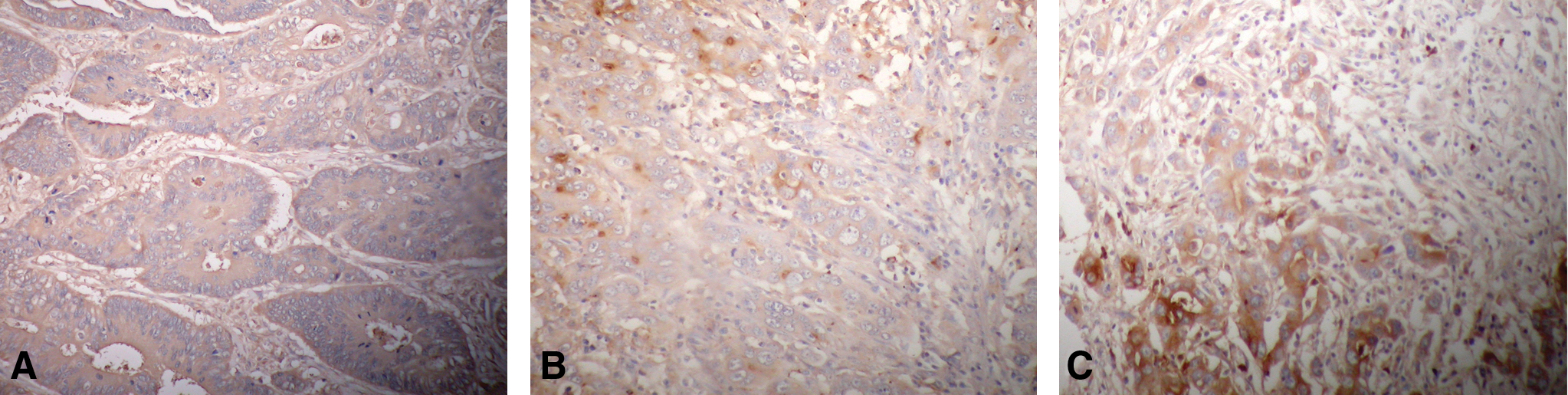
Immunohistochemical expression of MC3-Ag in colorectal cancer tissue. (
Immunohistochemical expression of MC5-Ag
As shown in Table 2, the expression of MC5-Ag was higher in moderate-differentiated (56%, 50/89) and poor-differentiated (65%, 47/72) colorectal cancer tissue than in well-differentiated colorectal cancer tissue (42%, 13/31) (Fig. 2). The expression of MC5-Ag in colorectal cancer tissue without metastasis (55%, 95/172) was found to be significantly less than that in tissue accompanied with metastasis (75%, 15/20).
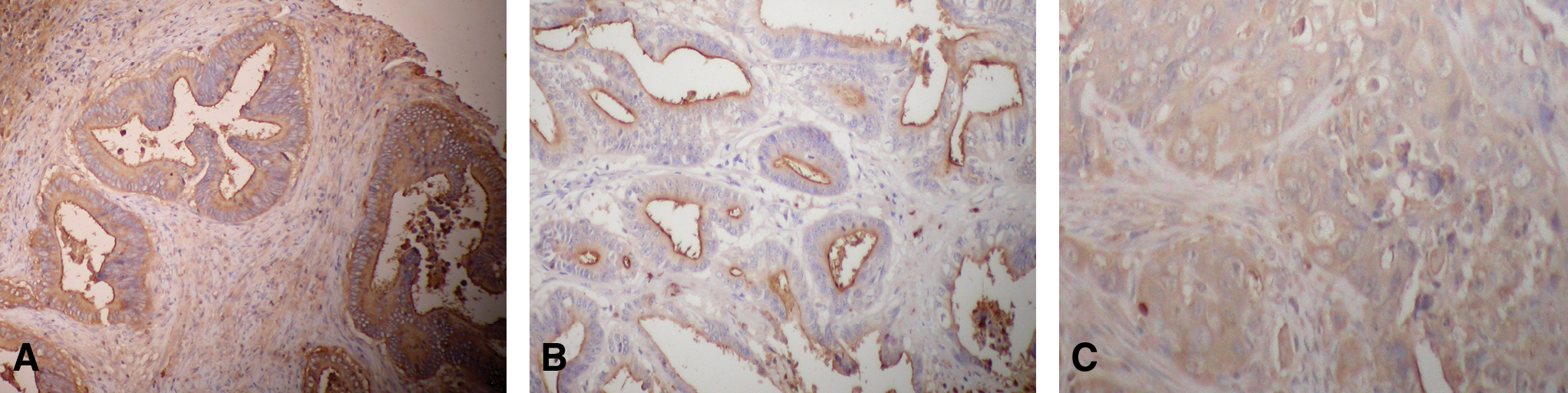
Immunohistochemical expression of MC5-Ag in colorectal cancer tissue. (
Discussion
Colorectal cancer is presently the fourth most prevalent malignancy in China. The high mortality rate found with colorectal cancer is largely a result of the lack of early detection methods. Serial combined testing for tumor markers including carcinoembryonic antigen, CA 50, CA 19–9, etc., plus colonoscopy, is the most commonly used strategy for early diagnosis.(6–9) However, the non-specificity and low sensitivity of these cancer biomarkers have hampered their use in cancer detection. Therefore, a biomarker specific to colorectal cancer would be beneficial for early diagnostic screening.
Using a homogenate preparation of colon cancer tissue, Fan and colleagues developed a series of colorectal cancer-specific MAbs. Among these antibodies, MC3 and MC5 are the most specific and sensitive antibodies.(4) Here we have found that the expression of MC3-Ag and MC5-Ag was higher in moderate-differentiated and poor-differentiated colorectal cancer tissue than in well-differentiated colorectal cancer tissue by immunohistochemistry. The expression of MC5-Ag in colorectal cancer tissue without metastasis was found to be significantly less than that in tissue accompanied with metastasis. The expression of MC3-Ag in colorectal cancer tissue was not significantly associated with metastasis.
The results clearly showed that MC3-Ag and MC5-Ag was highly detected in colorectal cancer, indicating that they might play important roles in colorectal carcinogenesis. Thus, MC3 and MC5 could be used as a tool for screening colorectal cancer.
Footnotes
Acknowledgment
This study was supported in part by grants from the National Scientific Foundation of China (30770958 and 30871141) and grants (09KY25, CBSKL200911, 2009CB521705, 30371760, and 2006BAI02A14).