




Abstract
We have devised a method for the purification of human basophils from umbilical cord blood by FCM. Umbilical cord blood was collected from six healthy, full-term deliveries. After separation of red blood cells, mononuclear cells were isolated by Ficoll-Hypaque. Six samples followed by positive selection using flowcytometry (FCM) for CD203c+CD45int+ cells. Purity and recovery of cells were measured. Purity and recovery of basophils by FCM with CD203c+CD45int+ markers were 95.02 ± 2.94% and 61.42 ± 5.95%. Cell sorting for CD203c+CD45int+ cells by FCM is an improved method for obtaining pure umbilical cord blood-derived basophils. We established cord blood-derived basophil purification technique as a source from which active basophils can be isolated for immunochemical characterization.
Introduction
Materials and Methods
Materials
Dextran and Ficoll were purchased from Amersham Pharmacia Biotech (Uppsala, Sweden). CD45 monoclonal antibody (MAb) was obtained from BD Biosciences (San Jose, CA). The MAb FR3-16A11 (CD203c) was obtained from Milteny Biotec (Bergisch Gladbath, Germany).
Purification of basophils by Ficoll density centrifugation
After receiving informed consent and approval from the Ethical Review Board at the Xinhua Hospital (Shanghai Jiaotong University School of Medicine, China), umbilical cord blood (60–100 mL) was collected from six normal, full-term deliveries. Heparinized cord blood was mixed 5:1 with 6% dextran and incubated for 30 min at 37°C after which any erythrocytes in samples were deposited. The cells were washed twice with 50 mL of PBS (pH 7.2, 0.01 M)/10 mM EDTA. The cells were diluted with PBS/EDTA to 4 mL, layered over the Ficoll-Hypaque (4 mL 1.065 g/mL), and centrifuged at 400 g for 30 min at 25°C. Basophils were concentrated at the 1.065 g/mL Ficoll interface, harvested, and washed twice with 50 mL of PBS/EDTA.
Flow cytometry
Mononuclear cells were pre-incubated with PBS/EDTA/0.5% BSA for 20 min at 4°C. Thereafter, cells were suspended in PBS/EDTA/BSA at a concentration of 107 per 80 μL, and incubated with 20 μL of FcR Blocking Reagent and 10 μL PE-labeled CD203c and 10 μL Percp-labeled CD45 per 107 cells for 10 min at 4°C with gentle shaking. The cells were then washed with PBS/EDTA/BSA. Basophils were sorted as a CD203c+CD45int+ cell population.
Percentage purity and recovery was determined by Alcian blue staining of cells both before and after sorting.(5) Viability was assessed by Trypan blue exclusion.
Statistical analysis
Statistical significance was assessed by SAS6.12.
Results
Percentage of purity and recovery of basophils
After removing erythrocytes by dextran, the human umbilical cord blood basophils obtained were only 0.26 ± 0.17% pure. The yield of basophils by this method was 90.08 ± 1.04%. Human basophils obtained by density gradient centrifugation over Ficoll-Hypaque were 2.97 ± 0.36% pure. The recovery was 75.43 ± 1.99%. Basophils purified by flow cytometry sorting resulted in a high purity of 95.02 ± 2.94% and a significantly high recovery 61.42 ± 5.95% (Table 1). Viability as assessed by Trypan blue exclusion was 99.5 ± 0.89%.
Expression of CD45 and CD203c on umbilical cord blood basophils
Basophils were identified as low side scatter (SSC), CD203c-PE+, and CD45-Percp intermediate+ cells (group P5) (Fig. 1).
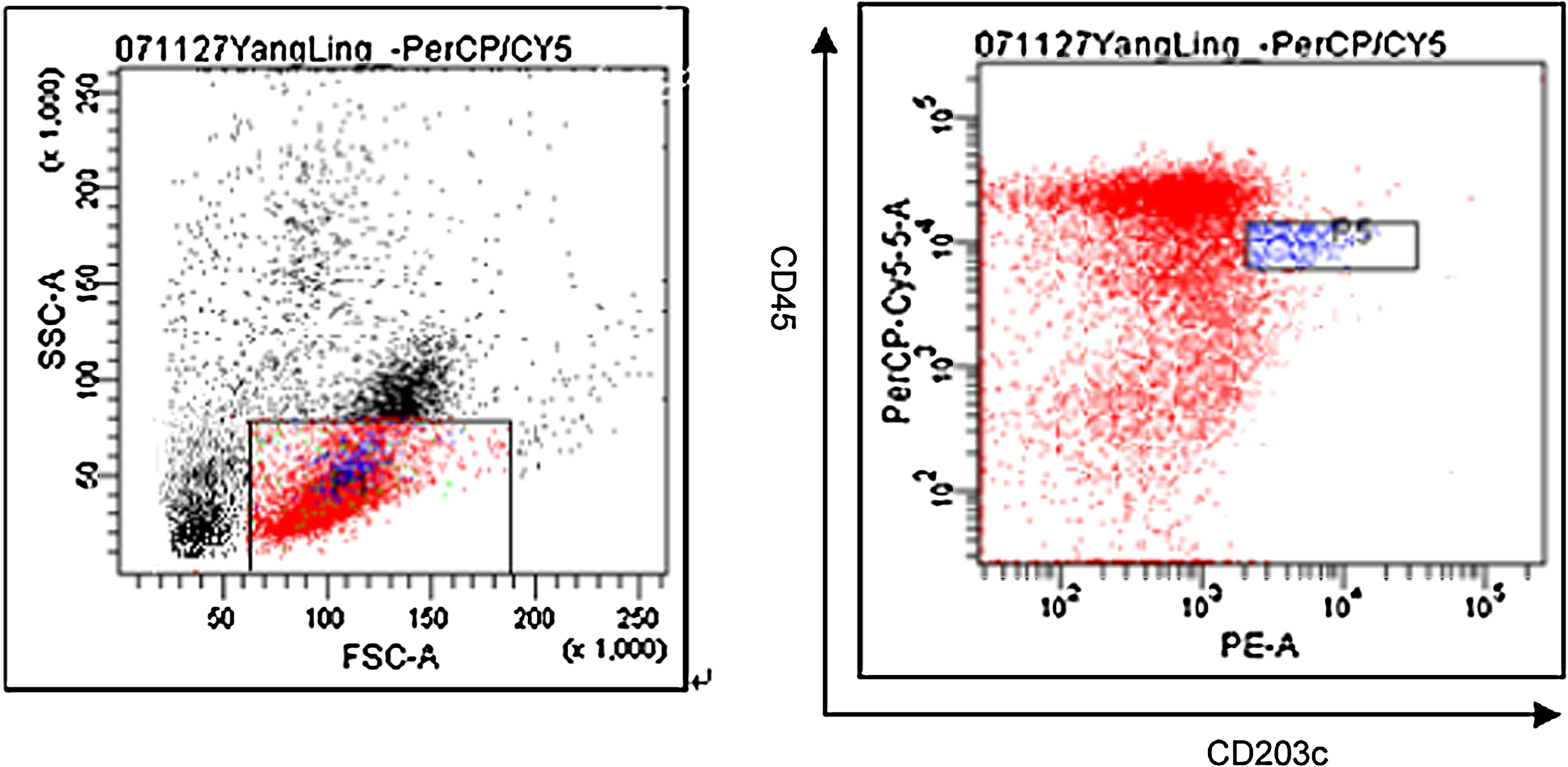
Basophils were identified as low SSC, CD203c-PE+, and CD45-Percp intermediate+ cell (group P5).
Purified basophils stained with Alcian blue
Morphology of purified basophils was shown through cytospin preparation with Alcian blue staining. Magnification 40 × (Fig. 2).
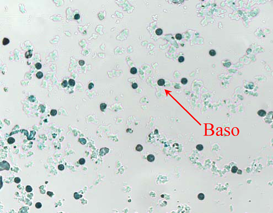
Purified basophils stained with Alcian blue (×400).
Discussion
A major obstacle in the thorough research of basophils has been the difficulty in obtaining sufficient numbers of pure basophils, as well as an almost complete absence of functional mRNA in peripheral blood basophils. Two tumor cell lines with basophil origin have already been described, KU812 and LAMA84,(6–8) but they display characteristics of multiple lineages(7) and have a very low degree of granulation, which means that they are not optimal for studies of lineage-specific granule proteins. Jensen and colleagues' research indicates that IgE does not induce intracellular signals in KU812 (i.e., tyrosine-phosphorylation or Ca2+ release).(9) The storage conditions considerably affect basophil IL-4 release. The above data confirm that the KU812 cell line does not share characteristics with human basophils and is inconsistent with the cellular models that have been used for studies of the mechanism responsible for triggering allergic inflammation.(10)
Immunocytes in the umbilical cord blood have not been stimulated directly by outside antigen, so it can be used as a perfect sample to study pathogenesis of allergic diseases.
A high degree of basophil purity is often required for functional studies of basophils, especially when mediator secretion from contaminating cells masks what is released by basophils. Pure basophil populations are also mandatory when investigating either intracellular signaling events or to exclude the possibility of priming cytokines (e.g., IL-3, GM-CSF), derived from cellular contaminants, to influence basophil function.(1) Therefore, several protocols for basophil purification have been published over the past decade.
One of these protocols is a three-step procedure that consists of a Ficoll density gradient step, counterflow elutriation, and negative selection by MACS. The mean purity of basophils was 97.6 ± 3.96% with a viability of 99.6 ± 0.83%. The recovery rate was 49.7 ± 15.6%.(11) A new method was devised by Gibbs and colleagues(12) in which heparinized or K3-ethylenediaminetetraacetic acid blood samples were first subjected to centrifugation in HetaSep, directly followed by negative selection using immunomagnetic beads. Using this protocol, basophils were enriched close to homogeneity in most cases with a mean purity of 99.34 ± 0.88% and a mean recovery of 75.6%. Basophil viability following purification was 99.6 ± 0.89% using Trypan blue exclusion.(12) These are good purification methods for peripheral blood. However, the component of cord blood is different from that of peripheral blood. Cord blood cells include a proportion of immature basophils that have not fully developed their granules.(13) Inasmuch as the cell surface expression of immature basophils may differ from the mature ones, purification protocols for basophils in the cord blood are different from those in the peripheral blood. In early cultures, almost 60% of the CD203c+ cells co-express human leukocyte antigen (HLA)-DR, a marker that is absent on mature circulating basophils. The presence of HLA-DR on basophil precursors may explain the low recovery (24 ± 5.2%) obtained after isolation of cultured basophils when using a conventional basophil isolation kit that removes HLA-DR+ cells.(14) To obtain active immature basophils, a purification method was developed. Umbilical cord blood cells were cultured in the presence of interleukin (IL)-3 and a two-step cocktail of antibodies against selected markers, which resulted in both high purity (95 ± 0.5%) and recovery (59 ± 1.5%) of cultured basophils. A drawback of this methods was that the cells had to be cultured for up to 9 days,(14) making the manipulation both time-consuming and complicated.
In Willheim and colleagues' study, combinations of negative and positive selection techniques were applied in order to purify normal human blood basophils. In his report, basophils (n = 9) were purified from peripheral blood (350–450 mL) by current counterflow elutriation followed by depletion of monocytes with CD14 MAb conjugated to magnetic beads, and subsequent cell sorting by flow cytometry for CDw17+ cells. Basophil purity was 99.5 ± 0.4% (range 98.7–99.9%). The yield of purified basophils was only 8–25%.(15)
In a past study, we established an original method, including the removal of erythrocytes by dextran, discontinuous density gradient centrifugation over Ficoll-Hypaque, and negative selection by MACS while using a cocktail of magnetically labeled antibodies directed against CD2, CD14, CD16, and CD19. Using this protocol, umbilical cord blood-derived basophils were enriched close to homogeneity in most cases with a mean purity of 75.8% and a mean recovery of 77.4%. Finally, we present a novel purification method for optimal recovery and purity of in vitro-derived basophil precursors.
A common surface marker on human basophils, mast cells, and their CD34+ progenitors was recently described and designated CD203c. This surface marker could become a very useful marker in allergy diagnosis as activated basophils upregulate their surface expression of CD203c.(16) The research of Reimer and colleagues showed that CD203c is indeed a marker expressed very early during basophil differentiation; the group therefore considers this marker a good alternative to FcɛRI for identifying basophil precursors.(14)
CD45 is a marker expressed on basophils and absent on mast cells. Here we describe a method based on positive selection optimal for the purification of umbilical cord blood-derived basophils, which resulted in both high purity (95.02 ± 2.94%) and recovery (61.42 ± 5.95%) with a viability of 99.5 ± 0.89%.
In conclusion, we have established an efficient and cost effective cord blood-derived basophils separation technique from which active basophils can be isolated for biochemical characterization.
Footnotes
Acknowledgment
We would like to thank Chayim Goldberg for editing the English syntax in this article.