




Abstract
To set up an immunoassay-based method to detect Bisphenol A (BPA), we generated a monoclonal antibody (MAb) using a specially designed carboxyl derivative of BPA as the immunogen. BPA-HS was synthesized by reaction using BPA and succinic anhydride. The mice were immunized with the BPA-HS-BSA conjugate. The MAb was obtained from a hybridoma. In addition, we showed that the MAb was highly specific for BPA. The limit of detection was approximately 0.05 ng mL−1 (ppb) in assay buffer and 0.1 ng mL−1 (ppb) in water samples. The recoveries of BPA for water samples were from 90.8% to 114%, and coefficients of variation were from 15.6% to 39.4%. Thus, the ELISA method is a rapid and high throughput screening tool to detect BPA in water products.
Introduction
While frequent use and heating can break down the plastic and cause BPA to leach from plastic products into food, the higher the temperature the sooner the speed of releasing.(4) As a result, BPA gets into the human body and increases the plasma level of BPA, which has been identified as a potential estrogenic substance. It can stimulate cell proliferation of the male and female sexual organs,(5) causing reproductive abnormalities in wildlife and disrupting endocrine function in animals and humans.(6–8) In addition, BPA is postulated to cause various kinds of cancers, such as prostate, testicular, and breast cancer, and has diverse pleiotropic effect on the brain and cardiovascular system.(9) Thus, a simple, selective, and sensitive analytical method for the detection of a trace amount of BPA in the environment has become a significant issue.
Recently, the United States, Canada, and France have banned the use of BPA on food packaging and contact materials; China also forbade the import of polycarbonate baby bottles containing BPA. To date, the methods most frequently used for the determination of BPA are high performance liquid chromatography (HPLC),(10) gas chromatography coupled with mass spectrometry (GC/MS),(11) liquid chromatography with electrochemical detection (LC–ED),(12) and liquid chromatography coupled with mass spectrometry (LC-MS).(13,14) Although these methods are quantitative, they take a long time, need large sample volumes, generate large amounts of waste, and require bulky and expensive instruments. Kaddar and colleagues(15) developed a radioimmunoassay using a polyclonal anti-BPA antibody (PAb), while the results show that the PAb is of low sensitivity and weak specificity. Marchesini and colleagues(16) established biosensor immunoassays for the detection of bisphenol A by employing anti-BPA antibodies (polyclonal [PAbs] and monoclonal [MAbs]). However, this assay is too sensitive to be disrupted and the binding capacity of the immobilized MAbs decrease fast. Therefore, a rapid and high throughput screening method to detect BPA in liquid storage containers is highly desirable.
In this study, we report the generation of a monoclonal antibody specifically recognizing BPA. Using the antibody, we designed an enzyme-linked immunosorbent assay (ELISA) for detecting BPA in liquid storage containers in a simplified and sensitive way. This method has the potential to be developed into an alternative tool for BPA detection.
Materials and Methods
Chemicals and reagents
All chemicals, unless otherwise stated, were analytical reagent grade. Bisphenol A, urea hydrogen peroxide, dicyclohexylcarbodiimide (DCC), Freund's adjuvant (Freund's complete adjuvant and Freund's incomplete adjuvant), bovine serum albumin (BSA), ovalbumin (OVA), horseradish peroxidase (HRP), polyethylene glycol 1450 (PEG 1450, 50%, w/v) and N-hydroxysuccinimide (NHS) were from Sigma-Aldrich (St. Louis, MO). 3,3′,5,5′-tetramethylbenzidine (TMB), dimethyl sulphoxide (DMSO), dimethylformamide (DMF), Tween-20, succinic anhydride, and pyridine were from Sinopharm Chemical Reagent Co. (Shanghai, China). Hypoxanthine/aminopterin/thymidine (HAT), hypoxanthine/thymidine (HT), and DMEM with l-glutamine culture medium were bought from Gibco (Invitrogen, Carlsbad, CA). Fetal calf serum was from Guangzhou Zhanchen Bio-tech Co. (Guangzhou, China). Peroxidase-labeled goat anti-mouse IgG (H+L) (HRP-IgG) was from Jackson Immunoresearch Laboratories (West Grove, PA).
Instruments
Microtiter plates were from Nunc (Roskilde, Denmark). The microplate reader was Synergy-HT from Biotek (Winooski, VT). Cell culture plates (24 and 96 wells) and culture flasks were from Costar (Cambridge, MA). Water was purified by a MilliQ purification system from Millipore (Billerica, MA). The LC-MS system model API 3000 was from Applied Biosystems (Bedford, MA).
Buffers and solutions
The following buffers were used in the experiments: (1) phosphate-buffered saline (PBS; 10 mmol L−1 sodium phosphate, 137 mmol L−1 NaCl, 2.7 mmol L−1 KCl [pH 7.5]) for the dilution of antibodies and the preparation of standard solutions; (2) stock solution of BPA at a concentration of 1 mg mL−1 was prepared in methanol and diluted to standard solutions with PBS; (3) the coating buffer was 0.1 M carbonate buffer (pH 9.6); (4) the blocking buffer was 1% OVA (m/v) in PBS; (5) phosphate buffer saline (PBS) with 0.8% (w/v) NaCl (pH 7.2); (6) PBST, a PBS buffer containing 0.05% Tween-20 (v/v), was used for washing; (7) the substrate buffer was 0.1 M citrate (pH 5.5); (8) the peroxidase substrate solution was prepared by mixing 200 μL 1% (w/v) TMB in DMSO with 64 μL 0.75% (w/v) to 20 mL substrate buffer; (9) the enzymatic reaction was stopped with 2 M H2SO4.
BPA hapten derivatives (BPA-HS) for conjugation with proteins
BPA (4.5 g) dissolved in 10 mL pyridine in a conical flask was mixed by slowly adding 2 g succinic anhydride. The mixture solution was then heated to 70°C and stirred for 3 h. Thin-layer chromatography of the reaction mixture demonstrated formation of a product (elution in 10% methanol in chloroform). After removal of pyridine by evaporation, the resulting mixture was crude BPA-HS, which was further purified by recrystallization in methanol.
Synthesis of BPA-HS-protein conjugates
BPA-HS was conjugated to bovine serum albumin (BSA) by an active ester method.(17–19) The carboxylic acid on the hapten was activated with DCC and NHS to produce an active ester, which then reacted with the amine groups on BSA or OVA to form amide bonds. BPA-HS (32.8 mg), NHS (11.5 mg), and DCC (20.6 mg) were each dissolved in 0.5 mL 1,4-dioxane. NHS, followed by DCC, was added slowly to the BPA-HS solution. This activation reaction and carried out overnight at room temperature with continuous stirring. The reaction mixture was centrifuged (2000 g, 10 min) and the supernatant was added very slowly to a BSA solution (340 mg BSA dissolved in 5 mL PBS). The mixture was stirred overnight at 4°C to complete the conjugation reaction. The mixture was then dialyzed against phosphate buffer (0.1 mol L-1 [pH 7.4]) for 3 days (two changes of buffer per day). BPA-HS-BSA was freeze-dried and stored at −20°C. Similarly, BPA-HS-OVA was synthesized as a coating conjugate. The complete antigen BPA-HS-BSA was identified by the ultraviolet and infrared spectrophotometers.
BPA-HS-HRP tracer
BPA-HS was conjugated to HRP by an active ester method.(20) BPA-HS (1.64 mg), NHS (0.58 mg), and DCC (1.03 mg) were dissolved in DMF (0.5 mL). The mixture was stirred at room temperature overnight. The activated hapten was centrifuged (2500 rpm/10 min) and 113.3 μL of supernatant was added dropwise under stirring to 3.4 mg of HRP in 0.13 mol L−1 NaHCO3 (600 μL) in order to obtain molar ratio 5:1 (hapten-HRP). The conjugation mixture was stirred at room temperature for 5 h, and the mixture was then dialyzed against phosphate buffer (0.1 mol L-1 [pH 7.4]). The tracer obtained was diluted with the same volume of glycerol and stored at −20°C until use.
Immunization of mice and production of monoclonal antibodies
Three Balb/c female mice, 8–10 weeks old, were immunized by intraperitoneal injection. BPA-HS-BSA (50 μg) immunogen was diluted in 100 μL sterile PBS and emulsified in Freund's complete adjuvant for the primary immunization. Booster doses of BPA-HS-BSA (50 μg) in Freund's incomplete adjuvant were then injected into the above mice once every 2 weeks for three times. Blood was obtained via retro-orbital venous plexus at day 12 after the third booster immunization and stored overnight at 4°C. Then antisera were collected from the blood by centrifugation at 4000 rpm for 10 min. The mouse that exhibited the highest titer by indirect ELISA was immunized with a final dose of BPA-HS-BSA (50 μg) for fusion experiment.
Anti-BPA monoclonal antibodies were produced using the hybridoma technique. The splenocytes from the immunized mice were fused with the SP2/0 myeloma cells. Hybridomas were selected in a HAT medium (DMEM medium containing 15% FBS). The cultures in 96-well plates were maintained in a 5% CO2 incubator at 37°C. When hybridoma colonies appeared, they were expanded in the HT medium. Hybridoma culture supernatants were screened by an indirect competitive ELISA (icELISA) with BPA as the competitor. BPA-HS-OVA conjugate was used as the coating conjugate. The hybridoma that secreted antibodies specific to BPA was subcloned by the serial dilution method. Colonies of interest were propagated, frozen (overnight at −80°C) in culture medium containing 10% dimethyl sulphoxide, and stored in liquid nitrogen.
For ascites production, two Balb/c female mice were used. The mice were treated by intraperitoneal injection with 0.5 mL of Freund's incomplete adjuvant. After 10 days, each mouse was intraperitoneally injected with 1 × 10⁷ hybridoma cells. After 15 days, a total of 7.4 mL of ascitic fluid was collected from the two mice, incubated overnight at 4°C, and centrifuged at 4°C for 10 min. The supernatant was finally obtained and stored at −20°C.
ELISA procedure
Indirect ELISA was carried out as follows: (1) 100 μL of coating conjugate BPA-HS-OVA diluted with the coating buffer at 0.1 μg mL−1 were added into a microtiter plate and incubated at 4°C overnight. (2) Plates were washed three times using 300 μL/well of the washing buffer and then 100 μL of antibody diluted with PBS were added to each well. (3) After incubation for 30 min at 37°C, the unbound compounds were washed away. (4) 100 μL of HRP-IgG (working concentration recommend 1:5000) were then added to each well and incubated for 30 min at 37°C. (5) After washing four times, 100 μL of substrate solution was added into each well and incubated for 15 min at 37°C. (6) The enzymatic reaction was stopped by the stopping solution (50 μL/well) and then absorbance at 450 nm was measured. The procedure for icELISA was similar to that of indirect ELISA, except for adding 50 μL of BPA standard solution (competitor) and 50 μL of antibody solutions to every well after washing in step 2.
Direct competitive ELISA (dcELISA) tests were carried out using the following procedure: 100 μL of antibody diluted with coating buffer was pipetted into a microtiter plate and incubated at 4°C overnight. Plates were washed three times using 300 μL/well of washing buffer solution and 50 μL of standard solution or sample, and 50 μL of tracer solution were added to each well. Unbound compounds were removed by washing solution after incubation for 30 min at 37°C. 100 μL of the substrate solution were then added to each well and the enzymatic reaction was stopped after 15 min incubation at 37°C by the addition of 50 μL/well of stopping solution. Absorbance values were measured at 450 nm and the concentration of the analyte in a sample was calculated from the calibration curve.
Monoclonal antibody titer and sensitivity
The monoclonal antibody titer was measured using the indirect ELISA method. The ascites titer with an absorbance of 2.0 was defined as the maximum dilution. The sensitivity to free BPA was determined using the dcELISA. To prepare a standard curve, 50 μL of BPA standard solution (prepared in PBS at 0, 0.1, 0.3, 0.9, 2.7, and 8.1 ng mL−1 [ppb]) was added in the dcELISA. The signals obtained in the presence of various BPA concentrations and without competitor (maximal signal) were referred to as B and B0, respectively. The inhibition ratio was obtained by dividing B with B0 (B/B0). A linear standard curve was prepared by plotting log (BPA concentration) versus the inhibition ratio. The limit of detection (LOD) was calculated as the standard deviation from the mean signal measured from the blank wells three times.
Assessment of monoclonal antibody specificity
The cross-reactivity of monoclonal antibody to various dyes and their related derivatives was tested using the dcELISA described, which was determined by measuring their IC50 values in the dcELISA. The IC50 values (50% inhibition levels) were used for calculation of cross-reactivity (CR50%) using the formula: CR50% = 100 × IC50 value of BPA/IC50 value of competitor.
Sample preparation
The following commercial samples were purchased in hypermarket: mineral (or purified) water in liquid storage containers (all samples deriving from different batches). Each kind of sample where BPA was demonstrated to be absent was considered as the blank and used for validation purposes.
Samples were fortified at this stage by addition of BPA standard solution in PBS buffer to give fortified calibration standards equivalent to 0, 0.1, 0.3, 0.9, 2.7, and 8.1 ng BPA per mL sample (the water in liquid storage containers). The result was analysed by the direct competitive ELISA. The recovery and coefficient of variation of BPA in sample were performed.
ELISA analysis and validation
The blank water samples were used for validation study using the dcELISA protocol to assess the range of matrix interferences in the assay and to provide data for determination of false positive rates. Coefficients of variation (CV) were determined by the analysis of the above samples fortified with BPA at 0.1, 1, and 10 ng mL−1 (ppb). The recovery (%) of the fortified BPA was calculated using the equation: conc. measured/conc. fortified × 100. Repeatability of the method was established using fortified duplicate blanks at the levels 0.1, 1, and 10 ng mL−1 (ppb).
Results and Discussion
Hapten design and synthesis of BPA conjugates
BPA are not able to initiate an immune response, so BPA-HS-BSA was used to produce antibody in this study. Since the absence of a known coupling group such as NH2, COOH, and SH on BPA, a BPA derivative (BPA-HS) (Fig. 1) was synthesized following derivatization with succinic anhydride (HS) and identified by mass spectrum (MS) after purification. A solution of the BPA-HS (∼100 μg mL−1 in methanol) was scanned in both positive and negative modes of LC-MS electrospray to confirm its identity. In positive mode, the protonated molecular ion [M+H]+ m/z 329.1 and the molecule peak m/z 328.2 were seen. In negative mode, the molecule peak m/z 327.1 and the deprotonated molecular ion m/z 226.3, 274.2 were seen. In negative mode, the relation between the primary deprotonated molecular ion of BPA-HS and structure of BPA-HS molecule is shown in Table 1.
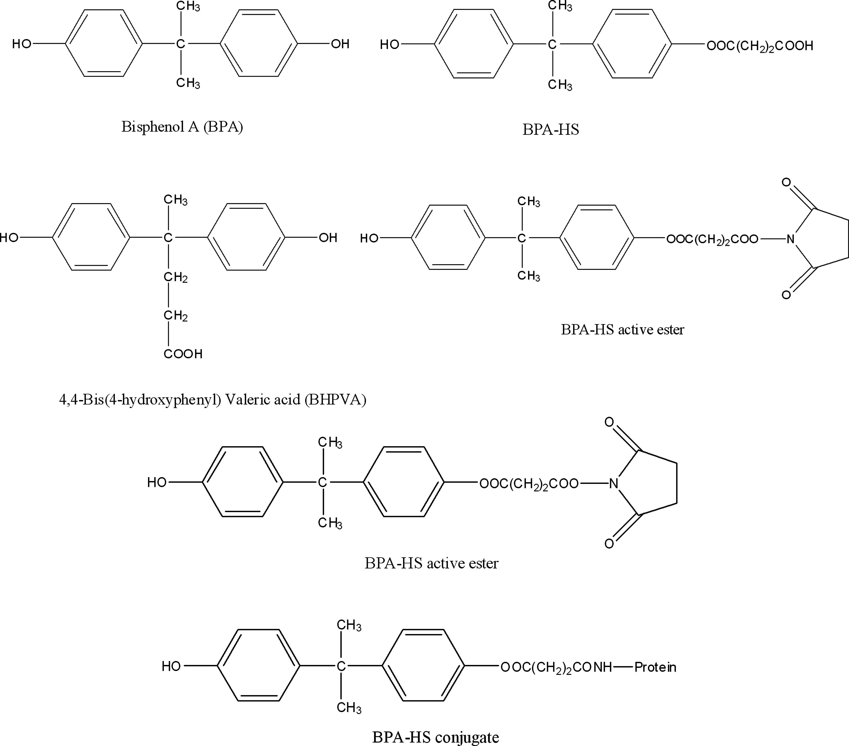
The structures of BPA, BPA-HS, BHPVA, BPA-HS active ester and BPA-HS conjugated with protein.
By an active ester method, the carboxylic acid group on BPA-HS was conjugated with the amino groups on BSA under the effect of NHS and DCC, forming amide bonds. It is different from selected BHPVA (Fig. 1) as hapten, producing BHPVA-HS with amide bond on a different site.(21)
Monoclonal antibody titer and sensitivity
Two fusion experiments were carried out to obtain the specific monoclonal antibody. The titer of the monoclonal antibody was determined by checkerboard titration. The optimal conditions of ELISA were chosen when the OD value was about 2.0. Likewise, the optimal concentration of the coating conjugate was found to be 0.2 μg mL−1. Under the optimal conditions, we found that the titer of the ascites antibody was 1 × 10⁵.
Representative ELISA curves for BPA were presented in Figure 2. The ratios of B/B0 refer to the standard dose responses. B0 ranged from 1.5 to 2 for both ELISA methods. As a result, the assays showed a high sensitivity. The LOD for the dcELISA was approximately 0.05 ng mL−1(ppb), with IC50 of 0.7 ng mL−1 (ppb) for BPA.

Representative dose response curves for BPA in dcELISA for 3C7 ascites antibody. B/B0 is the normalized response relative to the zero standard. Coating monoclonal antibody dilution = 1:4 × 10³, BPA-HS-HRP dilution = 1:2000. The regression curve equation is as follows: y = −36.678 × +44.322, with a correlation coefficient of 0.9943.
Monoclonal antibody specificity and isotypes
Specificity of the MAb in optimized assays was tested by measuring the cross-reactivity using BPA-related compounds. The cross-reactivity was determined by dcELISA described above. The chemical compounds and their corresponding cross-reactivity are shown in Table 2. The data showed that the immunoassays for BPA are highly selective. We found a high cross-reactivity with BPA-HS (Table 2), but only low cross-reactivity with BHPVA (18.9%). No cross-reactivity was detected with other related compounds shown in Table 2.
Through identifying the heavy and light chain isotypes of antibodies by SBA Clonotyping System/HRP kit, the MAb belongs to IgG1 type.
ELISA validation
Twenty different samples (identical matrix) from retail outlets were used as blank samples for the determination of LOD and detection capability of the ELISA procedure at different false positive and negative rates. The determination of LOD was based on 20 blank samples accepting no false positive rates (average + 3 SD). LOD in different samples are listed in Table 3. The LOD was approximately 0.1 ng mL−1 (ppb) in the two matrices. The accuracy and precision were evaluated (5 replicates) by spiking BPA at 0.1, 1, and 10 ng mL−1 (ppb). The recoveries of BPA in water samples were from 90.8 to 114% and coefficients of variation were from 15.6 to 39.4% (Table 3).
N = 5.
Conclusion
We have generated a monoclonal antibody specific for BPA. This antibody is useful for determining the concentrations of BPA within the curve range shown in Figure 2. The dcELISA using this antibody is sensitive enough to detect those substances at a very low concentration level reaching to ppb in water samples. Currently, HPLC and/or LC-MS/MS methods are used to detect the presence of BPA. However, our data showed that the dcELISA method is as sensitive as the LC-MS/MS method and is more sensitive than the HPLC method. Thus, using the monoclonal antibody developed in this study, the dcELISA method is a sensitive and simplified alternative for the detection of BPA in water samples.
Footnotes
Author Disclosure Statement
The authors have no financial conflicts to declare.