




Abstract
The transport of proteins in and out of the nucleus plays important roles in major cellular processes, such as signal transduction and regulation of cell cycle. Proteins that contain a nuclear localization signal (NLS) are recognized by an importin α/β heterodimer and targeted to the nucleus. Here, we report the generation of a rat monoclonal antibody (MAb) that recognizes a novel importin α family member, importin α8, which is expressed during oocyte maturation and early embryonic development. Immunoblot and immunolocalization analyses showed that this MAb was specific for mouse importin α8 and not other importin α family members. These data suggest that this MAb is useful for analyzing molecular functions of importin α8.
Introduction
To date, many importin α homologs have been identified: importin α1 (karyopherin α2, PTAC58), importin α3 (karyopherin α4, Qip1), importin α4 (karyopherin α3, Qip2), importin α5 (karyopherin α1, NPI-1), importin α6 (karyopherin α5), and importin α7 (karyopherin α6).(7) Additionally, very recently, a novel importin α isoform (importin α8, karyopherin α7) was identified(8) and found to be required for normal fertility and fecundity.(9) However, the biological roles of importin α8 have not been clarified.
Here we report the production and characterization of a rat monoclonal antibody (MAb) that specifically recognizes mouse importin α8. This antibody would be useful in immunoblotting and immunohistochemical analyses, and should aid in the elucidation of the physiological function of this protein.
Materials and Methods
Construction of expression plasmids
Full-length mouse importin α8 was isolated from a cDNA library of mouse adult ovary and subcloned into pFLAG-CMV-2 (Sigma, St. Louis, MO), pEGFP-C1 (Takara Bio, Shiga, Japan), and pGEX-6P-1 vector (GE Healthcare, Uppsala, Sweden).
Purification of importin α8 protein
Mouse importin α8 was expressed in Escherichia coli BL21 grown in LB medium containing 25 mg/mL ampicillin induced with 0.2 mM isopropyl β-D-1-thiogalactopyranoside (IPTG) overnight at 18°C. Bacteria were lysed in lysis buffer (50 mM Tris-HCl [pH 7.0], 150 mM NaCl, 1 mM EDTA, 2 mM DTT, 0.2 mM phenylmethylsulfonyl fluoride [PMSF]), sonicated, and clarified by centrifugation. Supernatant was collected and the fusion protein was adsorbed onto glutathione-Sepharose beads (GE Healthcare), and then the beads were washed five times. Importin α8 was obtained by separation of the GST-tag from proteins with PreScission Protease (GE Healthcare) according to the manufacturer's instructions.
Production of monoclonal antibody
An anti-importin α8 rat MAb was generated based on the rat lymph node method established by Kishiro and colleagues.(10,11) A 10-week-old female WKY/Izm rat (SLC, Shizuoka, Japan) was injected via the hind footpads with 200 μL of an emulsion containing importin α8 recombinant protein and Freund's complete adjuvant. After 3 weeks, the cells from the medial iliac lymph nodes of the immunized rat were fused with mouse myeloma SP2 cells at a ratio of 10:1 in a 50% polyethylene glycol (PEG 4000, Merck, Darmstadt, Germany) solution. The resulting hybridoma cells were plated onto 96-well plates and cultured in HAT selection medium (Hybridoma-SFM [Invitrogen, Carlsbad, CA]; 10% fetal bovine serum [FBS]; 5% BM-condimed H1 [Roche, Mannheim, Germany]; 100 mM hypoxanthine; 0.4 mM aminopterin; 16 mM thymidine). The hybridoma supernatants were screened by means of an enzyme-linked immunoadsorbent assay (ELISA) against the GST-importin α8 recombinant protein on day 10 post-fusion. Positive clones were subcloned and rescreened by ELISA and immunoblotting.
ELISA
Recombinant GST-importin α8 protein in TBS-T (20 mM Tris-HCl [pH 7.5], 150 mM NaCl, and 0.05% Tween-20) was adsorbed on the surface of 96-well Nunc immunoplates (Nunc, Roskilde, Denmark) by overnight incubation at 4°C. The plates were blocked with 1% bovine serum albumin (BSA) in TBS-T to limit non-specific binding. The hybridoma supernatants were incubated for 1 h at room temperature, and then washed three times with TBS-T. The plates were incubated for 30 min at room temperature with an alkaline phosphatase-conjugated anti-rat IgG antibody (Jackson ImmunoResearch Laboratories, West Grove, PA). After washing three times with TBS-T, immunoreactivity was visualized using a pNPP phosphatase substrate system (KPL, Gaithersburg, MD).
Oocyte collection
Adult C57BL/6 female mice (Crea Japan, Tokyo, Japan) were superovulated by intraperitoneal injection with 5 IU of PMSG (Serotropin, ASKA Pharmaceutical, Tokyo, Japan) and induced to ovulate after 48 h with 5 IU of hCG (Gonatropin, ASKA Pharmaceutical). Thirteen hours after hCG treatment, oocytes were collected from the oviducts of the mice.
Cell culture
The human epithelial adenocarcinoma cell line, HeLa, and African green monkey kidney fibroblast cell line, COS7, were cultured in Dulbecco's modified Eagle's medium, supplemented with 10% FBS in a humidified atmosphere of 5% CO2 at 37°C.
Immunoblotting
Whole cell extracts of COS7 cells transfected with pFlag-importin α were lysed with RIPA buffer (10 mM Tris-HCl [pH 7.2], 150 mM NaCl, 0.1% SDS, 1.0% Triton X-100, 1.0% sodium deoxycholate, 5 mM EDTA, 10 μg/mL each of leupeptin, pepstatin, and aprotinin, and 1 mM PMSF). Cell lysates were loaded in each lane for SDS-PAGE and then transferred onto Immobilon-P polyvinylidene fluoride (PVDF) transfer membranes (Millipore, Schwalbach, Germany). The membrane was blocked with 3% skim-milk in TBS-T (20 mM Tris-HCl [pH 7.5], 150 mM NaCl, and 0.05% Tween-20), and then incubated with anti-importin α8 MAb 3F2 or anti-Flag rabbit antibody (Sigma). Then the membrane was incubated with horseradish peroxidase (HRP)-conjugated anti-rat IgG antibody (Jackson ImmunoResearch Laboratories), and proteins were visualized using ECL Western Blotting Substrate according to the manufacturer's protocol (GE Healthcare).
Immunofluorescence
HeLa cells transfected with pEGFP-importin α8 were grown on coverslips in culture medium. The cells and mouse oocytes were fixed with 3.7% formaldehyde in PBS for 15 min followed by permeabilization with 0.5% Triton X-100 in PBS for 5 min. After treatment with a goat serum blocking buffer (2% goat serum, 1% BSA, 0.1% gelatin, 0.1% Triton X-100, and 0.05% Tween-20), the cells were incubated with anti-importin α8 MAb 3F2 and the antibody was detected with Alexa 568-conjugated goat anti-rat IgG (Invitrogen).
Results and Discussion
The recombinant mouse importin α8 was expressed and purified from E. coli BL21 as an antigen. A 10-week-old WKY/Izm rat was immunized via the hind footpad with a single injection. At three weeks post-immunization, lymphocytes were collected from the enlarged medial iliac lymph nodes of the rat, and fused with mouse myeloma SP2 cells. The resulting hybridomas were screened by ELISA for production of MAbs that recognize the recombinant importin α8 protein. Additionally, hybridomas that expressed anti-importin α8 MAbs by ELISA were further examined by immunoblotting for specificity to the recombinant importin α8 protein. Three positive clones were obtained. One of these antibodies, designated as MAb 3F2, was selected for further study, because it yielded the strongest immunostaining signals.
In order to further investigate the specificity of MAb 3F2 to importin α8, we transfected COS7 cells with flag-tagged importin αs and then conducted immunoblotting analysis (Fig. 1). The result revealed that MAb 3F2 bound specifically to importin α8 and did not recognize the other importin αs. It has previously been shown that importin α8 is only detected during oocyte maturation and early embryonic development(8,9); therefore this antibody was characterized by immunofluorescence staining of HeLa cells transfected with GFP-tagged mouse importin α8. The MAb 3F2 specifically recognized GFP-tagged mouse importin α8, which was predominantly nuclear, indicating that this monoclonal antibody would be very useful in immunolocalization analysis of importin α8 (Fig. 2A). Furthermore, this antibody was characterized by the immunofluorescence staining of mouse unfertilized oocyte. As shown in Figure 2B, we found that importin α8 protein was especially localized to the spindle, confirming the previously reported localization of the protein in mouse MII oocyte.(9)
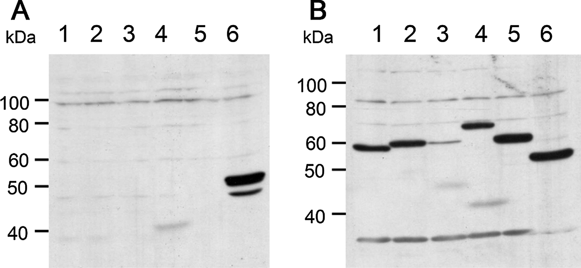
Production of an anti-importin α8 monoclonal antibody. The rat anti-importin α8 monoclonal antibody was generated based on the rat lymph node method. COS7 cell extracts transfected with Flag-importins α1 (lane 1), α3 (lane 2), α4 (lane 3), α5 (lane 4), α7 (lane 5), or α8 (lane 6) were subjected to immunoblotting using MAb 3F2 (
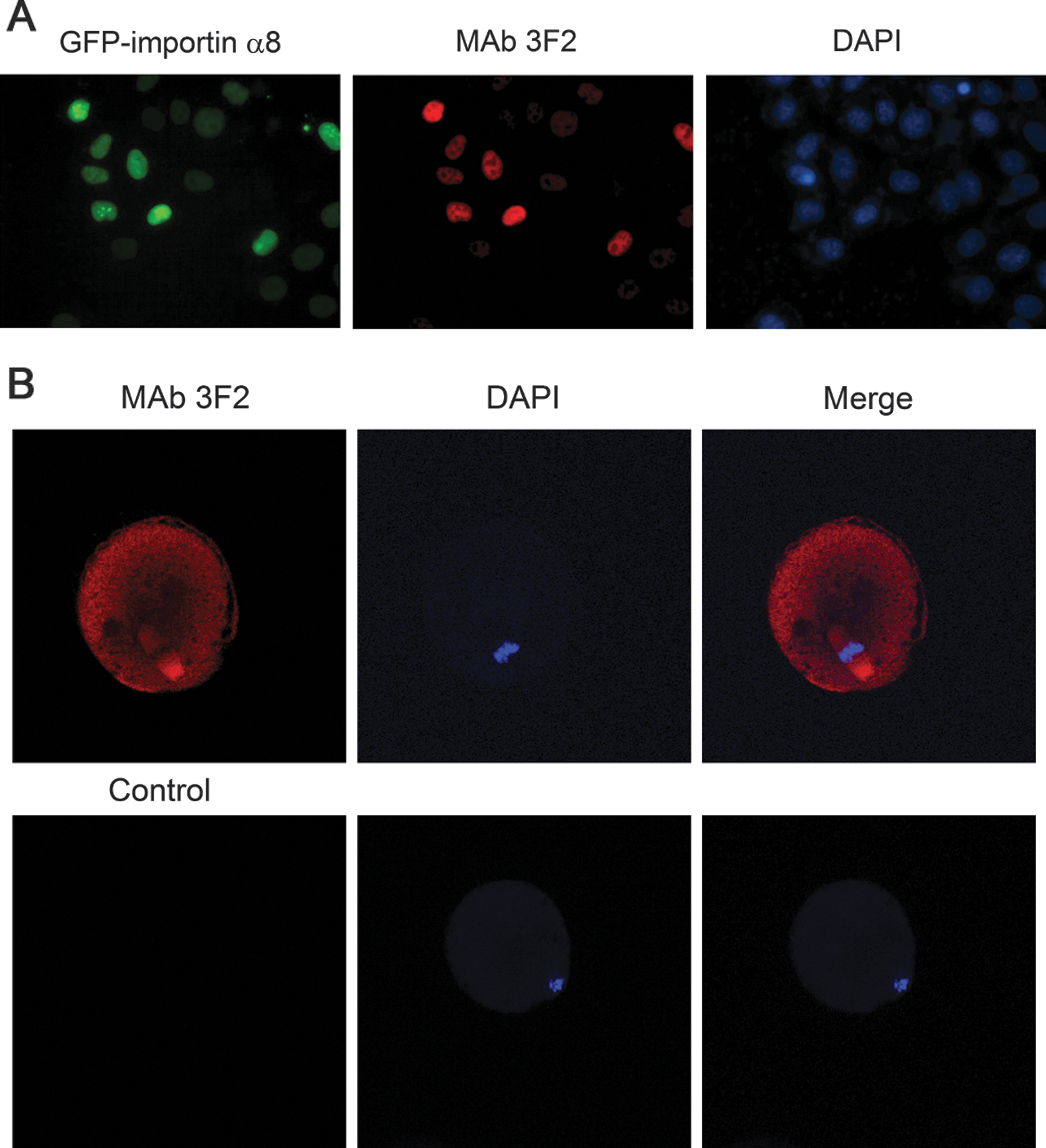
Indirect immunofluorescence of Hela cells transfected with GFP-importin α8 and mouse egg using MAb 3F2. (
In summary, we report here on the production and characterization of a monoclonal antibody that specifically recognizes mouse importin α8. We propose that MAb 3F2 will be beneficial for the study of the function of importin α8.
Footnotes
Acknowledgments
We thank Dr. T. Tachibana (Osaka City University, Osaka, Japan) for technical advice. This work was supported, in part, by the Ministry of Education, Culture, Sports, Science and Technology of Japan and the Takeda Science Foundation.
Author Disclosure Statement
The authors have no financial interests to disclose.