
Abstract
Many nuclear proteins are transported into the nucleus via the importin α/β-mediated pathway. Importin α comprises a multigene family. In this study, we generated and characterized a rat monoclonal antibody (MAb) 3F8 to importin α7. The antibody was generated by the hybridization of mouse myeloma cells with lymph node cells from an immunized rat. The MAb 3F8 specifically recognized importin α7 among importin α isoforms as evidenced by immunoblotting analysis. Furthermore, MAb 3F8 detected exogenous importin α7 in COS-7 cells by immunofluorescence. This MAb will be useful in the analysis of the isoform-specific function of importin α7.
Introduction
The budding yeast Saccharomyces cerevisiae has a single importin α gene; in contrast, importin α constitutes a multigene family in mammals, which can be classified into three distinct subgroups(11): α1 (Rch1); α3 and α4 (Qip-1 and 2); α5; and α7 (NPI-1 and 2). The subgroups of importin α proteins have approximately 50% sequence identity, and within each subgroup the proteins have at least 80% sequence identity.(12) Even though all members of importin α are able to mediate nuclear import of substrates containing the NLS, some substrates are recognized by a specific isoform of importin α. For example, the extracellular signal-dependent nuclear import of STAT1 is preferentially mediated by importin α5.(13) Furthermore, U69 protein kinase of human herpesvirus 6 is more efficiently transported to the nucleus by importin α7 than importin α1 or importin α3.(14) In addition, the switching of importin a subtypes regulates cell differentiation.(15)
In the present study, we describe the generation and characterization of a monoclonal antibody against importin α7. The monoclonal antibody (MAb) 3F8 specifically recognized importin α7 and would be useful for immunoblotting and immunolocalization studies. The MAb 3F8 will aid in the elucidation of the physiological function of importin α7.
Materials and Methods
Immunization of rat and generation of monoclonal antibody
The specific sequence peptide of importin α7 (CSLLMDSYVSSTTGE) was conjugated with KLH (keyhole limpet hemocyanin) carrier protein and used for immunization. The anti-importin α7 rat monoclonal antibodies were acquired based on the rat lymph node method established by Kishiro and colleagues with minor modifications.(16–18) In brief, a 10-week-old female WKY/IZM rat was injected via the hind footpads with 200 μL of an emulsion containing importin α7 peptide and Freund's complete adjuvant. After 3 weeks, cells from the lymph nodes of an immunized rat were fused with mouse myeloma Sp2/0-Ag14 cells at a ratio of 10:1 in a 50% polyethylene glycol (PEG4000, Merck, Darmstadt, Germany) solution. The resulting hybridoma cells were plated in 96-well plates and cultured in HAT selection medium (Hybridoma-SFM complete DPM [Invitrogen, Eugene, OR], which included 10% FBS [fetal bovine serum], 10% BM condimed H1 [Roche, Indianapolis, IN], 10 mM hypoxathine, 0.4 mM aminopterin, and 1.6 mM thymidine). The hybridoma supernatants were screened by an enzyme-linked immunoadsorbent assay (ELISA) against recombinant peptides of the specific sequence of importin α7. Positive clones were subcloned and rescreened by ELISA, then tested by immunoblotting and immunostaining on COS-7 cells overexpressing N-terminally EGFP-tagged importin α7.
ELISA
The specific sequence peptide of importin α7 in PBS (1.37 mM NaCl, 27 mM KCl, 15 mM KH2PO4, 81 mM Na2HPO4) was adsorbed to the surface of 96-well flexible microplates (Nunc, Roskilde, Denmark) for 1 h at 37°C. To prevent non-specific binding, the plates were blocked with 1% bovine serum albumin (BSA) in T-TBS (20 mM Tris-HCl [pH 7.5], 150 mM NaCl, 0.05% Tween-20). The hybridoma supernatants were incubated for 1 h at room temperature and then washed twice with T-TBS. The plates were incubated for 30 min at room temperature with alkaline phosphatase-conjugated anti-rat IgG antibody. After washing with T-TBS three times, immunoreactivity was then visualized by means of a pNPP phosphatase substrate system (KPL, Gaithersburg, MD).
Immunoblotting
COS-7 cells were transfected with each isoform of FLAG-importin α. The cell lysates were separated by 10% SDS-PAGE and then electrophoretically transferred to a nitrocellulose transfer membrane (GE Healthcare, Buckinghamshire, United Kingdom). The membrane was blocked for 1 h at room temperature with blocking solutions (3% skim milk in TBS), and then incubated overnight with hybridoma supernatants of the anti- importin α7 rat antibody. After washing with T-TBS, the membrane was incubated for 30 min with HRP-conjugated anti-rat IgG (Jackson, ImmunoResearch Laboratories, West Grove, PA). After washing with T-TBS, the membrane was developed with ECL Western Blotting Detection Reagents (GE Healthcare).
Immunofluorescence
COS-7 cells transfected with pEGFP-importin α7, which express the EGFP-tagged importin α7, were fixed with 3.7% formaldehyde in PBS for 15 min and permeabilized with 0.1% Triton X-100 in PBS for 5 min at room temperature. After treatment with blocking solutions (1% BSA, 2% goat serum, 0.1% gelatin, 0.1% Triton X-100, and 0.05% Tween-20 in PBS), the samples were incubated with the primary antibody (MAb 3F8) diluted in blocking solution overnight. After washing with PBS, the cells were incubated with Alexa568-conjugated goat anti-rat IgG (Invitrogen) as a secondary antibody (1:500 diluted in blocking solution) for 30 min and washed with PBS. DAPI was applied and incubated for 10 min to visualize cell nuclei.
Results and Discussion
The specific sequence peptide of importin α7 conjugated to KLH was used for immunization. A 10-week-old female WKY/IZM rat was immunized via the hind footpads with only single injection. In this method, a single injection of antigen is sufficient for immunization. Supernatants from 21 hybridoma clones were found to be positive by ELISA, and were examined by immunoblotting for specificity using the lysate of COS-7 cells overexpressing FLAG-importin α7. The other isoforms of importin α (α1, α3, α4, and α5) were used as negative controls (Fig. 1).
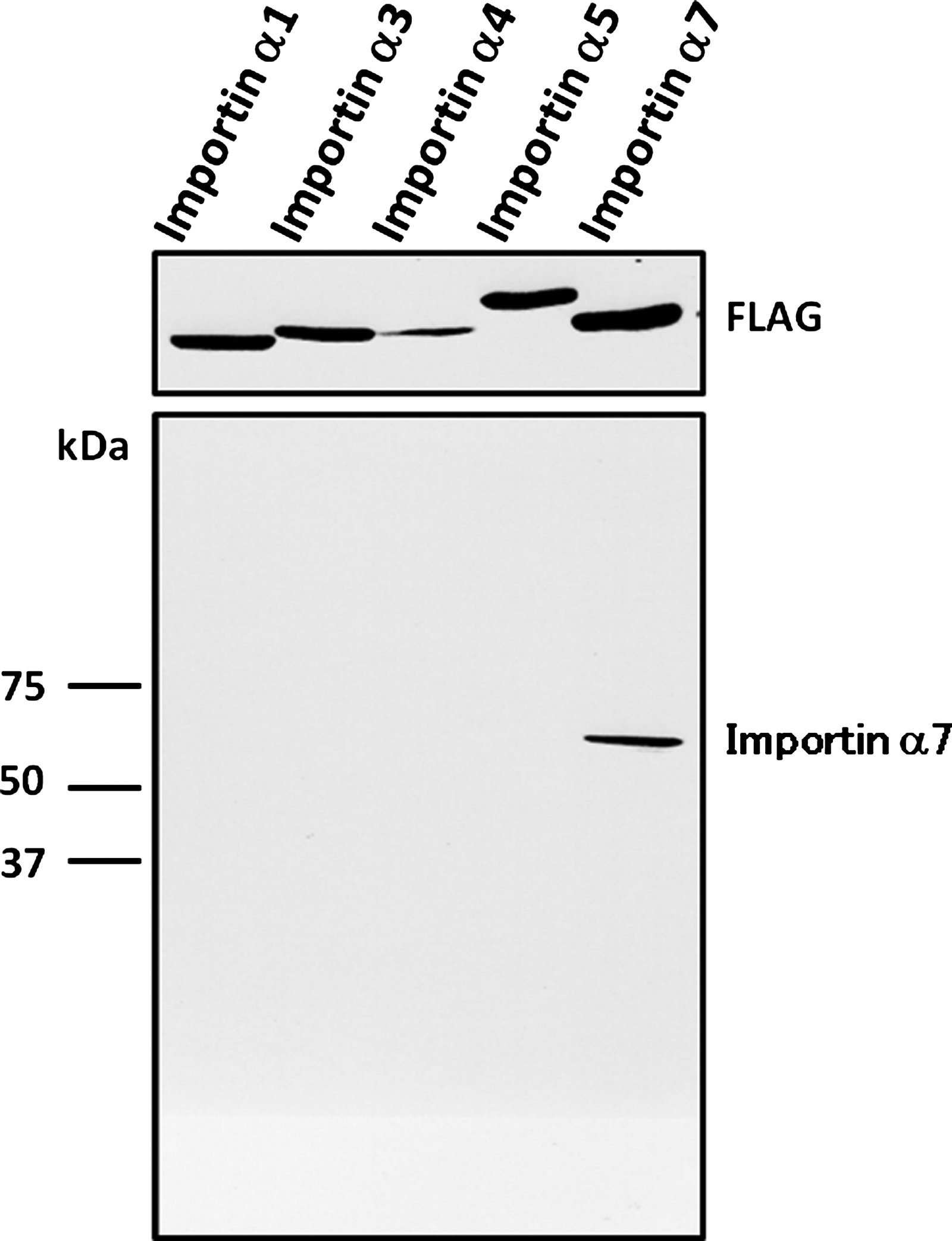
Specificity of MAb 3F8 to importin α7. COS-7 cells were transfected with FLAG-importin α1, FLAG-importin α3, FLAG-importin α4, FLAG-importin α5, and FLAG-importin α7 (lanes 1–5, respectively). Immunoblot analysis of COS-7 whole cell lysates was performed using anti-FLAG or anti-importin α7 (MAb 3F8) antibodies.
One of these antibodies, designated as MAb 3F8, was selected for further study. MAb 3F8 detected only importin α7, but not the other importin α isoforms, as a single band (Fig. 1). This antibody was further characterized by immunostaining of COS-7 cells overexpressing N-terminally EGFP-tagged importin α7. Indirect immunostaining revealed that the staining pattern by MAb 3F8 corresponded to the localization of EGFP-importin α7 (Fig. 2). Since the cellular function of importin α7 remains largely unknown, the anti-importin α7 specific monoclonal antibody (MAb 3F8) developed in this study would serve as an aid in elucidating its functional roles.
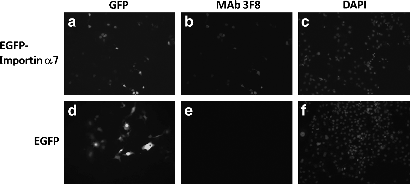
Indirect immunofluorescence of COS-7 cells using anti-importin α7 (MAb 3F8) antibody. HeLa cells were transfected with EGFP-importin α7 (
Footnotes
Acknowledgments
We are deeply grateful to Dr. Tachibana (Osaka City University, Osaka, Japan) for technical advice. This work was supported, in part, by the Ministry of Education, Culture, Sports, Science and Technology of Japan and the Takeda Science Foundation.
Author Disclosure Statement
The authors have no financial interests to disclose.