
Abstract
Immunological detection of viruses and their components using monoclonal antibodies (MAbs) is a powerful diagnostic method. Here we report a detailed method for the establishment of MAbs against severe acute respiratory syndrome coronavirus (SARS-CoV). To express and purify the nucleocapsid protein (N protein) of SARS-CoV and generate MAbs against the N protein, gene encoding N protein was separated into two parts according to the prediction of epitopes and cloned into pET32a(+), respectively. Expression of the target proteins were induced by M isopropyl-β-thio-D-galactopyranoside (IPTG) and purified by a single-step affinity chromatography on a Ni-NTA column. BALB/c mice were immunized with the purified recombinant proteins to prepare MAbs by hybridoma technique. The reactivity and specificity of the MAbs were analyzed by ELISA and Western blot analysis. Seven MAbs against N1 and two MAbs against N2 were obtained. In the present study, recombinant SARS-CoV N protein was expressed and purified and nine specific MAbs against SARS-CoV N protein were obtained successfully. This panel of anti-N MAbs may be used as a tool for rapid and specific diagnosis of SARS-CoV.
Introduction
SARS-CoV is a single-stranded plus-sense RNA virus. Its genome is about 30 kb in length and contains 23 putative open reading frames. Like other coronaviruses, it has four main structure proteins: the spike (S), membrane (M), envelope (E), and nucleocapsid (N).(2) In mature virions, N protein binds to virus RNA and forms a nucleocapsid complex by interacting with M and E proteins. The N protein may be implicated in many functions such as genome RNA replication and subgenomic RNA transcription and translation.(3) Compared with other viral proteins, N protein has a higher expression level and a stronger antigenicity, which make it a preferred candidate antigen for development of a vaccine and diagnostic antibody.(4)
Early diagnosis of SARS requires a highly sensitive test that can detect low levels of viral genome or proteins since the level of virus excretion is comparatively low during the initial phase.(5) Several molecular assays based on PCR have been designed(6); however, PCR methods need special devices and high qualifications with somewhat complex protocols and may bring false-positive results for contamination. A specific antibody or antigen detection test would be technologically simpler and less expensive. In this study, we expressed and purified the fragments of the N protein of SARS-CoV and produced MAbs against this protein. The characteristics of the antibodies were further studied by Western blot analysis. The generation of an antibody against SARS-CoV N protein provides a basis for the development of a clinical diagnostic kit in the future.
Materials and Methods
DNA synthesis and plasmid construction
The DNA fragments corresponding to the nucleocapsid protein (Genebank ID AY274119) were synthesized by Sangon (Shanghai, China). To facilitate the expression of the long protein, the full gene was separated into two fragments according to the results of online epitope prediction. One is called N1 with sequence 1–549 bp nucleotide of N gene and the other is N2 with the 496–1267 bp nucleotide of N gene. The nucleoside digestion enzyme sites of BamH I and Hind III were added to the 5′ frank and 3′ frank for insertion into the vector plasmid, respectively. The enzyme sites in the N gene were modified by nonsense mutations. The two DNA fragments were inserted into the pET32a(+) expression vector and the inserted fragments were confirmed by DNA sequencing.
Expression and purification of N protein
For expression in Escherichia coli, the recombinant plasmids were used to transform E. coli BL21(DE3). Bacteria transformed with the corresponding plasmids were grown in Luria-Bertani medium containing 60 μg/mL ampicillin at 37°C. When an A600 of 0.6 was reached, 1 mM isopropyl-β-thio-D-galactopyranoside (IPTG) was added, and cultures were grown for an additional 4 h. Cells were harvested by centrifugation at 8000 rpm, and pellets that were not used immediately were frozen at − 80°C. The expressions of target proteins were observed by gradient sodium dodecyl sulfate polyacrylamide gel electrophoresis (SDS-PAGE). For the scale-up purification procedure, frozen pellets from 4 L of BL21 cells overproducing N1/N2 were thawed and resuspended in denaturing lysis buffer (50 mM Tris-HCl [pH 7.5], 100 mM sodium phosphate, 8 M urea, 0.1% Emulphogen [polyoxyethylene 10-tridecyl ether; Sigma-Aldrich, St. Louis, MO], 10 mM imidazole, and 0.2 mM phenylmethylsulfonyl fluoride) containing 1 M NaCl and were equilibrated at pH 8 with 10 N NaOH. Cells were lysed by ultrasonication. The crude extract was centrifuged at 50,000 g for 30 min at 4°C, and the supernatant was purified using a nickel-nitrilotriacetic acid-agarose matrix column according to the manufacturer's instructions (Qiagen, Hilden, Germany). The apparent molecular mass of each polypeptide was determined by SDS- PAGE. The concentration of proteins was determined by the Bradford method.
Immunization of mice
BALB/c mice (8 weeks old) were subcutaneously immunized with 10, 20, or 30 μg of N1 or N2 proteins resuspended in phosphate-buffered saline (PBS) plus equal volumes of complete Freund's adjuvant (Sigma-Aldrich) and boosted twice at 2-week intervals. Preimmune sera were collected before starting the immunization, and antisera were collected 7 days after each boost. Sera were kept at 4°C before use. The mouse with the highest antibody titer was subjected to a special booster injection with 50 μg of antigen protein in PBS prior to hybridoma fusion.
Generation of anti-N monoclonal antibodies
Hybridoma cells producing anti-N1 or anti-N2 MAbs were generated using standard protocols, as previously described. In brief, the splenocytes from the N1/N2 protein immunized mice were harvested and fused with SP2/0 myeloma cells. Cell culture supernatants from the wells containing hybridoma colonies were screened by enzyme-linked immunosorbent assay (ELISA) using the purified N1 or N2 as a coating antigen. Cells from positive wells were expanded and retested. Cultures that remained positive were subcloned to generate stable hybridoma cell lines by limited dilution methods.
ELISA assay of antibodies
The reactivity of mouse sera or hybridoma supernatants was determined by ELISA. Briefly, 10 μg/mL purified recombinant protein was used to coat 96-well microtiter plates in 0.1 M carbonate buffer (pH 9.6) at 4°C overnight. After the plates were blocked with 2% non-fat milk, serially diluted mouse sera or cell culture supernatants were added and incubated at 37°C for 1 h, followed by three washes with PBS containing 0.1% Tween-20. Bound antibodies were detected with horseradish peroxidase-conjugated goat anti-mouse IgG (Sigma-Aldrich) at 37°C for 1 h, followed by washes. The reaction was visualized by addition of the substrate 3,3’,5,5’-tetramethylbenzidine, and absorbance at 450 nm was measured by an ELISA plate reader. The preimmune sera or hybridoma supernatants that did not produce MAb served as negative control and a value of P/N (the OD of positive samples over the OD of negative control) above 2.1 was regarded as positive.
Isotyping of MAbs
Isotypes of MAbs were determined with a mouse monoclonal antibody isotyping kit (Sigma-Aldrich) according to the manufacture's protocol.
Western blot analysis of MAbs
Briefly, 1 μg of each purified His6-tagged recombinant proteins were loaded to 12% SDS-PAGE gel, and subsequently electroblotted to a polyvinylidene difluoride membrane (Millipore, MA). The blot was cut into strips and was first blocked with PBS containing 0.1% Tween-20 and 5% non-fat dry milk at room temperature for 2 h and then incubated separately with the supernatants of hybridoma cells or mice sera diluted 1000-fold with washing buffer (PBS with 0.1% Tween-20). Strips were washed three times and incubated with 1:10,000 dilution of horseradish peroxidase-conjugated goat anti-mouse immunoglobulin polyclonal antibody (Sigma-Aldrich). Antigen and antibody interactions were detected by a peroxidase reaction using a tetramethylbenzidine-hydrogen peroxide solution as the substrate.
Results
Epitope analysis of N protein
According to the online epitope analysis, the antigenic determinants are mainly located in the C terminus of N protein. The strongest antigenic determinants lie in the domains of 219∼288 aa, 390∼399 aa, and 157∼167 aa. The N1 peptide includes 1–183 aa from the N terminus while the N2 peptide includes 166–422 aa.
Expression and purification of N protein
His6-tagged recombinant N1 or N2 peptides of SARS-CoV N protein were highly expressed in E. coli and purified from the soluble fraction of the bacterium extracts. As shown in Figure 1, a special protein band at about 39 or 48 kDa appeared in the lysates of induced BL21 cells transformed with the recombinant plasmids of pET32(a)-N1 or pET32(a)-N2, respectively, but was not present in the non-induced cells. Densitometric scanning of stained gels showed that the induced proteins were approximately 30 and 40%, respectively, of the total protein in the transformed E. coli by IPTG induction for 4 h. Purified by Ni-TNA, a high protein yield was obtained and the target protein is above 90% of the product. As shown in Figure 2, a nearly single band was obtained for each protein. The dried protein (stored in mannitol) was about 10 μg/mg for N1 and 1 μg/mg for N2 as measured by Bradford's method.

SDS-PAGE analysis of the proteins expressed in BL21(DE3) E. coli transformed with recombinant plasmids. Lane M, protein molecular mass markers; lane 1, pET32a-N1-BL21 without IPTG induction; lane 2, pET32a-N1-BL21 induced by IPTG for 4 h; lane 3, pET32a-N2-BL21 without IPTG induction; lane 4, pET32a-N2-BL21 induced by IPTG for 4 h.
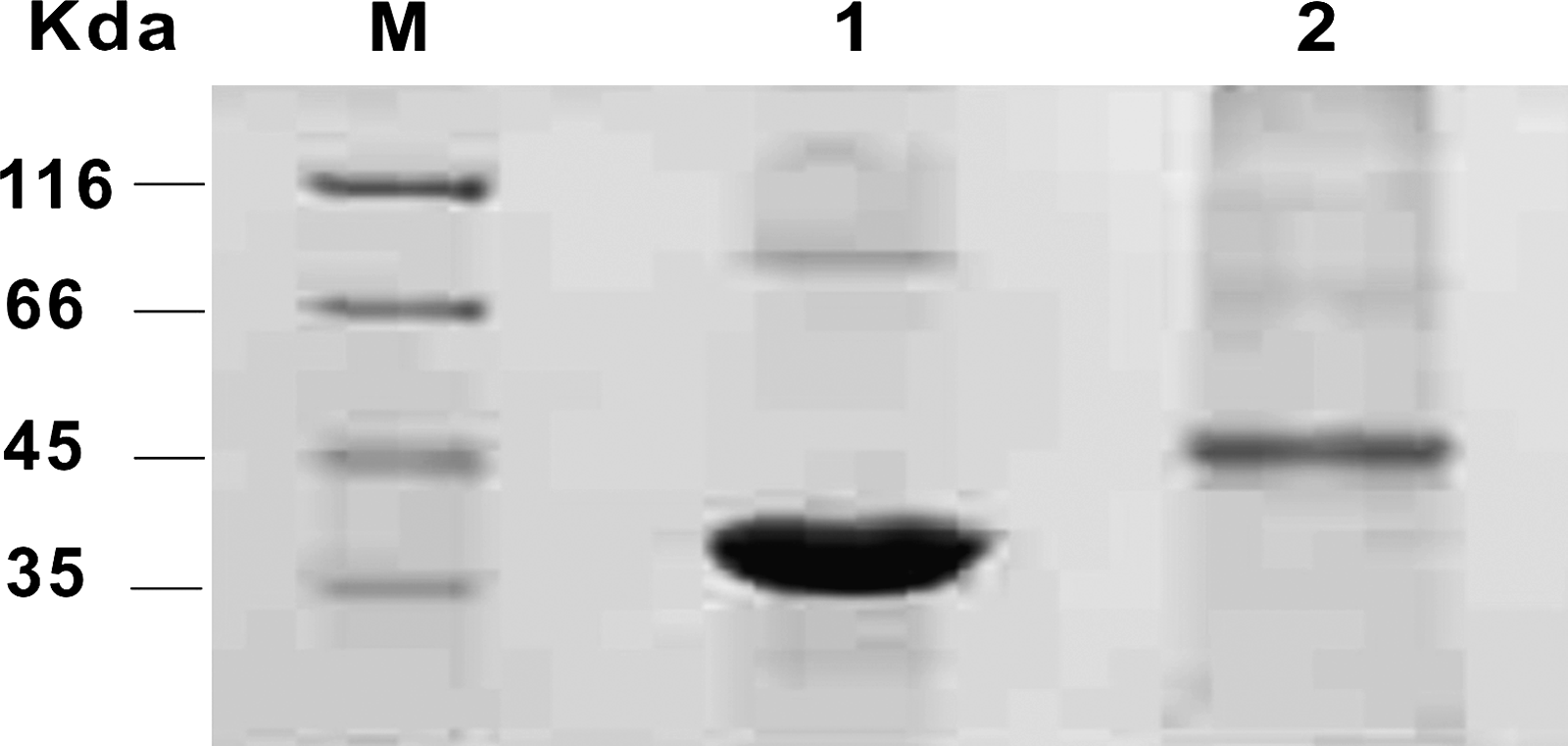
SDS-PAGE analysis of the purified N1/N2 protein. Lane M, protein molecular mass markers; lane 1, N1 protein; lane 2, N2 protein.
Recombinant proteins induced high titers of antibodies
The purified proteins were used to immunize mice, and the antibodies were analyzed by ELISA and Western blot analysis. All immunized mice produced significant antibody response against the corresponding immunogen after the first boosting immunization, and their serum reactivity apparently increased with subsequent boosts. As shown in Table 1, the reactive titers reached 1:100,000 after the third boost. The mice of N1B1, N1C1, N2B1, and N2C1 produced the strongest immune response with antisera titers above 1:1,000,000. The specificity of antibodies was further confirmed by Western blot as shown in Figure 3. The antibodies reacted with the purified proteins to produce a single band on the membrane. These results suggested that prokaryotic expressed recombinant proteins have highly immunogenic probability in mice.
Western blot analysis of polyclonal antibodies from immunized mice. (Top) The purified prokaryotic N1 protein was used as antigen to detect the antibodies from mice immunized with N1 protein. Sera from unimmunized mice (NC), immunized mice of N1B1 and N1C1 were detected. (Bottom) The purified N2 protein was used as antigen to detect antibodies from mice immunized with N2 protein. Sera from unimmunized mice (NC), immunized mice of N2B1 and N2B2 were detected.
+, − Results above and below the cutoff (P/N = 2.1), respectively.
NC, negative control, sera from unimmunized mice.
Establishment of monoclonal antibody secreting hybridoma
Fusion of spleen cells from immunized BALB/c mice with SP2/0 myeloma cells produced several hybridoma clones secreting MAbs against N1 or N2 proteins. Seven stable clones secreting MAbs against N1 and two clones secreting MAbs against N2 were generated and designated as N1.1A6, N1.1B1, N1.1D7, N1.1H2, N1.2C5, N1.2D3, N1.2F6, N2.1C4, and N2.1H6, respectively. The antibodies were confirmed by Western blot using the purified N1 or N2 proteins as antigens (Fig. 4).

Western blot analysis of the MAbs from hybridoma cells. (Top) The purified prokaryotic N1 protein was used as primary antigen. Supernatants of hybridoma cells of N1.1A6, N1.1B1, N1.1D7, N1.1H2, N1.2C5, N1.2D3, and N1.2F6 were detected. Serum from N1 immunized mice and unimmunized mice were used as positive control (PC) and negative control (NC), respectively. (Bottom) The purified prokaryotic N2 protein was used as primary antigen. Supernatants of hybridoma cells of N2.1C4 and N2.1H6 were detected. Sera from N2 immunized mice and unimmunized mice were used as positive control (PC) and negative control (NC), respectively.
Isotype of MAbs
The supernatants of stable hybridoma cells were collected for isotyping analysis. The results demonstrated that six MAbs were IgG1 class (N1.1D7, N1.1H2, N1.2C5, N1.2D3, N1.2F6, N2.1H6), two MAbs were IgG2b (N1.1B1 and N2.1C4), and N1.1A6 was IgG3.
Discussion
SARS-CoV is the first serious new threat to the global human population in the twenty-first century. Accurate laboratory diagnostic tests are essential for appropriate individual patient management, local infection control, and public health measures to halt virus spread. A molecular detection method with high sensitivity for SARS-CoV RNA by RT-PCR or other techniques such as aptamer-based detection were developed soon after global spread of the virus.(6,7) However, the RT-PCR tests should adopt strict criteria for probable contamination and false positive results. Furthermore, they require specific laboratories and expertise in molecular diagnosis. Thus, other simpler detection methods such as serological tests should be in demand for rapid diagnosis in a routine laboratory.
Antibodies against SARS-CoV are produced early, at 7 days after infection, in response to the course of infection; detection of antibody may be a valuable tool for the rapid diagnosis of acute virus infection.(2) To develop a serological test kit, it is essential to choose a good antigen and produce a special antibody with high affinity. The N protein of SARS-CoV plays an important role in the replication of the virus. It is abundantly released in the patient's blood in the course of early infection, which suggests that the N protein is a suitable candidate for diagnostic applications.(8) In this study, the N protein was expressed in two fragments according to the results of epitope analysis for overcoming the difficulty of expressing a large protein.(9) The DNA sequence was synthesized by chemical method, which excludes the possibility of virus contamination. The peptides were expressed with a tag of His6 in a prokaryotic vector and purified with a high purification. Both the N1 and N2 proteins reacted with SARS-CoV positive sera with a high specificity.(10) Purified proteins were used to immune mice to produce MAbs. The titers of polyclonal antibodies in a sera sample of mice immunized by the purified proteins reached 1:1,000,000 by ELISA assay. By hybridoma technology, seven cell lines secreting antibody against N1 protein and two cell lines secreting antibody against N2 protein were established. By isotyping assay, the monoclonal antibodies are all IgG, including several subtypes such as IgG1, IgG2b, and IgG3. All of the antibodies reacted with the purified prokaryotic expressed proteins. The special reactivity of the achieved monoclonal antibodies indicated that they may be used for the development of serological detection kits in the future.
After the emergency outbreak of SARS-CoV, several studies dealing with the detection of specific antigens or antibodies were reported. Most of the studies used N protein or S protein or a cocktail of both to develop a serological test.(11,12) Although these diagnostic methods showed good sensitivity and specificity, there are still several problems in their application, such as cross-reaction, low predictive value, and so on.(13–15) To overcome these shortages, different MAbs with high specificity are needed.(16,17) In this study, nine MAbs targeting N protein were obtained by hybridoma technique and their characteristics were illustrated. These MAbs would potentially be ideal candidates for developing early and sensitive serological detection tests against SARS-CoV.
Footnotes
Author Disclosure Statement
The authors have no financial interests to disclose.