
Abstract
The entire pig aminopeptidase N (pAPN) gene was amplified by RT-PCR using total RNA extracted from intestinal brush border membrane of a newborn piglet. The amplified products of the pAPN gene were cloned into the vector pMD18-T, generating a recombinant plasmid pMD18-T-pAPN. The C subunit of pAPN (pAPN-C) produced by PCR from the plasmid pMD18-T-pAPN was expressed in Escherichia coli using vector pET-32a with His tag. After confirming reactivity of the recombinant protein pAPN-C to antibody against native pAPN, polyclonal antibody against the recombinant protein pAPN-C was prepared in rabbit using purified protein as immunogen. In Western blot analysis, the antibody elicited by the recombinant protein pAPN-C could recognize the native pAPN. These data demonstrate that the pAPN-C recombinant protein and its polyclonal antibody can provide some basis for further receptor antagonist.
Introduction
Both TGEV and PEDV belong to the member of the genus Coronavirus, family Coronaviridae, order Nidovirales, and they show a high similarity in virus particle structure and pathogenicity.(10,11) Piglet diarrhea disease caused by TGEV and PEDV has frequently broken out in many swine-raising countries and has led to heavy economic losses, notably in China.(12) In the past few decades, many anti-virus studies, including attenuated vaccines, genetically engineered vaccines, shRNA, TGEV-based vector vaccine, and so on, have been reported for TGEV and PEDV.(13–15) However, TGEV and PEDV still exist and frequently cause the occurrence of piglet diarrhea in some countries and regions.
In the current situation, development of a novel anti-virus strategy for TGEV and PEDV is still essential. Taking into account the characteristics of cellular receptor pAPN, pAPN-C-based receptor antagonist will have a potential use in developing an effective inhibitor for TGEV and PEDV. In this study, we describe a simple and efficient approach for generating the recombinant protein pAPN-C and its polyclonal antibody with immunological activity.
Materials and Methods
Cloning of pAPN cDNA
Total RNA extraction from intestinal brush border membrane of a newborn piglet was carried out using TRIzol Reagent (Invitrogen, Carlsbad, CA) according to the manufacturer's instructions. The specific primers for RT-PCR of the pAPN gene, pAPN-F (5′-ACCATGGCCAAGGGATTCTACATTTC-3′) and pAPN-R (5′-CTATTAGCTGTGCTCTATGAACCAAT-3′), were designed based on the reference sequence of the pAPN gene (GenBank accession no. NM_214277). The first-strand cDNA of the pAPN gene was synthesized by M-MLV reverse transcriptase reagent kit (Promega, Madison, WI) using the primer pAPN-R. The pAPN gene was amplified by PCR using the cDNA as a template. Subsequently, the PCR products were cloned into vector pMD18-T (TaKaRa, Dalian, China). Nucleotide sequence of the pAPN gene was confirmed by automated sequence analysis in Sangon Biotech Co. (Shanghai, China). The recombinant plasmid of the pAPN gene was designated as pMD18-T-pAPN.
Cloning and expression of pAPN-C
Using the plasmid pMD18-T-pAPN as templates, the pAPN-C gene was amplified by PCR using a pair of specific primers, pAPN-C-F (5′-AAC
Purification of pAPN-C
The inclusion body of the recombinant protein pAPN-C was extracted from the lysate of the IPTG-induced host bacteria processed by supersonic waves. The extracted inclusion body protein was treated with 2×SDS loading buffer and then separated by 12% SDS-PAGE. After electrophoresis, the gel was stained with 0.3 M of cold potassium chloride (KCl) solution until the recombinant protein pAPN-C in gel appeared white. The white protein band of the recombinant protein pAPN-C was cut from the stained gel. The gel-cutting band was washed with sterile deionized water for 20 min at room temperature for removing KCl. The washed gel was ground into small particles and then soaked in an appropriate amount of phosphate buffer solution (PBS, pH 7.4) overnight at 4°C. After centrifugation at 15,000 g for 20 min, the supernatant containing the purified pAPN-C protein was collected. The concentration of the purified protein was determined using Bradford assay (Bio-Rad) with bovine serum albumin as a standard.(16) The His-tag protein as a control was prepared and purified by the same procedure.
Western blot analysis of recombinant protein pAPN-C
The purified recombinant protein pAPN-C was separated by 12% SDS-PAGE. The separated recombinant protein pAPN-C was transferred to nitrocellulose membrane (NC) using the semi-dry transfer apparatus DYCP-40C (Beijing Liuyi Instrument Factory, Beijing, China). The NC membrane was blocked using 5% (W/V) non-fat dry milk in PBS at 37°C for 1 h, and then incubated with 100 μL of polyclonal antibody against pAPN (1:200 dilution in PBS) at 37°C for 1 h. After washing three times with PBS, the NC membrane was incubated with 100 μL of goat anti-mouse IgG IRDye 700 conjugated (1:10,000 dilution in PBS) at 37°C for 1 h. After washing three times with PBS, the NC membrane was scanned and the captured image of the signal was processed and quantified with Odyssey™ Infrared Imaging System. In this section, polyclonal antibody against the pAPN was prepared in BALB/c mice using the native pAPN from porcine kidney (Sigma-Aldrich, St. Louis, MO) as immunogen.
Generation of polyclonal antibody against recombinant protein pAPN-C
A New Zealand rabbit was subcutaneously injected with 200 μg of the purified recombinant protein pAPN-C emulsified in an equal amount of complete Freund's adjuvant (Sigma-Aldrich). At 3 week intervals, two boosters of 200 μg of the purified recombinant protein pAPN-C emulsified in an equal amount of incomplete Freund's adjuvant (Sigma-Aldrich) were administered. After each inoculation, the blood sample was taken from the immunized animals and the serum was tested for the presence of specific antibodies by ELISA using the purified recombinant protein pAPN-C as coating antigen. The immunized rabbit was bled 1 week after the last booster and then antiserum was obtained by centrifugation at 1000 g for 10 min.
Western blot analysis of polyclonal antibody against recombinant protein pAPN-C
The Western blot procedure was similar to that used for the recombinant protein pAPN-C as previously described, and only showed a little difference. In this section, the native pAPN was separated by 12% SDS-PAGE. The polyclonal antibody against the recombinant protein pAPN-C was used to detect the native pAPN transferred to nitrocellulose membrane (NC), and conjugated goat anti-rabbit IgG IRDye 700 was used as the second antibody in the Western blot.
ELISA of polyclonal antibody against recombinant protein pAPN-C
ELISA plate wells were coated overnight in 0.1 mol/L carbonate buffer (pH 9.6) at 4°C with 100 μL of 10 μg/mL purified recombinant protein pAPN-C and native pAPN, respectively, and blocked with 5% skimmed milk at 37°C for 1 h. After washing three times with PBST, 100 μL of immune serum or preimmune serum (1:100 dilution in PBST) were added to the wells and incubated at 37°C for 1 h. After washing three times with PBST, the plates were incubated with 100 μL of HRP-conjugated sheep anti-rabbit IgG (1:8000 dilution in PBST) at 37°C for 1 h. Coloration was carried out by the substrate TMB solution, and the reaction was stopped by 50 μL of 2 M H2SO4. The absorbance at 450 nm was measured.
Results
PCR amplification of genes pAPN and pAPN-C
The complete open reading frame of the pAPN gene was amplified by RT-PCR from intestinal brush border membrane of a newborn piglet (Fig. 1A). The amplified pAPN gene is composed of 2892 bp encoding 963 amino acids (GenBank accession no. HQ824547), and shows 99.7% homologous identity with the reference sequence (GenBank accession no. NM_214277). On that basis, the C subunit gene of pAPN (pAPN-C) was generated using the recombinant plasmid pMD18-T-pAPN as templates by PCR. The size of the amplified pAPN-C gene was found to be 1200 bp in length (Fig. 1B).

PCR amplification of genes pAPN and pAPN-C. (
Expression and purification of recombinant protein pAPN-C
In order to obtain recombinant protein, the pAPN-C gene was cloned into prokaryotic expression vector pET-32a, and then the recombinant plasmid pET-32a-pAPN-C was transformed into Escherichia coli BL21(DE3) gold strain. Expression of the recombinant protein was induced by addition of IPTG to a final concentration of 0.8 mM. The pAPN-C gene was expressed at a high level in Escherichia coli. The expression protein was found in an inclusion body form and its molecular weight was approximately 63 kDa (Fig. 2A). The recombinant protein pAPN-C was purified from inclusion body form by gel-cutting purification (Fig. 2B). The concentration of the purified recombinant protein pAPN-C was tested as 0.5 mg/mL.
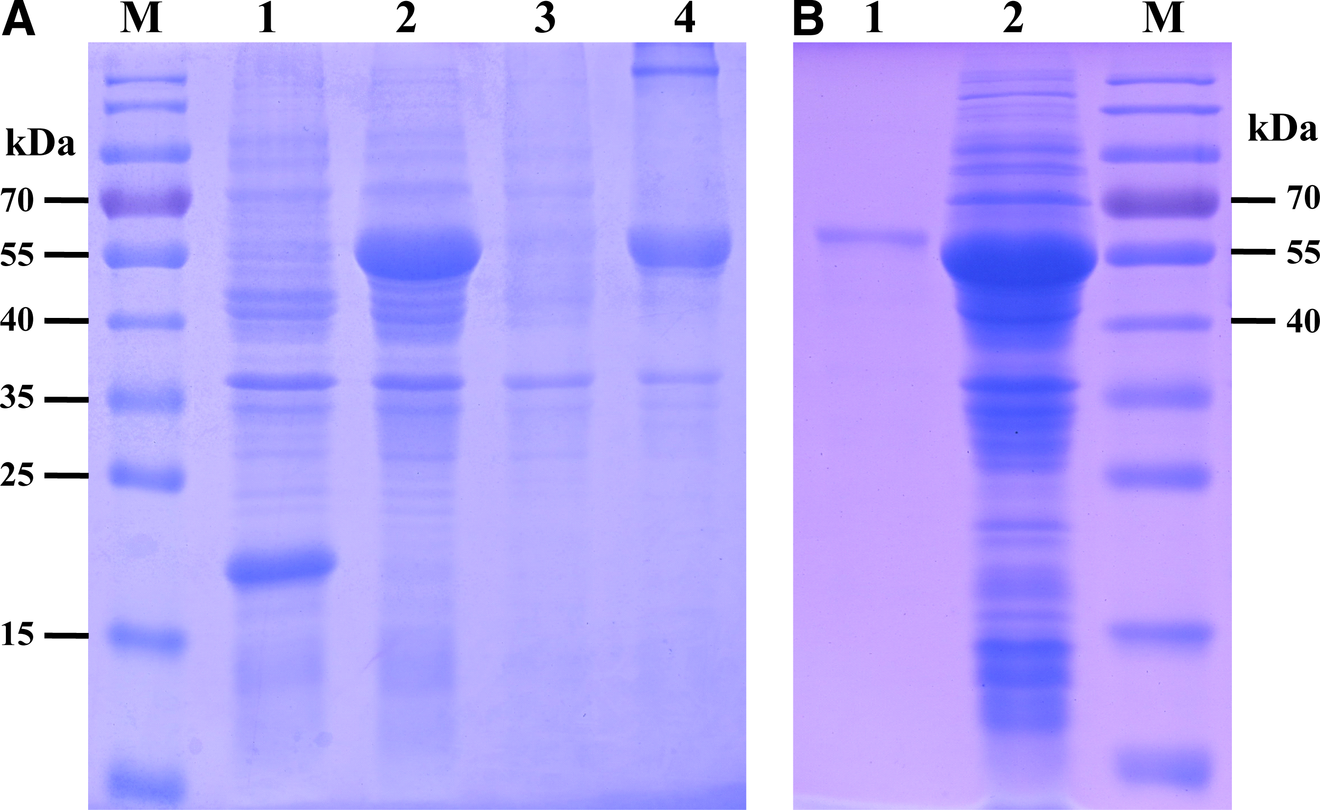
Expression and purification of recombinant protein pAPN-C. (
Western blot analysis of recombinant protein pAPN-C
To identify antigenicity of the recombinant protein pAPN-C, polyclonal antibody against pAPN was prepared using the native pAPN as immunogen in BALB/c mice. The reaction of the recombinant protein pAPN-C with antibody against the native pAPN was detected by Western blot. Western blot analysis indicated that one specific band was found in the lane of the recombinant protein pAPN-C rather than the His-tag protein control generated from the empty vector pET-32a (Fig. 3).
Western blot analysis of recombinant protein pAPN-C. Lane M, PageRuler Prestained Protein Ladder (10–170 kDa); lane 1, purified His-tag control; lane 2, purified recombinant protein pAPN-C.
Western blot analysis of polyclonal antibody against recombinant protein pAPN-C
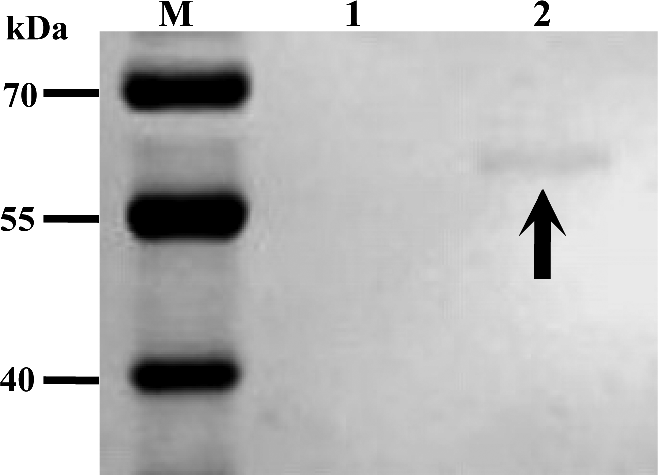
To detect immunogenicity of the recombinant protein pAPN-C, a New Zealand rabbit was immunized using the His fusion protein pAPN-C. After immunization three times, the reaction of polyclonal antibody against the recombinant protein pAPN-C with the native pAPN was determined by Western blot analysis. Results demonstrated that both the entire native pAPN of 150 kDa and its C subunit of 50 kDa could be recognized by the polyclonal antibody induced by the recombinant protein pAPN-C (Fig. 4).

Western blot analysis of polyclonal antibody against the recombinant protein pAPN-C. Lane M, PageRuler Prestained Protein Ladder (10–170 kDa); lane 1, native pAPN.
ELISA of polyclonal antibody against recombinant protein pAPN-C
In order to further validate immunogenicity of the recombinant protein pAPN-C, an ELISA test was performed for analyzing reactivity of anti-serum against pAPN-C using the recombinant protein pAPN-C and native pAPN as coating antigens, respectively. The result revealed that polyclonal antibody against the recombinant protein pAPN-C showed positive reaction with the recombinant protein pAPN-C and native pAPN in ELISA (Fig. 5).
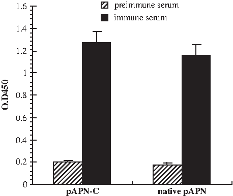
ELISA of polyclonal antibody against recombinant protein pAPN-C.
Discussion
Aminopeptidase N is a 150-kDa type II transmembrane glycoprotein with metalloprotease activity that is expressed on the apical membranes of epithelial cells in the respiratory and enteric tracts, endothelial cells, and kidney cells. APN has been shown to act as the major receptor for the group 1 coronaviruses that infect a single species, including swine transmissible gastroenteritis virus, porcine epidemic diarrhea virus, porcine respiratory coronavirus (PRCoV), feline infectious peritonitis virus (FIPV), feline enteric coronavirus (FCoV), human respiratory coronavirus strain 229E (HCoV229E), and canine coronavirus (CCoV).(1,2,17–19) The determinant regions of APN for entry of the group 1 coronaviruses have been revealed by chimeras between APN proteins of different species. Amino acids (aa) 717-813 of pAPN are necessary for TGEV receptor activity and aa 670-840 of feline APN are necessary for FCoV receptor activity, while aa 643-841 of canine APN are necessary for CCoV receptor activity.(3,8,9) In recent studies pAPN without signal peptide has been successfully expressed in Escherichia coli, and the anti-serum against recombinant pAPN protein can block TGEV infection in vitro. Further study reveals that the major viral binding regions of pAPN are located in regions aa 36-223, aa 349-591, and aa 592-963.(7,20) These studies suggest that the C subunit (about 50 kDa), which is located in the C-terminus of APN, holds significant potential for development of virus receptor-targeting antagonist.
In this study, the entire open reading frame of the pAPN gene was cloned from intestinal brush border membrane of a newborn piglet. Although several point mutations are found in the cloned gene, the pAPN gene displays a high level of homology (99.7%) at the nucleotide level with the unique reference sequence (GenBank accession no. NM_214277). Considering the potential use for other research, the nucleotide sequences of the cloned pAPN gene were submitted to GenBank with accession no. HQ824547. After amplification of the C subunit of pAPN from recombinant plasmid pMD18-T-pAPN, the prokaryotic expression vector pET-32a with T7 promoter was applied to drive expression of recombinant protein, fused to His-tag at N-terminus. The molecular weight of the recombinant protein pAPN-C is approximately 63 kDa, consists of 18 kDa His-tag protein, and 45 kDa pAPN-C protein predicted by the EditSeq procedure of DNAstar software.
Currently, a series of protein purification methods have been reported, including affinity chromatography, ion exchange chromatography, and so on.(21,22) Nevertheless, purification of the recombinant proteins could not achieve the desired results in most cases, especially purification of inclusion body proteins. In our study, gel-cutting purification, a simple, time-saving, and much less expensive means, was chosen for purification of the recombinant protein pAPN-C. In this way the recombinant protein pAPN-C was obtained in high purity from inclusion bodies. After validation of antigenicity, polyclonal antibody against the recombinant protein pAPN-C was generated in rabbit. In Western blot analyses, two specific bands were found in the lane of the native pAPN, one band located in size of 150 kDa, and another located in size of 50 kDa. Generally, the native pAPN protein extracted from porcine kidney is composed of the entire pAPN (150 kDa), the B subunit (95 kDa), and the C subunit (50 kDa). The result indicated the polyclonal antibody elicited by the recombinant protein pAPN-C could recognize the entire pAPN and its C subunit. The reactivity of anti-serum against the recombinant protein pAPN-C was further verified in the ELISA test.
In conclusion, we cloned the complete-length porcine aminopeptidase N gene and expressed the C subunit of pAPN in Escherichia coli. The recombinant protein pAPN-C shows reaction with polyclonal antibody against the native pAPN, and can elicit the formation of polyclonal antibodies combined with the native pAPN in rabbit. These properties make the expressed pAPN-C protein a potential candidate for use in further anti-virus research.
Footnotes
Acknowledgments
This work was supported by a grant from the National Natural Science Foundation of China (no. 31001081), and the Technological Innovation Team Building Program of University of Heilongjiang Province (no. 2010td05).
Author Disclosure Statement
The authors have no financial interests to disclose.