
Abstract
The human ADAMTS-18, a disintegrin and metalloproteinase with thrombospondin type-1 modules 18, is a secreted Zn-metalloproteinase. The C-terminal 385-amino acid fragment of ADAMTS-18 (AD18C) is highly effective at promoting platelet thrombus dissolution in vivo. Therefore, polyclonal antibody (pAb) against AD18C fragment should be able to keep platelet thrombus stability, which has direct clinical relevance. In this report, pAb against AD18C fragment was generated from rabbit immunized with AD18C recombinant protein (rAD18C). The pAb showed specific binding with rAD18C and natural ADAMTS-18 protein by ELISA and Western blot assay. It shortens the mouse tail bleeding time in a dose-dependent manner. Thus, anti-AD18C pAb contributes to the regulation of platelet thrombus stability.
Introduction
Recent studies have revealed that ADAMTS-18, which are mainly produced in endothelial cells,(5,6) is a physiologic ligand of platelet GPIIIa49-66. The active AD18C fragment clusters the β3 integrins and induces oxidative platelet fragmentation as we previously described anti-GPIIIa49-66 Ab.(7) It protects against FeCl3-induced carotid artery thrombosis, as well as cerebral infarction in a post-ischemic stroke model.(7) Therefore, production of pAb against active AD18C may have direct clinical relevance, as it would attribute to the regulation of platelet thrombus stability. In this study, we first report a highly efficient procedure of preparation of pAb against active AD18C fragment.
Materials and Methods
Materials
All reagents were obtained from Sigma (St. Louis, MO), unless otherwise designated. Escherichia coli strains BL21 (DE3) PlysS, plasmid pET-29a, and Ni-NTA agarose resin were from Novagen (Nottingham, United Kingdom). BALB/C mice were raised in our laboratory. All animal experiments were approved by the Institutional Animal Care and Use Committee of the East China Normal University.
Preparation of active C-terminal ADAMTS-18 fragment
Active C-terminal ADAMTS-18 fragment was prepared as described previously.(7) Briefly, E. coli BL21 (DE3) PlysS cells transformed with the expression vector pGEX4T2-AD18C plasmid were cultured in 1 L LB medium containing ampicillin (50 μg/mL) and chloramphenicol (34 μg/mL) with shaking at 37°C until the optical density (600 nm) was 0.4–0.6. Production was induced by the addition of 1 mM isopropyl-β-D-thiogalactopyranoside (IPTG) and the cells were allowed to incubate at 37°C for 4 h with shaking. Refolding and purification of recombinant protein were performed at 4°C, as previously described.(8)
Production of anti-AD18C polyclonal antibody
The polyclonal antibody was acquired as described previously.(9) Briefly, two New Zealand rabbits were inoculated with 1 mL purified rAD18C protein (1 mg/mL) emulsified with equal amounts of Freund's complete adjuvant via subcutaneous injection. The immunization of the rabbit was boosted seven times by inoculation of the same antigen (1 mL) mixed with equal volume of Freund's incomplete adjuvant at 2-week intervals. Anti-serum was isolated from peripheral blood of the immunized rabbit.
ELISA
The specificity of anti-AD18C pAb was determined using ELISA. Briefly, purified rAD18C protein (10 μg/mL) in phosphate-buffered saline (PBS) was adsorbed to a 96-well ELISA plate at 4°C overnight, and blocked with blocking buffer (3% BSA in PBS-Tween [0.1%]) at room temperature (RT) for 2 h. Primary antibody was added at room temperature for 1 h, followed by horseradish peroxide (HRP)-conjugated secondary antibody for 1 h at RT. Plates were washed six times with Tris-buffered saline (0.1%) Tween-20 at each step in the procedure. The ABTS reagent used to develop the HRP color reaction at OD405 nm using an ELISA reader.
Immunoblotting
Samples were separated on 8% SDS–PAGE under reducing conditions and then electroblotted onto a PVDF membrane. The membrane was blocked in blocking buffer containing 5% skim milk in PBS-Tween (0.1%), and then incubated with purified anti-AD18C pAb (1:1000 dilution) for 90 min at RT. After washing, the membrane was incubated with HRP-conjugated goat anti-rabbit IgG (1:5000 dilution) for 60 min at RT. The immunoreactive bands were visualized with ECL Western Blot kit (Amersham Biosciences, Hillerod, Denmark).
Bleeding time
To determine the bleeding time, the mouse tail vein was severed 2 mm from its tip and blotted every 30 s on a circular sheet of filter paper to obtain an objective measurement. Termination of the bleeding time was recorded after absence of blood on the filter paper.
Results and Discussion
Preparation and characterization of polyclonal antibody
The rAD18C protein was expressed and purified from Escherichia coli (data not shown), and used as antigen to immunize rabbits for generation of pAb. ELISA indicated the titer of antibody was about 1:3200 (data not shown). To achieve a highly specific pAb only directed to C-terminal ADAMTS-18 fragment, affinity purification with rAD18C fragment was further performed. The specificity of pAb was determined by ELISA. It was demonstrated that pAb could bind to rAD18C in comparison to control protein BSA and GST (Fig. 1). Western blot analysis showed that pAb could detect rAD18C level as low as 50 ng (data not shown). We next examined the reactivity of pAb with natural ADAMTS-18 protein by Western blot assay. It was shown that the pAb specifically recognized the natural ADAMTS-18 protein from ECV304 lysis compared to negative control 293T and B16 whole cell lysis (Fig. 2).
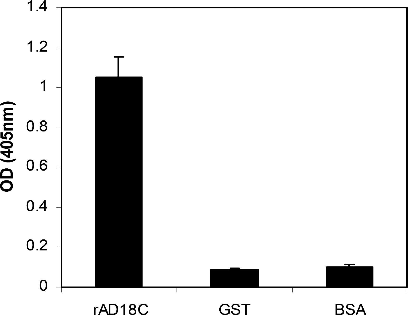
The specificity analysis of polyclonal antibody by indirect ELISA. Different antigens were coated onto the bottom of 96-well ELISA plates, and the purified polyclonal antibody was used for the specificity assay. The recognizing ability of the antibody to the antigens is indicated by comparison OD405 values. The data are expressed as mean OD of duplicates±SE.

Specific recognition of natural ADAMTS-18 protein with purified polyclonal antibody. The whole cell lysis from different cells was transferred on a PVDF membrane, and immunoblotting was performed using purified polyclonal antibody as primary antibody. Lane 1, 293T; lane 2, B16; lane 3, ECV304. The molecular weight of the natural ADAMTS-18 protein is approximately 135 kDa.
Effect of polyclonal antibody on mouse bleeding time
Because previous studies have revealed that C-terminal fragment of ADAMTS-18 was involved in dissolving platelet thrombus in vivo,(7) we postulated that anti-AD18C pAb would contribute to platelet thrombus formation. It was found that a dose-dependent shortening of the mouse tail vein bleeding time after intravenous injection of a different dose of anti-AD18C pAb (Fig. 3), confirming our previous observations.(7)
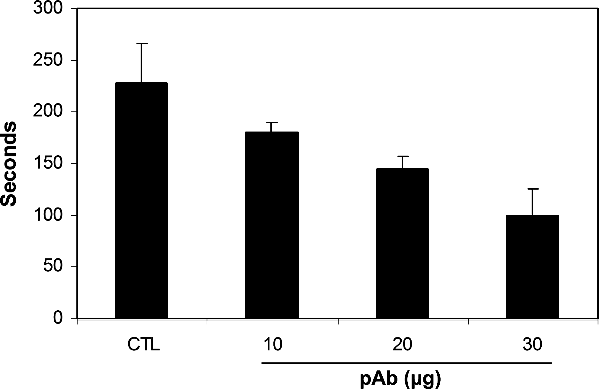
Effect of polyclonal antibody on mouse bleeding time. BALB/C mice were injected intravenously with different doses of anti-rAD18C pAb (from 10 to 30 μg, respectively) or preimmune IgG, and bleeding time was monitored 1 h later (n=8).
These data support the hypothesis that anti-AD18C pAb is a positive regulator of platelet thrombus stability. The protein and the specific antibody may be used as therapeutic reagents for research of arterial thrombosis-related diseases.
Footnotes
Acknowledgments
This work was supported by grants from ECNU (77202203) in China. We thank Dr. Bi-Sen Ding at Weill Cornell Medical College (New York, NY) for his critical reading of the manuscript.
Author Disclosure Statement
The authors have no financial conflict to disclose.