
Abstract
Epithelial cell adhesion molecule (Ep-CAM) is a 40 kDa transmembrane glycoprotein overexpressed in majority of tumor epithelial cells and has a major morphoregulatory function, relevant not only to epithelial tissue development, but also in carcinogenesis and tumor progression. Since Ep-CAM localizes at the cell surface of most carcinomas, the molecule is an attractive target for immunotherapy and several strategies have been deployed to treat cancer using Ep-CAM targeting, including MAb therapy. For improving effective targeting of this protein for diagnostics in various clinical samples, we generated and characterized an anti-Ep-CAM MAb (C4) using recombinant Ep-CAM protein, comprising the highly immunogenic domain. The specificity of C4-MAb was characterized in Ep-CAM positive cell lines (PC3 and MCF-7) by flow cytometry and immunofluorescence. The immunohistochemistry analysis in clinical tissue samples showed specific detection of epithelial antigens in breast, colon, stomach, and prostate carcinomas. Thus, this Ep-CAM MAb (C4-MAb) could be used for both diagnostic and therapeutic applications due to its specificity.
Introduction
Due to its frequent and high-level expression, Ep-CAM has gained interest as a potential diagnostic target and for antibody-based immunotherapy to combat a spectrum of malignancies, most notably colorectal carcinoma.(8) The use of Ep-CAM specific monoclonal antibody (MAb) has been successfully demonstrated by increased survival in colon and breast cancer patients, with minimal residual disease.(13) These antibodies have been reported to exhibit potential for tumor cell destruction by activating an array of endogenous cytotoxic mechanisms, including antibody-dependent complement-mediated cytotoxicity.(12,14)
Currently, Ep-CAM-specific antibodies such as ING-I, adecatumucab, and edrecilomab developed and immunotoxin containing Ep-CAM as target domain are also used in clinical trials.(15–20) Therefore, it is imperative to understand the variety of human cancers that are amenable to Ep-CAM-specific immunotherapy based on Ep-CAM expression with respect to intensity, frequency, and stage of disease. Several monoclonal antibodies have been developed against different domains of Ep-CAM such as extracellular domain encompassing EGF-like repeats, transmembrane region, and cytoplasmic tail. It has been reported that the mature peptide of Ep-CAM carries the immunodominant epitope.(21) Hence, MAb developed specifically against the mature Ep-CAM protein devoid of the signal peptide could be used for efficient targeting and therapy.
Since the immunodominant epitopes of antigens give rise to high affinity antibodies, we used the mature protein encompassing the immunodominant extracellular domain of Ep-CAM for the production of specific MAb in this study. The MAbs exhibited specific binding to Ep-CAM protein in tumor cell lines, as well as various Ep-CAM expressing tissues such as breast, colon, stomach, and prostate. Our study shows the use of such MAbs for a broad range of applications including targeted therapies, diagnosis, and research purpose.
Materials and Methods
Cell lines and culture conditions
Human breast carcinoma (MCF-7), human promyelocytic leukemia cells (HL-60), human leukemic monocyte lymphoma cell line (U937), human prostate carcinoma (PC3), and SP2/0 (mouse myeloma cells) were procured from NCCS (Pune, India). MCF-7 and PC3 cells were maintained in DMEM, and HL-60 and U937 were maintained in RPMI 1640 and SP2/0 cell line in IMDM supplemented with 10% fetal bovine serum (FBS), 2 mM glutamine, 100 IU penicillin, and 100 μg/mL streptomycin at 37°C in 5% CO2. All cell culture reagents were procured from Invitrogen (Grand Island, NY).
Expression and purification of recombinant Ep-CAM protein
The DNA sequence coding for the mature protein of human Ep-CAM (70-999) was cloned in Hind III and Xho I sites of pET28b (+) plasmid using the primers 5'CCCAAGCTTCAGGAATGTCTCTGT3' (forward) and 5'CCGCTCGAGTGCATTGAGTTCCCTATGCA3' (reverse). The recombinant protein was expressed in Escherichia coli BL21 (DE3) induced with 1 mM IPTG (BioBasic, Markham, Canada) for 4 h. Expression was confirmed by 12% SDS-PAGE, and rEp-CAM protein was purified by Ni-NTA (Qiagen, Hilden, Germany) affinity chromatography at 300 mM imidazole. The protein was refolded by on-column method using decreasing concentrations of urea (Sigma-Aldrich, St. Louis, MO) buffer.(22) Purity was confirmed with 12% SDS-PAGE and visualized by Coomassie-brilliant (SRL, Mumbai, India) blue staining.
Generation and purification of MAb
To generate MAb against mature Ep-CAM, BALB/c mice were immunized with purified protein mixed with Freund's adjuvant by intraperitoneal injection. Booster doses were given four times at 2-week interval. After confirming the hyperimmune response in mice using double immuodiffusion assay, polyethylene glycol-mediated fusion method was used for cell fusion using spleen cell and myeloma cells (SP2/o).(23) After 14 days of fusion, supernatant from hybridomas were screened for anti-Ep-CAM antibodies by ELISA. The positive clones showing the highest reactivity were used for clonal selection and for further characterization. Protein G affinity columns were used to purify antibodies. All reagents were procured from Sigma-Aldrich.
Western blot analysis
For Western blot analysis, the whole bacterial cell lysate was electrophoresed on 12% SDS-PAGE and electroblotted on to nitrocellulose membrane (Bio-Rad, Hercules, CA) at 100 V for 2 h. After blocking for 2 h at room temperature with 5% BSA (BioBasic) in TBST (0.02 M Tris HCl [pH 8], 0.5 M NaCl, 0.05% Tween-20), the membrane was incubated overnight at 4°C with anti Ep-CAM antibody purified from selected hybrid clone at a dilution of 1:100 in TBST with 3% BSA. Membrane was washed thrice using TBST and incubated with goat anti-mouse IgG-HRP conjugate (1:10,000; Sigma Aldrich) for 1 h. After washing, the membrane was developed with TMB/H2O2 (Gene I, Bangalore, India) for 5–10 min.
Antibody titer determination by enzyme-linked immunosorbent assay
To determine the antibody titer, the purified anti-Ep-CAM MAbs from the selected positive hybrid were tested at different dilutions for their reactivity with recombinant protein. Briefly, 96-well ELISA plate was coated with 100 μL of 200 ng Ep-CAM protein diluted in sodium bicarbonate buffer and incubated overnight at 4°C. After three washes with TBST, the wells were blocked using 100 μL of 5% bovine serum albumin for 1 h at 37°C. After further washes, each well was incubated for 3 h with 100 μL of purified anti-Ep-CAM MAbs at different dilutions (1:50 to 1:32,000). Later the wells were incubated at 37°C for 1 h with 50 μL of horseradish peroxidase (HRP)-conjugated with goat anti-mouse immunoglobulin (IgG, 1:10,000; Sigma-Aldrich). After washing, 100 μL of tetra methyl benzidine (TMB) and hydrogen peroxide (H2O2) substrate were added to each well for color development. The reaction was stopped after 10 min by adding 50 μL of 2 N H2SO4 and absorbance was measured at 450 nm using a Tristar LB 941 plate reader (Berthold, Bad Wildbad, Germany).
Immunofluorescent staining
For confirming the specificity of the antibody, MCF-7 and PC3 cells, which show high expression of Ep-CAM, were chosen as positive controls. HL-60 and U937 cell lines that do not express Ep-CAM served as negative controls for the study. Cells were washed and fixed with 4% paraformaldehyde (Himedia, Mumbai, India) for 30 min at room temperature. About 200 μL of purified anti-Ep-CAM MAb was added at 1:100 dilution to wells containing fixed cells and incubated for 3 h at 37°C. After washing, the cells were incubated for 1 h at 37°C with FITC-conjugated goat anti-mouse IgG at 1:100 dilution. After two successive washes with wash buffer, cells were examined under fluorescent microscope (Eclipse Ti, Nikon, Tokyo, Japan).
Flow cytometry analysis
Flow cytometry analysis was carried out in all the cell lines mentioned above. A total of 1×10⁵ cells was collected for each analysis. The cells were washed once with DPBSA and incubated at 4°C for 30 min with anti-Ep-CAM antibody (C4) or commercially available Ep-CAM antibody (Santa Cruz Biotechnology, Santa Cruz, CA) as reference. A FITC-conjugated goat–anti-mouse IgG (Sigma-Aldrich) was used to detect the binding of anti-Ep-CAM MAbs. Cells were analyzed within 2 h after staining using flow cytometer (FACSCalibur, BD Biosciences, San Jose, CA). Appropriate controls were used for every cell line.
Immunohistochemistry
A total of 25 tumor samples of routinely processed paraffin-embedded infiltrating duct breast, prostrate, stomach, and colon carcinoma were included in this study (received from Dr. Almpgibms, Department of Pathology, Chennai, India). Three μM thick sections were stained by hematoxylin/eosin (H&E, Himedia) for histopathological evaluation of histological type and grade of the tumor and visualized under microscope (TI 21, Olympus, Hamburg, Germany). Grading was assessed according to the WHO guidelines for the corresponding tumor types. All the tumor samples were tested according to ICH guidelines. The sections were mounted on poly-
Tissue sections were subjected to antigen retrieval by pressure cooker method using 0.01 mol/L sodium citrate (pH 6.0). After blocking with 15 mL/L normal goat serum for 20 min, immunohistochemistry assay was carried out using an immuno-peroxidase method with a commercial kit (Vector Laboratories, Burlingame, CA). The MAb against Ep-CAM was used at a dilution of 1:20 for 1 h at room temperature. Diaminobenzidine (DAB) was added and counterstaining was carried out with Harris' hematoxylin. Appropriate controls were included in the study. For each sample, staining intensity (negative, weak, moderate, strong) and percentage of positive tumor cells were estimated. Positivity was assessed using the following scoring system. A sample was considered strong positive (3+) if the antibody detected >70% positive cells, moderately positive (2+) if the antibody detected 30–69%, weakly positive if the antibody detected 10–29% (1+), and negative (0) if <10% cells were stained. Staining in cytoplasm alone was considered non-specific, as Ep-CAM is localized on the cell membrane.
Results
Expression and purification of recombinant Ep-CAM protein
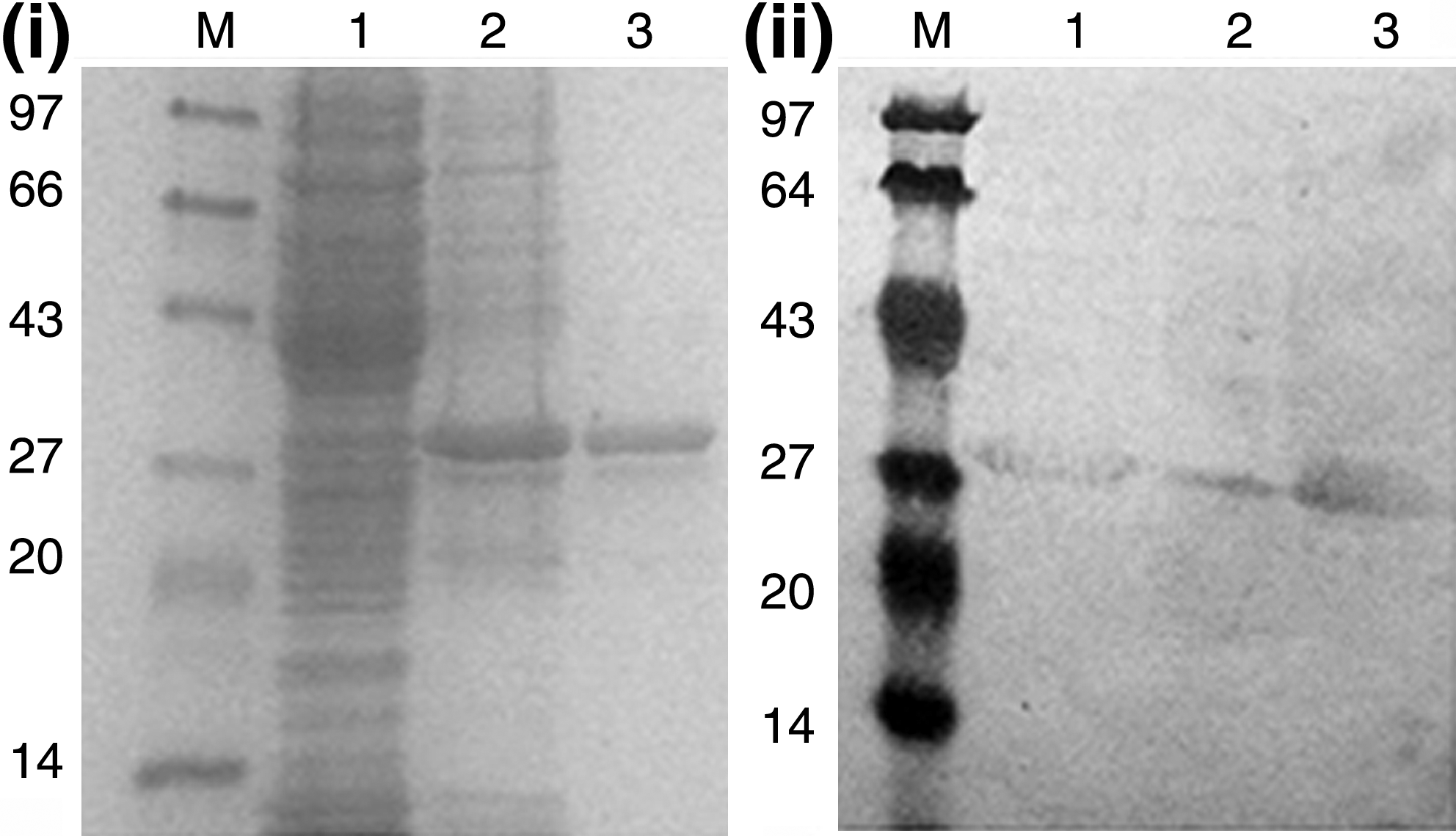
The coding region of Ep-CAM mature protein was PCR amplified and was cloned between Hind III and XhoI sites of pET28b (+) vector in order to express recombinant Ep-CAM in E. coli with N-terminal histidine tag. A 900-bp DNA fragment coding for a mature Ep-CAM protein was confirmed by sequencing and restriction digestion. Recombinant Ep-CAM protein was expressed in E. coli BL21 (DE3). SDS-PAGE analysis confirmed a protein band corresponding to the expected molecular weight of 30 kDa. The expressed rEp-CAM protein was purified from insoluble fraction by Ni-NTA affinity column chromatography and refolded (Fig. 1i).
(
Generation of monoclonal antibody against Ep-CAM and isotyping
The purified protein was used to immunize BALB/c mice. Hybridomas were produced by fusion of SP2/0 mice myeloma cells with spleen cells from mice hyper-immunized with recombinant Ep-CAM protein. Hybridomas secreting anti-Ep-CAM MAbs were selected based on positive reactivity in ELISA. Eight clones showing the highest reactivity by indirect ELISA with antigen were further selected and expanded. Among them, the clone denoted as C4-MAb showed highest binding affinity to Ep-CAM protein and was sub-cloned. The culture supernatant was purified using protein G column and isotyped. It was found that C4-MAb belonged to IgG2 isotype (data not shown).
Reactivity of C4-MAb with rEp-CAM
The specificity of the purified antibody was demonstrated by Western blot analysis of denatured Ep-CAM protein using anti-Ep-CAM MAb. Purified anti-Ep-CAM antibody was able to detect the denatured rEp-CAM protein expressed in bacteria (Fig. 1ii).
Antibody titer
The selected C4 MAb was purified from the hybridoma culture supernatant using Protein-A affinity column. The titer of the purified anti-Ep-CAM MAb (C4 MAb) was evaluated by indirect ELISA with the dilutions ranging from 1:50 to 1:32,000. The highest antibody titer was observed at 1:800 dilution.
Immunocytochemistry
Ep-CAM expression was qualitatively characterized on cell lines by immunofluorescence staining using anti-Ep-CAM MAb and anti-mouse IgG-FITC conjugated secondary antibody. Immunostaining with Ep-CAM C4 MAb showed surface expression of Ep-CAM in PC3 and MCF-7 cells, whereas in HL60 and U937 cells, staining was not detectable (Fig. 2) due to the absence of Ep-CAM antigen.

Immunofluorescent staining of Ep-CAM expression on MCF-7, HL-60, PC3, and U937 cells with C4 MAb. Cells were fixed with 4% paraformaldehyde and immunostained with C4 MAb. MCF-7 and PC3 cells were used as positive controls. HL-60 and U937 cells were used as negative controls. Left panel (1) represents images in bright field; middle panel (2) images of Ep-CAM-FITC receptor expression; and right panel (3) represents images of merged figures of anti Ep-CAM MAb FITC and nuclei stained by Hoechst.
Flow cytometry analysis of C4-MAb binding with Ep-CAM
Ep-CAM expression was also studied by flow cytometry analysis using C4 MAb in PC3, MCF-7, HL-60, and U937 cells. While anti-Ep-CAM C4 MAbs showed negligible detection in HL-60 and U937 cells, the results showed significant peak shift in Ep-CAM expressing positive cells (Fig. 3b,e). The C4 MAb (Fig. 3b) showed similar peak shift compared with the commercially available reference antibody (Fig. 3a) in PC3 cells. The results of immunostaining and flow cytometry analysis proved that anti-Ep-CAM C4 MAb specifically binds to the native cell surface antigen.

Flow cytometry analysis of MCF-7, HL-60, PC3, and U937 cells with C4 MAb. Cells were incubated with primary antibody (C4 MAb) followed by incubation with a FITC-conjugated secondary antibody and analyzed by flow cytometry. Staining of PC3 cells using commercially available Ep-CAM antibody (
Immunohistochemistry staining of clinical samples
To evaluate the potential application of anti-Ep-CAM C4 MAb in clinical diagnosis, the MAb was tested for specificity on various cancerous tissues including human breast, colon, prostate, and stomach, along with normal breast tissue. Immunostaining of the normal breast acini ducts showed 0–1+ positivity of the cell membrane, indicating weak positivity of Ep-CAM expression. Out of the 25 the breast carcinoma samples studied, 10 were found to be grade II infiltrating duct carcinoma (IDC) no specific type (NOS) and 15 were grade III. In breast carcinoma, the grade III tumors showed staining of cell membrane with 3+ score. The grade II tumors exhibited 1+ to 3+ staining. All 25 samples of moderately differentiated colon carcinoma were stained with anti-Ep-CAM MAb and staining was localized to tumor cell membrane with a score of 3+. Among those, 21 tissue samples (grade II tumors) showed 1+ staining score whereas the remaining grade II tumors had a score of 3+. Out of the 25 samples of stomach adenocarcinoma included in this study, 9 samples were poorly differentiated and 16 were moderately differentiated (histopathogical grade). All 25 stomach adenocarcinoma tumors samples showed neoplastic cells with a score of 3+. Among the prostate adenocarcinoma cases, 12 samples were of grade I tumors and showed staining of neoplastic cell membrane with a score of 1+. Remaining 13 samples were of grade III tumors showing 2+ to 3+ positivity (Fig. 4, Table 1).
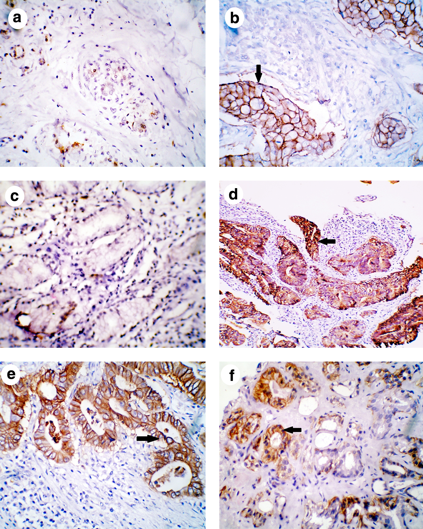
Immunohistochemical staining of Ep-CAM in breast, colon, stomach, and prostate tumor tissues. (
n=number of cases.
Ep-CAM overexpression was evaluated by calculating the total immunostaining score as the product of the proportion score and the intensity score.(35) The proportion score described the estimated fraction of positive-stained tumor cells [strong positive, >70% positive cells (3+); moderately positive, 30–69% (2+); weakly positive, 10–29% (1+); negative, <10% cells (0)].
Discussion
Ep-CAM expression has been widely studied in several carcinomas including breast,(24–26) colon,(27) prostate, and stomach.(28) These carcinomas have high Ep-CAM expression and have been reported as a marker to distinguish epithelial neoplasias from neoplasias derived from non-epithelial tissues. Ep-CAM-positive tumors are derived from epithelial cells, whereas Ep-CAM-negative tumors may originate from non-epithelial as well as epithelial tissues.(8)
Ep-CAM antigen is anchored to the cell surface via its transmembrane region and cytoplasmic domains.(29) The extracellular domain is exposed, which can be targeted for binding with suitable ligand. Most of the known antibodies are raised against whole Ep-CAM antigen and have been used for inhibition or targeting studies.(21) Here, we have produced the recombinant Ep-CAM coding for mature protein, devoid of its signal peptide, and used as an immunogen for developing hybridomas for the production of Ep-CAM specific MAbs.
Based on quantitative and qualitative studies of specific clones producing MAb, a high titer clone, C4, was chosen, which was further characterized and its applications were analyzed in four different tumor samples including breast, colon, stomach, and prostate. Initial characterization was carried out in Ep-CAM positive and negative cell lines followed by detection in clinical samples. We have shown that the C4 MAb was able to distinguish between positive and negative cell lines using immunofluorescent assay. Anti-Ep-CAM MAbs detected Ep-CAM expression exclusively throughout the cell surface of PC3 and MCF-7 cells (Fig. 2). However, the immunostaining of the receptor expression was found to be significantly low in HL-60 and U937 cells (Fig. 2). This is due to the decreased surface expression of Ep-CAM in these cells, whereas the receptor has been known to be overexpressed in PC3 and MCF-7 cells. Flow cytometry analyses corroborated the binding specificity of C4 MAbs quantitatively in PC3 and MCF-7 cells, as observed by the clear peak shift (Fig. 3), whereas there was negligible shift with HL-60 and U937cells. All these observations clearly validate that the generated MAb distinctively binds to the cell membrane and also reflects its efficiency to bind to the native surface antigen.
The immunohistochemistry analysis in breast, colon, prostrate, and stomach carcinoma tissues specifically detected Ep-CAM antigen. Infiltrating duct carcinoma of the breast grade III and adenocarcinoma of colon showed 3+ positivity of tumor cell membrane for Ep-CAM antigen. Needle biopsy of prostate adenocarcinoma grade III samples exhibited 2+ positivity, while the mucin secreting adenocarcinoma stomach tumor poorly differentiated type tumor cells showed 3+ positivity for Ep-CAM. The normal tissues were negative for Ep-CAM, which clearly indicates the ability of the C4 MAb to specifically detect and differentiate the epithelial tumors (Fig. 4).
Thus, the C4 MAb could be used to detect biomarker for determining the receptor expression on tumor samples during early prognosis of the disease. Further, C4 MAb could be used as a valuable tool for the study of Ep-CAM expression in clinical samples of different stages of epithelial malignant tumors and its conformational changes in response to various stages of different carcinoma.(30) Recently, Ep-CAM has been used as a target for biological therapy and as a tool in surgical pathology.(11,31–33) The emerging observation from Ep-CAM targeting immunotherapies also depicts the wide application of Ep-CAM antigen. In the present study, we used different clinical samples to prove the specificity of the C4 MAb by immunohistochemistry, which showed strong staining pattern in the membrane of all the studied tumor samples. Our results also confirmed that C4 MAb was able to detect Ep-CAM antigen in formalin-fixed paraffin-embedded carcinoma tissue sections. The staining pattern of colon was the same and consistent with the previous report.(34) These data suggest high specificity of these anti-Ep-CAM MAbs and their potential use in clinical diagnosis as well as in targeted therapies. The C4 MAb would be suitable for detecting a broad range of carcinomas by immunohistochemistry.
In summary, our data indicates that anti-Ep-CAM C4 MAb, specifically binds to the cell surface of different malignant epithelial cells. The anti-Ep-CAM C4 MAb might have potential application in both determining the Ep-CAM expression and quantifying the expression level in different carcinomas. Further studies with large numbers of clinical epithelial malignancies would be helpful in elucidating the diagnostic and prognostic utility of this antibody. Finally, our observation that C4 MAb binds with less intensity to lower histological grades of carcinoma points toward a potential use of this antibody in prognostic assessment of malignant epithelial tumors.
Conclusion
In conclusion, we have generated anti-Ep-CAM MAb directed against the mature protein and performed extensive clinical characterization to prove its broad application in diagnosis in various epithelial carcinomas. The present study may contribute towards the development of new therapeutic strategies employing Ep-CAM as a target antigen. Therapy using C4 MAbs with specific binding efficiency combined with chemotherapeutic agent can be used for the management of a wide variety of cancers.
Footnotes
Acknowledgment
This work is partially supported by a grant from the Department of Biotechnology, New Delhi, India (BT/PR7968/MED/14/1206/2006 to R.S.V.).
Author Disclosure Statement
The authors have no financial interests to disclose.