
Abstract
The G10P[15] rotavirus CC0812-1 isolated from a diarrheal woman in Wuhan, China, in 2008 is phylogenetically close to the Lanzhou lamb rotavirus (LLR) of a monovalent human rotavirus vaccine produced by the Lanzhou Institute of Biological Products, China, and rotavirus Lamb-NT. This rotavirus can be used as the backbone of the attenuated rotavirus reassortant as a rotavirus vaccine candidate. In this study, rotavirus CC0812-1 was purified from the culture supernatant of CC0812-1-infected MA104 cells and used as antigen to immunize BALB/c mice. Four hybridoma clones were developed secreting antibodies that reacted with CC0812-1, designated as 1B1, 1B8, 1F11, and 1G10, respectively. Western blot analysis indicated that the four monoclonal antibodies (MAbs) were all specific for VP4 of rotavirus CC0812-1. Isotyping revealed that MAbs 1B1, 1B8, and 1G10 belonged to the IgM class, while MAb 1F11 belonged to the IgG1 subclass. A neutralization test demonstrated that the four MAbs all had the capacity to neutralize rotavirus CC0812-1. The neutralizing titers of the BALB/c mice ascites were 1:2048, 1:1024, 1:512, and 1:512 for MAbs 1B1, 1B8, 1F11, and 1G10, respectively.
Introduction
In China, RotaVac (Lanzhou Institute of Biological Products, Lanzhou, China) is composed of a live-attenuated Lanzhou lamb RV LLR, which was licensed in 2000. The strain LLR (G10P[15]) was isolated from a local lamb with diarrhea in 1985.(9) Later, a G10P[15] RV, designated as strain CC0812-1, was isolated from a diarrheal women in Wuhan, China, in 2008. Full genomic analysis of RV CC0812-1 revealed that it is phylogenetically close to RV LLR and Lamb-NT (JH Yang, unpublished data). Although the G and P genotypes of RV CC0812-1 are not among the five most prevalent G/P-type combinations of human RVs, it can be used as a backbone of attenuated RV reassortants with the most common G and P genotypes of human RVs. In this study, we report the establishment of monoclonal antibodies against VP4 of RV CC0812-1.
Materials and Methods
Cultivation of RV CC0812-1
MA104 cells were routinely cultured in DMEM (Gibco, Life Technologies, Carlsbad, CA) supplemented with 10% fetal calf serum (Gibco) and 1% L-glutamine (Amresco, Cleveland, OH) in a 5% CO2 incubator at 37°C. The cultivation procedure of RV CC0812-1 was carried out as described by Urasawa and colleagues.(10) Briefly, 1 mL of RV CC0812-1 (1.0×10³ TCID50) was treated with 15 μg/mL trypsin for 30 min at 37°C and inoculated to MA104 cells cultured in a 25 cm2 flask. Following adsorption to the MA10 cells for 1 h at 37°C, the virus inoculums were pipetted off. After being washed three times with Dulbecco's phosphate-buffered saline (DPBS), the cells were maintained in DMEM containing 2 μg/mL trypsin. The culture supernatant of RV-infected MA104 cells was harvested at 96 h post-infection.
Purification of RV CC0812-1
The harvested culture supernatant of RV-infected MA104 cells were frozen and thawed three times and clarified by centrifugation at 7000 rpm for 30 min. The polyethylene glycol (PEG) 6000 (Merck, Darmstadt, Germany) was added to the supernatant to a final concentration of 5%. The supernatant was then kept in a 4°C refrigerator overnight to allow virus precipitation. The precipitated RVs were collected by centrifugation at 15,000 rpm for 30 min and were resuspended in DPBS. The clarified suspension was subjected to Sepharose CL-4B gel for filtration chromatography and eluted with DPBS. The elutions were inspected by ultraviolet spectrophotometer at the wavelength of 280 nm. The RV fractions were collected and combined; the concentration of RVs was determined by the ultraviolet spectrophotometer method. The purified RV CC0812-1 was negative stained by phosphotungstenic acid and was observed by electron microscopy (Hitachi H-7000FA).
Mice immunization
Six-week-old female BALB/c mice were purchased from the Medical Laboratory Animal Center of Hubei Province and were maintained and bred in the laboratory animal house of Huazhong Normal University (Wuhan, China) under specific pathogen-free conditions and 22±1°C standard with a regular 12 h light/12 h dark cycle and allowed free access to standard laboratory food and water. Animal studies were approved by the Science and Technology Department of Hubei Province. Female BALB/c mice were injected intraperitoneally four times with 100 μg purified RV CC0812-1 at 1-week intervals. Complete Freund's adjuvant (Sigma-Aldrich, St. Louis, MO) was used in the first immunization and incomplete Freund's adjuvant was used in the subsequent three booster shots. A final immunization with 100 μg purified RV CC0812-1 only was given intravenously 3 days before cell fusion.
Mice bearing myeloma
The sp2/0 cells were maintained in RPMI-1640 with 10% fetal calf serum (Gibco) and 1% L-glutamine (Amresco, Solon, OH) in a 5% CO2 incubator at 37°C. After being washed three times with DPBS, 5–10×106 sp2/0 cells were intramuscularly implanted into the back of BALB/c mice to form a solid tumor of mice myeloma 10–14 days before cell fusion.
Cell fusion and cloning
Cell fusion was performed using the modified methods described by Campbell.(11) Spleens were dissected from BALB/c mice immunized with purified RV CC0812-1, and splenocytes were prepared in serum-free RPMI-1640 medium (Invitrogen, Carlsbad, CA). The myeloma sp2/0 cells were isolated from the sp2/0 solid tumor of BALB/c mice-borne myeloma. For hybridoma preparation, the splenocytes were mixed with mouse myeloma sp2/0 cells at a ratio of 5–10:1 in serum-free RPMI-1640 medium at room temperature and were centrifuged at 1000 rpm for 10 min. The mixture pellet of splenocytes and myeloma cells was resuspended in 1 mL 50% PEG 1450 (Sigma-Aldrich) over 1 min, followed by the serial addition of 3 mL RPMI-1640 over 3 min and 10 mL RPMI-1640 over 1 min, with gentle stirring. Fused cells were centrifuged at 1000 rpm for 5 min and resuspended in RPMI-1640 medium with 20% fetal calf serum and HAT medium (hypoxanthine, aminopterin, and thymidine, Sigma-Aldrich) at 2.5×10⁶ splenocytes/mL. The fused cells were then dispensed at 100 μL/well into 96-well cell culture microplates (Greiner Bio-one, Frickenhausen, Germany) previously plated with BALB/c mice macrophage feeder cells of 104 cells/well. Cells were incubated at 37°C with 5% CO2. The medium was half-replaced with fresh selection medium 7 days later. The culture supernatants were pipetted off for antibody screening by ELISA. Antibody-producing hybridoma clones were cloned three times in RPMI-1640 medium supplemented with 20% fetal calf serum and HT medium (Gibco) by limiting dilution method.
Screening ELISA
For screening antibody-producing hybridoma clones, 96-well microplates were coated with 100 μL purified RV CC0812-1/well at 8 μg/mL diluted in carbonic acid buffer (coating buffer, 33.7 mM NaHCO3, 15 mM Na2CO3 [pH 9.6]) and incubated at 4°C overnight. The microplates were blocked by incubation with 10 mg/mL bovine serum albumin (BSA) in DPBS containing 0.05% Tween-20 (DPBS-T) for 2 h at 37°C and washed three times with DPBS-T. Then 100 μL culture supernatant was added and incubated for 1–2 h at 37°C. After being washed three times with DPBS-T, 100 μL of rabbit anti-mouse IgG HRP conjugate (1:5000 diluted in DPBS-T with 0.5 mg/mL BSA) were added and incubated for 1–2 h at 37°C. After being washed three times with DPBS-T, ELISA analyses were developed using 100 μL of 0.04 mg/mL of freshly prepared OPD (o-phenylenediamine) substrate, stopped with 50 μL of 2 M H2SO4, and read at 492 nm on the microplate reader (SpectraMax M5/M5e, Molecular Devices LLC, Sunnyvale, CA).
Isotyping of MAbs
The immunoglobulin class and subclass of MAbs were determined with an indirect ELISA assay using a mouse immunoglobulin isotyping kit (Proteintech Group, Chicago, IL) according to the manufacturer's instructions.
Western blot analysis
The specificity of the MAbs was evaluated by Western blot analysis using purified RV CC0812-1. Western blot analysis of MAbs was carried out as described by Towbin.(13a) Purified RV CC0812-1 were dissolved in protein sample buffer, immediately boiled for 10 min, and then separated with 10% SDS-PAGE. After electrophoresis, RV proteins were transferred to a nitrocellulose membrane (Immobilon-P, Millipore, Billerica, MA). The membrane was blocked by immersion in Tris-buffered saline (TBS) containing 5% nonfat milk at 37°C for 1 h, and washed three times with TBS containing 0.05% Tween-20 (TBS-T). The membrane was then incubated with MAbs of RV CC0812-1 at 37°C for 1 h; rabbit anti-mouse IgG HRP conjugate (1:500 diluted in DPBS-T with 0.5 mg/mL BSA) was added after washing three times with TBST and incubated at 37°C for 1 h. Finally the membranes washed three times with TBST were developed using an enhanced DAB reagent.
Neutralization test
For the neutralization test, MA104 cells were plated in 96-well plates. The MAbs serially two-fold diluted with PBS were added to an equal volume of 100 TCID50 trypsin-treated RV CC0812-1 and incubated for 1 h at 37°C. The serial reaction resultants of the MAb-RV were regularly inoculated to MA104 cells in 96-well plates. The infected MA104 cells were maintained in DMEM containing 2 μg/mL trypsin. The CPE on MA104 in the 96-well plates was checked daily and the neutralization titer was calculated using the Reed-Muench method.
Results
Morphology of purified RV CC0812-1
Calculated according to an optical density value of 260 and 280 nm, the concentration of RV CC0812-1 purified by PEG precipitation combined with Sepharose CL-6B gel filtration chromatography was up to 600 μg protein/mL. Under transmitted electron microscopy, most of the purified RV CC0812-1 were distinctive, intact, and triple-layered particles and exhibited a typical morphologic appearance of rotavirus virion resembling a wheel with short spokes and a well-defined, smooth outer rim (Fig. 1).

Morphology of purified RV CC0812-1 (10,000×).
Efficiency of cell fusion
Seventy-two hours after cell fusion, hybridoma cells were observed emerging in 133 wells of two 96-well plates and the efficiency of the cell fusion was up to 69%. Screening the supernatant of these 133 clones for RV antibodies, 57 clones were positive by indirect screening ELISA. After cloning three times by the limiting dilution method, four hybridoma cell lines, designated as 1B1, 1B8, 1F11, and 1G10, respectively, which stably secret antibodies against RV CC0812-1, were selected and established from these 57 positive clones.
Characterization of MAbs
The isotypes of four MAbs were determined by indirect ELISA. The indirect ELISA analyses revealed that MAbs 1B1, 1B8, and 1G10 belonged to the IgM class, and MAb 1F11 belonged to the IgG1 subclass.
Specificity of the MAbs was further analyzed by Western blot. The Western blot analysis demonstrated that brown bands at approximately 88 kDa, consistent with the expected molecular weight of VP4 of RV CC0812-1, were all developed on nitrocellulose membranes for MAbs 1B1, 1B8, 1F11, and 1G10 (Fig. 2). These four MAbs were all directed against the structural protein VP4 of RV CC0812-1.
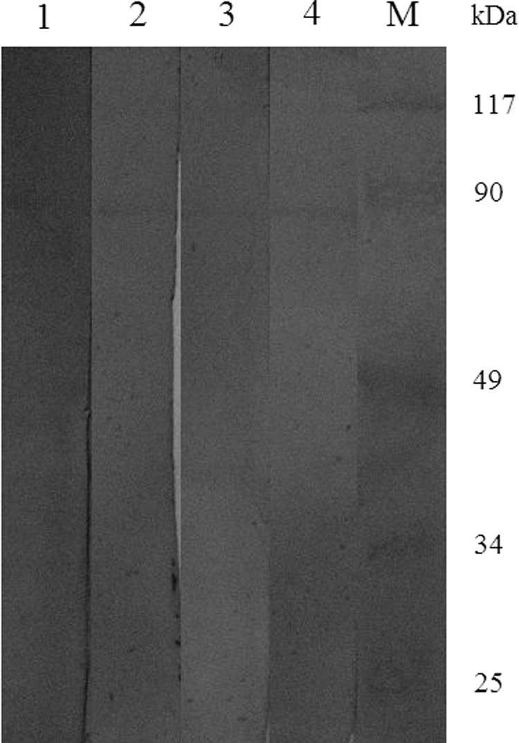
Western blot analysis of MAbs secreted by four hybridoma cell lines. M, molecular weight marker of proteins; lane 1, 1-B1; lane 2, 1-B8; lane 3, 1-F11; lane 4, 1-G10.
The neutralizing activity of the MAbs was further investigated by neutralization test. The neutralization test showed that all four MAbs had the capacity of neutralizing RV CC0812-1; they were all neutralizing MAbs against VP4 of RV CC0812-1 although their neutralization titers were different. The neutralization titers of MAbs 1B1, 1B8, 1F11, and 1G10 in BALB/c mice ascetic fluids were 1:2048, 1:1024, 1:512, and 1:512 for RV CC0812-1, respectively.
Discussion
The monoclonal antibody technique or hybridoma technique was developed Köhler and Milstein in 1975.(12) These researchers found a way to combine the unlimited growth potential of myeloma cells with the predetermined antibody specificity of normal immune spleen cells by fusing myeloma cells with antibody-secreting cells from an immunized mouse. In conventional monoclonal antibody technique, the myeloma cells cultured in vitro were used for fusion with spleen cells. In our study the myeloma cells were intramuscularly inoculated into the back of BALB/c mice to form a solid tumor of myeloma. The solid tumor of myeloma was used to prepare myeloma cells. Our results revealed that sp2/0 cells prepared from BALB/c mice-borne myeloma exhibit higher fusion efficiency than sp2/0 cells cultured (data not shown). This may be due to the fact that sp2/0 cells grown in mice possessed higher viability and that macrophages of mice could eliminate probable contamination of mycoplasma in the sp2/0 cells.
VP4 and VP7 are two outer capsid neutralizing antigens of RVs that can independently induce neutralizing antibodies.(13,14) Usually RVs were classified by a binary system similar to that used for influenza viruses in which distinct serotypes/genotypes of VP4 and VP7 are recognized.(15) For VP7, 27G genotypes have been identified(3); a correlation between genotype and serotype has been established.(2) But for VP4, a lack of readily available typing antibodies to different VP4 types has hampered classification of VP4 (P) serotypes. Such a correlation is less clear for VP4, although 35 P genotypes of RVs were determined based on sequence analysis of VP4 genes.(2,3) The MAbs against VP4 of P[15] RV CC0812-1 might contribute to typing the antibodies pool for VP4 serotyping of RVs.
Thereafter, these MAbs of VP4 could be used for detection of P[15] RV. The generated MAbs will provide an effective way to detect rotavirus in farm livestock.
In 2006 Komoto and colleagues successfully established a helper virus-driven reverse genetics system based on transcription of exogenous RV VP4 gene in the cell cytoplasm.(16) In this system, the exogenous RV VP4 gene was transcribed by T7 RNA polymerase expressed by a recombinant vaccinia virus, the helper rotavirus provided backbone for reassortant RV, and the exogenous RV VP4 mRNA synthesized was packaged in place of its homologous counterpart into a helper RV. This reverse genetics system allowed for the generation of engineered RV reassortant with a single exogenous gene encoding antigenically distinct viral surface proteins. The system also required the use of specific neutralizing antibodies to eliminate wild-type helper rotaviruses. The RV CC0812-1 was phylogenetically close to attenuated lamb RV LLR of human RV vaccine Rotavac. Although its G and P genotypes are not among G/P types of the most prevalent human RVs, it could be used as a helper RV to produce attenuated RV reassortant by Komato's RV reverse genetics system. MAbs 1B1, 1B8, 1F11, and 1G10 all had the capacity of neutralizing RV CC0812-2; they could be used to produce attenuated RV CC0812-2 reassortant with the most prevalent P genotype in humans.
Footnotes
Acknowledgments
This work was financially supported by the Key Natural Science Foundation of Hubei Province, China (no. 2008CDA101); and by self-determined research funds of CCNU from the colleges' basic research and operation of MOE (no. CCNU11A02013).
Author Disclosure Statement
The authors have no financial interests to disclose.