
Abstract
Calreticulin (CRT) has various versatile functions. It is one of the family of heat shock proteins (HSPs), and is mainly located in the lumen of the endoplasmic reticulum (ER). In order to elucidate the functional and structural properties of CRT, we expressed and purified CRT protein; we then developed a monoclonal antibody (MAb) against mouse CRT by immunizing BALB/c mice with a specific region of the N and P domains of CRT (dCRT) as antigen, which was expressed in Escherichia coli. A stable hybridoma cell line was established by enzyme linked immunosorbent assay (ELISA) screening. The MAb was then prepared from mouse ascites after inoculating the hybridoma cells. Different methods were used to analyze the characterization of the MAb: ELISA, Western blot analysis, immunofluorescence, and flow cytometry. The dCRT protein was expressed and purified and a MAb cell line for CRT was established through immunization, fusion, and screening. ELISA and Western blot analysis indicated that the MAb specifically recognized CRT. In addition, immunofluorescence and flow cytometry demonstrated that the MAb exhibits excellent reactivity to the ecto-CRT when the cells were induced to apoptosis. This CRT MAb will be a valuable tool for further investigation of calreticulin functions.
Introduction
Materials and Methods
Reagents and cell lines
Freund's adjuvant, bovine serum albumin (BSA), polyethylene glycol 4000 (PEG 4000, 50%w/v), 3,3’,5,5’-tetramethylbenzidine (TMB) were purchased from Sigma-Aldrich (St.Louis, MO). Hypoxanthine/aminopterin/thymidine (HAT) and hypoxanthine/thymidine (HT) were from Roche (Indianapolis, IN), and DMEM with L-glutamine culture medium and fetal bovine serum were purchased from Gibco (Invitrogen, Carlsbad, CA). Peroxidase-labeled goat anti-mouse IgG(H+L) (HRP-IgG) was from Jackson Immunoresearch Laboratories (West Grove, PA).
Mouse melanoma B16-F1 cells, mouse lymphoma EL4 cells, and mouse myeloma cell line SP2/0 cells were cultured in RPMI-1640 medium (for B16-F1 and EL4 cells) or Dulbecco's modified Eagle's medium (for SP2/0 cells) supplemented with 20% fetal bovine serum (FBS), 100 μg/mL streptomycin, and 100 U/mL penicillin in a humidified 5% CO2 and 95% air incubator at 37°C.
Expression and purification of recombinant CRT protein
The construction of pET-28a(+)-dCRT plasmid has been described previously.(11) Escherichia coli BL21 (DE3) cells were transformed with pET-28a(+)-dCRT, and the recombinant protein was induced at 30°C for 12 h under 0.1 mM IPTG. The cells were harvested, resuspended in lysis buffer (PBS containing 0.5%Triton X-100 and 1 mM PMSF), and disrupted by sonication. The recombinant protein was purified by affinity chromatography using Ni-NTA agarose resin (Amersham Pharmacia Biotech, Piscataway, NJ) as recommended by the manufacturer. Protein concentrations were determined using a Bradford Assay kit, and purified proteins were stored at −20°C.
Immunization and preparation of monoclonal antibody
Anti-CRT mouse MAbs were generated based on the murine lymphocyte method. A 5-week-old female BALB/c mouse was injected via the hind footpads with 200 μL of an emulsion containing 100 μg of recombinant dCRT protein and Freund's complete adjuvant. Booster doses of dCRT protein (50 μg) in Freund's incomplete adjuvant were then injected into the above mouse once every 2 weeks for three immunizations. After the final booster immunization, the murine splenocytes immunized with an antigen were fused with murine myeloma Sp2/0 cells at a ratio of 5:1 in a 50% polyethylene glycol (PEG 4000 solution). The resulting hybridoma cells were plated onto 96-well plates and cultured in HAT selection medium. Hybridoma colonies were expanded in the HT medium at 10 days post-fusion. The hybridoma supernatants were screened by means of ELISA against the CRT-His recombinant protein. Positive clones were subcloned and rescreened by ELISA and immunoblotting. A total of ascitic fluid in BALB/c female mice was prepared for the interesting colonies.
ELISA screening
CRT protein (100 μL/well, 1.0 μg/mL) in 0.05 M carbonate buffer (pH 9.6) was coated on the surface of a 96-well flexible microplate (Nunc, Roskilde, Denmark) by overnight incubation at 4°C. To avoid unspecific binding, the plates were blocked with 1% bovine serum albumin (BSA) in PBST (a PBS buffer containing 0.05%Tween-20 [v/v]). Hybridoma supernatants (100 μL) were incubated for 30 min at 37°C and then washed with PBST three times. The plates were incubated for 30 min at room temperature with horseradish peroxidase (HRP)-conjugated anti-murine IgG antibody (diluted 1:3000). After washing with PBST three times, ELISAs were developed using 100 μL of TMB substrate, stopped with 50 μL of 2M H2SO4, and read at 450 nm.
Western blot analysis
The plasmid pcDNA3.1(+) or pcDNA3.1(+)-dCRT was transfected into B16-F1 cells with LipofectamineTM 2000 reagent, according to the manufacturer's instruction. B16-F1/pcDNA3.1(+) and B16-F1/pcDNA3.1(+)-dCRT cells were collected and lysed in SDS sample buffer. Lysates were boiled and equal amounts of proteins were subjected to 10% SDS-PAGE and electro-transferred onto a PVDF member, which was blocked with 5% non-fat milk in TBST overnight at 4°C. The membrane was then incubated at room temperature for 1 h with the prepared monoclonal 7C3; followed by HRP-labeled goat anti-mouse IgG for 1 h. The blot was developed using an ECL Plus Western Blotting Detection System (GE Healthcare, Pittsburgh, PA) and exposed to a piece of BioMax x-ray film (Kodak, New Haven, CT).
Immunofluorescence staining
For detection of CRT protein on the surface of drug-treated cells by immunofluorescence (CIF), B16-F1 cells were grown on cover slips in each culture medium and treated by the antitumor drug mitoxantrone (1 μmol/L) for 48 h. Then the cells were fixed with 4% paraformaldehyde in PBS for 15 min at room temperature and treated with a blocking solution (PBS containing 50 mM glycine and 1% BSA). The cells were incubated with monoclonal antibody 7C3 and detected with rhodamine-conjugated secondary antibody. 4′,6-Diamidino-2-phenylindole (DAPI) at 1 μg/mL was used to counterstain the nuclei. The samples were examined using an Eclipse TE2000-S inverted fluorescent microscope (Nikon, Tokyo, Japan).
Flow cytometry
CRT expression on the surface of the tumor cells was detected by flow cytometry (FCM) using the monoclonal antibody 7C3. The EL4 cells were treated by the antitumor drug mitoxantrone (1 μM/L) for 48 h, collected, and used for membrane CRT detection. The cells were incubated with the MAb 7C3 at room temperature for 30 min, followed by incubation with the FITC-conjugated anti-murine IgG antibody (1:1000) at room temperature for 1 h (avoid light). After washing, the cells were analyzed by EPICS XL-4 flow cytometer (Beckman-Coulter, Fullerton, CA) to identify CRT on the EL4 cell surface.
Results and Discussion
Expression and purification of recombinant protein CRT
In order to obtain the CRT antigen, we constructed pET28a(+)-dCRT and expressed and purified the dCRT protein. Figure 1 shows the SDS-PAGE results of purified 34 kDa dCRT, which was fused with a 6x His to the CRT C-terminal. The protein was identified by Western blot using a His antibody (Fig. 1B).

Expression and purification of mouse recombinant calreticulin in prokaryotic cells. (
Sensitivity and specificity determination of anti-CRT MAb
Monoclonal antibody 7C3 was prepared through standard immunization, cell fusion, and antibody screening. After purification and concentration, the interesting antibody 7C3 was evaluated for its affinity to CRT protein. Purified CRT antibody was diluted in a 2-fold series for sensitivity test by ELISA. The result showed that the sensitivity of MAb 7C3 was over 105 dilution (Fig. 2A). We also evaluated the ability of MAb 7C3 to recognize endogenous and transfected CRT by Western blot. MAb 7C3 recognized a 46 kDa protein that exists in a variety of eukaryotic cells (Fig. 2B) and a recombinant dCRT that was expressed in the transfected cells (Fig. 2C). This means that MAb 7C3 appears to have an affinity for natured CRT.
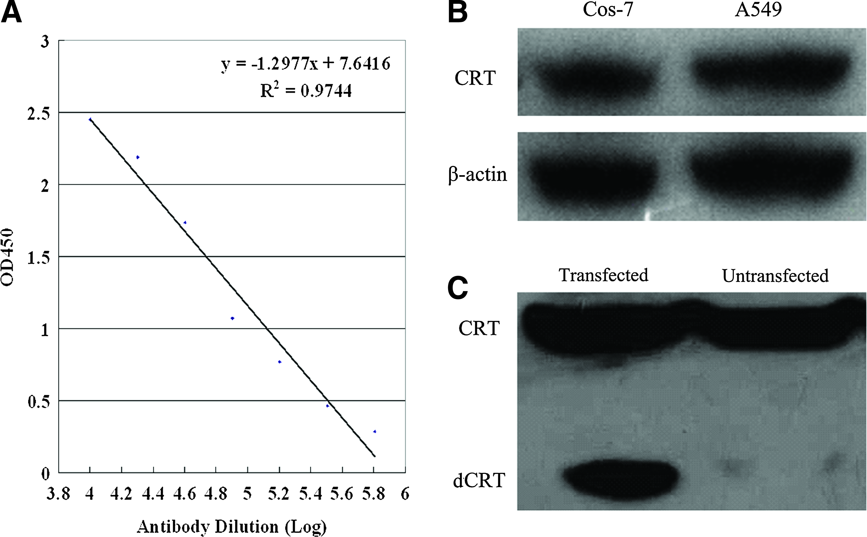
Evaluation of anti-CRT monoclonal antibody (MAb 7C3) to the endogenous and transfected CRT by ELISA and Western blot. (
Measurement of CRT expression on membranes of B16-F1 and EL4 cells
These antibodies were suitable for recognizing the membrane CRT by flow cytometry and immunofluorescence. Red fluorescence on the membrane of B16-F1 and EL4 cells was observed by immunofluorescence using the antibody 7C3 (Fig. 3), which was consistent with the previous report. When B16-F1 and EL4 cells (Fig. 3A,B) were treated by the antitumor drug mitoxantrone, apoptosis(12) was induced; at the same time, the CRT quickly relocated from the ER lumen onto the cell surface. In summary, we have generated a new monoclonal antibody, 7C3, that specifically recognizes CRT protein by Western blot analysis, flow cytometry, and immunofluorescence. Preparation of the 7C3 antibody will be helpful to elucidate CRT functions.
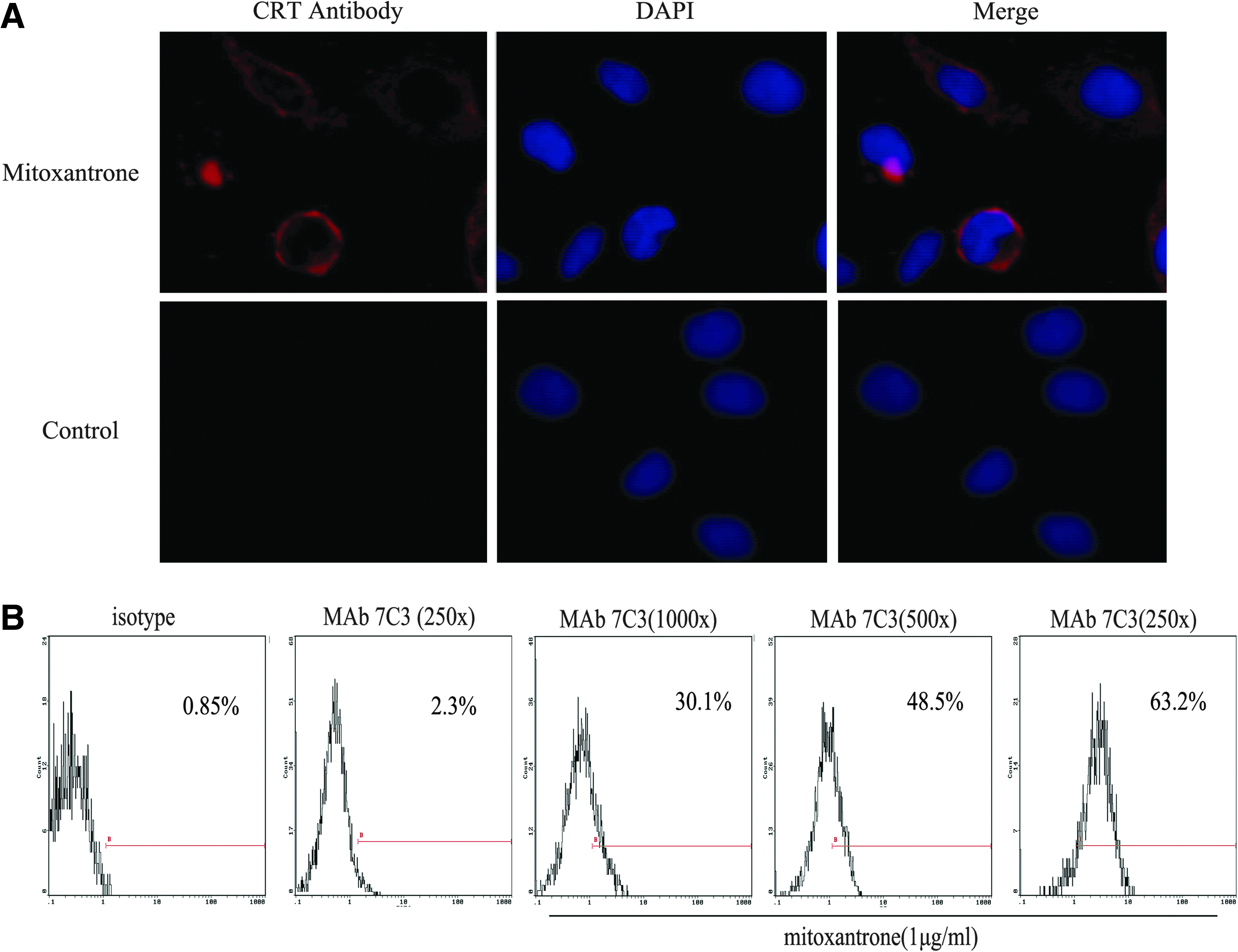
Reactivity of MAb 7C3 to ecto-CRT by immunofluorescent staining and flow cytometry. (
Footnotes
Author Disclosure Statement
The authors have no financial interests to disclose.