




Abstract
Glypican-3 (GPC3) has been reported as a novel serum and histochemical marker for hepatocellular carcinoma (HCC) by several groups. As an oncofetal protein, it is expressed abundantly in the fetal liver, inactive in the normal adult liver, and frequently reactivated in HCC. Immunology reagents are urgently needed to proceed with mechanism-related research, clinical validation, and application. In this report, monoclonal antibodies (MAbs) against GPC3 were made from hyperimmune BALB/c mice by injecting 100 μg of purified antigen intraperitoneally. Hybridomas were screened by indirect enzyme-linked immunosorbent assay (ELISA) using purified protein. Finally 13 mouse hybridomas producing MAbs to GPC3 were established. The MAbs obtained were fully characterized using Western blot analysis, immunofluorescence, and immunohistochemistry. The results showed that these antibodies could be used for preliminary application of the next step mechanism-related research and GPC3 expression level analysis.
Introduction
GPC3 has been reported to be a novel serum and histochemical marker for HCC by several groups.(4–7) GPC3 is a member of the heparan sulfate proteoglycan family. The GPC3 gene is located on the human X chromosome (Xq26) and encodes a 70 kDa core protein with 580 amino acids, which can be cleaved by furin to generate a 40 kDa amino (N)-terminal protein and a 30 kDa membrane-bound carboxyl(C)-terminal protein.(8) GPC3 is linked to the outer surface of the cell membrane through a glycosylphosphatidylinositol anchor(9) and released from the cell surface by a lipase called notum to regulate the signaling of Wnts, hedgehogs, fibroblast growth factors (FGFs), and bone morphogenetic proteins (BMPs).(10–14) Depending on the cellular context, its function can be stimulatory or inhibitory activity or signaling. GPC3 plays an important role in the embryonic and fetal periods of tissue, organ formation, and developmental stages, mainly playing a negative regulatory role, preventing tissue organs from growing too large and regulating body size overall.(15) In tissue GPC3 has been detected in the placenta and fetal liver, but not in other adult organs. During hepatic carcinogenesis, GPC3 appears in the HCC tissue and is released into serum; it was a potential tumor marker for HCC.(4–6)
An effective detection method can provide help for the early diagnosis of HCC. Therefore, a large quantity of monoclonal antibodies (MAbs) specific for GPC3 is needed. Several monoclonal and polyclonal antibodies against human GPC3 are commercially available from various manufacturers (e.g., Biomosaics, Burlington, VT; Santa Cruz Biotechnology, Santa Cruz, CA; and R&D Systems, Minneapolis, MN). However, the polyclonal antibody cannot be satisfied for the development of follow-up testing reagents, the monoclonal antibodies available are mainly for the C-terminus of the GPC3 (such as 1G12), and also lacking are paired MAbs for the development of follow-up testing reagents. Since the fragment GPC3 release into the blood is the N-terminal, the N-terminal fragment is the key target for a serum GPC3 diagnostic kit. The preparation of anti-GPC3 monoclonal antibody against the N-terminal for the development of follow-up testing reagents is necessary.(16–21)
In this study, in order to obtain antibodies for which we would have the independent intellectual property rights and that can be used to develop serological detection reagents, we have produced, characterized, and purified monoclonal antibodies specific for GPC3 that can be used for preliminary and clinical applications.
Materials and Methods
Materials
The PMD18T/GPC3, sp2/0 cell line was obtained from the Biotechnology Institute of Southern Medical University Agriculture (Guangzhou, China). BALB/c mice (6–8 weeks old, female) were obtained from the Experimental Animal Center of Southern Medical University in China. PHUE plasmid (a gift from Dr. Shu-hong LUO, University of Illinois) was reformed on the vector basis of PET15b, add a period of ubiquitin sequence between the His tag and the target protein. Ubiquitin can increase the solubility of the target protein. His and ubiquitin label can be removed from the purified recombinant protein to harvest, containing only the amino acid sequence of the target protein product.
Clone of GPC3 gene
As a template to PMD18T/GPC3, according to the sequence in GenBank, the primer sequences for amplification of the GPC3 gene were as follows: P1, 5′ TAATGAATTCATGCAGCCCCCGCCGCC 3′ with the EcoRI (GAATTC) restriction site; and P2, 5′ AGCGGTCGACTCACTTGGCATGGCGAACAACAA 3′ with the Sal I (GTCGAC), which have sites for EcoRI (GAATTC) and Sal I (GTCGAC) in forward and reverse primers. Gel images were obtained and the densities of the PCR products were quantified using densitometry methods.
Expression and purification of fusion protein in E. coli Rosseta
PCR fragments were digested by EcoRI and Sal I and then cloned into the EcoRI and Sal I digested PHUE vector. Recombinant plasmids were transformed into E. coli. Rosseta and soluble fusion protein GPC3 were produced after the induction of 0.1 mM isopropyl-1-thio-D-galactopyranside (IPTG) at 37°C and 220 rpm for 4 h. The expressed fusion protein was purified with affinity chromatography via its His-tag and then ion exchange chromatography via protein charge. Purified fusion proteins were used as immunogen and screening antigen in enzyme-linked immunosorbent assay (ELISA) for the identification of monoclonal antibody against GPC3.
Immunization, cell fusion, and screening
Three adult female BALB/c mice (8∼10 weeks of age) were injected intraperitoneally with 500 μL of a 1:1 GPC3 fusion protein (100 μg per mouse) and complete Freund's adjuvant (Sigma, St. Louis, MO) the first time and then boosted with the antigen in incomplete Freund's adjuvant at 2-week intervals. Before fusion, the mice were again boosted with 100 μg of the antigen in phosphate-buffered saline (PBS, pH 7.2). Fusion to SP2/0 myeloma cells was carried out using standard methodology. Fusion cells were resuspended in 120 mL RPMI 1640 containing 20% fetal calf serum and HAT. Then cell suspensions were plated on 96-well plates and incubated overnight at 37°C, 5% CO2, 95% humidity. Seven days later culture medium was replaced by RPMI 1640 containing 20% fetal calf serum and HT. At day 15, the wells were tested for positive colonies by indirect ELISA. 96-well polystyrene plates were coated with 30 μg fusion protein and incubated at 4°C overnight. The plates were washed three times with washing buffer (0.05% Tween-20 in PBS). Then 10% calf serum in PBS was added to the wells and the plates were incubated for 1 h at 37°C followed by washing as above. The culture supernatants of the hybridomas (100 μL/well) were added to the wells (GPC3 immune serum as a positive contrast, normal mouse serum as a negative control) and incubated for 1 h at 37°C. After washing, goat anti-mouse IgG-HRP conjugate was added and incubated for 1 h at 37°C. After treatment with washing buffer three times, 100 μL of the chromogenic reagent 3, 3′, 5, 5′-tetramethylbenzidine (TMB) were added. After 20 min, the reaction was stopped with 2 M H2SO4 and at 450 nm each well was measured by a microplate reader (Bio-Rad ELISA Reader, Tokyo, Japan).
Western blot analysis
For Western blot analysis, the proteins from the expression proteins and hepatoma cell lines Huh7, HepG2, L02, SW480, and Hela were used. After dealing with the protein sample, they were loaded per lane, electrophoresed in 12% SDS-PAGE, and blotted on a PVDF membrane. The membrane was blocked with 5% non-fat dried milk in TBST (10 mM Tris-HCl [pH 7.4], 150 mMNaCl) at 4°C overnight. After washing three times for 15 min in TBST, the membrane was incubated with hybridoma culture supernatant for 1 h at room temperature. Followed by washing as above, goat anti-mouse IgG-HRP was used as a secondary antibody at room temperature. The membrane was washed again and blots were visualized by ECL.
Production of monoclonal antibodies
In order to obtain a considerable number of antibodies, adult female BALB/c mice were injected with about 105 hybridoma cells in 0.5 mL of RPMI 1640. Then a large number of ascites were produced within 1–2 weeks. The ascites were centrifuged at 12000 rmp for 10 min, and the supernatant was collected.
Classification of MAbs
The class and subclass of MAbs were determined using mouse hybridoma subtyping kit (Boehringer Mannheim, Germany).
Purification of monoclonal antibodies
The ascites were diluted with PBS (pH 7.2) at least five to six times. They were then purified by Protein G-Sepharose CI-4B (Pharmacia, Uppsala, Sweden). The Hit rap Protein G column was equilibrated with at least two column volumes of starting buffer (0.01 M PBS, pH 7.4). Then the sample was pumped into the column and washed with the starting buffer for 5 column volumes and eluted with elution buffer (0.1 M Gly-HCl, pH 2.8) 1–3 column volumes. The purified antibodies were neutralized by a buffer (1.0 M Tris-HCl, pH 9.6). Flow rates of washing and equilibration were 2 mL/min, and rates of sample application and elution were 1 mL/min.
Immunofluorescence
After the collection of liver cancer cells Huh7, cells were spread on a glass slide and grown overnight. After washing in PBS, they were fixed with ice-cold paraformaldehyde and were blocked with 10% goat serum for 1 h. After washing three times with PBS, the cells were incubated with 7D11 for 1 h at room temperature with PBS as negative control. Followed by washing as above, fluorescent-labeled goat anti-mouse was used as a secondary antibody at room temperature. Stained cells were observed in the fluorescence microscopy.
Immunohistochemistry
After pre-incubation of the sample with 5% bovine serum albumin (BSA) for 10 min, anti-GPC3 primary antibody (1:1000) was added and incubated at 4°C overnight. Location of the primary antibodies was achieved by subsequent application of a biotinylated anti-primary antibody, an avidin-biotin complex conjugated to horseradish peroxidase, and diaminobenzidine (Dako, Glostrup, Denmark). The slides were counterstained by hematoxylin.
Results
Expression and purification of fusion protein in E. coli
Recombinant plasmid PHUE-GPC3 was established and transformed into competent E. coli Rosseta. Fusion protein was expressed in bacteria with the induction of 0.1 mM IPTG overnight at 37°C and 220 rpm (Fig. 1A). Expressed fusion proteins were purified using affinity chromatography via His-tag and achieved up to 95% purity determined by SDS-PAGE (Fig. 1B).

(
ELISA analysis for MAb against GPC3
Hybridomas were cultured in 96-well culture microtitration plates. In order to assess anti-GPC3 antibody production, the supernatants were assayed after cell fusion for 11 days. Sixteen hybridomas (1A1, 1E1, 2E11, 4A8, 4B8, 4E12, 5D6, 5G7, 7D10, 7D11, 7E9, 7H7, 8B2, 8E7, 8F10, 8G6) were selected because of their stability and specificity. Secretory hybridoma cells of antibodies were cloned and recloned three times to guarantee monoclonal behavior of the produced immunoglobulins. These stable hybridomas, designated as1A1, 2E11, 4A8, 4B8, 4E12, 5D6, 5G7, 7D10, 7D11, 7H7, 8B2, 8F10, and 8G6, were obtained after cell fusion and subcloning. All of the subclones were identified as IgG1a isotype and purified with protein G-Sepharose CI-4B resin under the condition of low salt and low pH (Table 1).
Western blot analysis of specific MAbs against GPC3
In order to test the specificity of MAb against GPC3, Western blot analyses of monoclonal antibodies were done using hybridoma supernatants as the primary antibody. Because the GPC3 protein used in immunization was a prokaryotic protein, its structure might be somewhat different with the GPC3 in eukaryotic cells. Therefore, the human liver cancer cell lines HepG2, Huh7, L02, SW480, and Hela were used to examine the binding ability of MAb7D11 with GPC3 in eukaryotic cells. The fusion protein GPC3, HepG2, and Huh7 cells were subjected to SDS-PAGE and Western blot analysis with 7D11 as the primary antibody. Western blotting results showed that the recombinant protein GPC3-His can be detected by anti-His antibody, the recombinant protein GPC3-GST can be detected by anti-GST antibody (Fig. 2A), and 7D11 had specific binding ability with fusion protein GPC3 (Fig. 2B) and GPC3 in eukaryotic cells (Fig. 2C).

(
Immunofluorescence analysis of specific MAbs against GPC3
The cells were observed in fluorescence after staining by monoclonal antibody 7D11 (Fig. 3A) and no fluorescence after staining by PBS (Fig. 3B). This result suggested that 7D11 could be used to detect GPC3 protein expression level in liver cells.
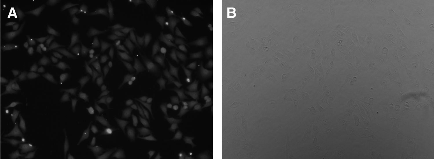
Immunofluorescence analysis of specific MAbs against GPC3.
Immunohistochemistry analysis of specific MAbs against GPC3
In immunohistochemistry assay, HCC tissue, HCC adjacent tissue, and liver abscess tissue were used to determine the reactivity of MAb 7D11. There was little immunoreactivity observed in HCC adjacent tissue and liver abscess tissue. Strong immunoreactivity was observed in HCC tissue.
Discussion
GPC3, a membrane heparan sulfate proteoglycan, regulates the development of tissues and organs in the embryo and fetus negatively. The abnormal expression of GPC3 in tumor development is closely related to HCC after the birth; GPC3 is highly expressed in the HCC state and can be detected in the early stage of liver cancer. In AFP negative hepatocellular carcinoma, GPC3 has a certain expression rate, so GPC3 can be used for the early diagnosis of liver cancer.
In 1997 Hsu and colleagues(22) first reported on the high expression of GPC3 mRNA in HCC, followed by more researchers confirming in GPC3 that both mRNA and protein were shown to be overexpressed in HCC and that this occurred in the early stage of HCC.(23) GPC3 expression was also found in the HCC cell lines HepG2, Hep3B, and HuH-7(24) and in more than 70% of HCC tumors, but not in normal liver tissue.(5,6) Studies by Di Tommaso and co-workers(25) have shown that sensitivity is 69%, with a specificity of 91% in the diagnosis of early-stage HCC by detecting GPC3.
The differences of GPC3 expression are obvious in HCC tissues and benign liver organizations. Llovet and colleagues(26) reported that GPC3 was expressed in 100% liver organization with the background of early liver cirrhosis in hepatitis C (<2 cm liver nodules) and was not expressed in other benign liver diseases and normal liver tissues. In comparing the GPC3 expression in HCC and ICC (intrahepatic cholangiocarcinoma) organization, Shirakawa and colleagues(27) found that GPC3 was expressed in 78.3% of HCC, but not expressed in ICC. The positive rate of serum GPC3 protein was 40.0% (16/40) in liver cancer patients, patients with cirrhosis, and chronic hepatitis, and healthy controls had negative expression (0/13, 0/34, 0/60).(6) The level of serum GPC3 was increased in 53% (18/34) of liver cancer patients, lower than the positive rate (72%) of HCC tissues. Only 1 of 20 patients with liver cirrhosis was high, while normal controls and hepatitis patients were negative.(5)
In AFP negative HCC, GPC3 also has a higher expression rate. Ding and colleagues(28) detected GPC3 mRNA in 41 AFP-negative HCC tissues and adjacent noncancerous tissues. In cancer tissue, the positive rate was 73.17%, and in adjacent tissue, it was only 9.76%. Moreover, GPC3 mRNA was weakly expressed; the expression rate was 79.31% in large hepatocellular carcinoma (>5 cm), 41.67% in small HCC (≤5 cm), 76.47% in poorly differentiated carcinoma (III–IV class), 42.86% in well-differentiated carcinoma (I–II level). Immunohistochemistry results showed that GPC3 was expressed in hepatoma cells, negative in normal liver cells, bile duct cells, endothelial cells, Ito cells, and fibroblasts. There are no significant correlations between serum GPC3 and AFP levels, which is to a certain extent compensatory for the lack of AFP diagnosing for liver cancer.(5,29) Further, GPC3 was found to be an absence protein expression in CCA, indicating that GPC3 may be potentially used as a diagnostic marker to distinguish between these two primary liver cancers too.(30)
GPC3 expression status was also related to the prognosis of patients with hepatocellular carcinoma. In the study by Shirakawa and associates, 107 cases of hepatocellular carcinoma patients were divided into GPC3 positive and negative groups by immunohistochemical staining. Five-year follow-up found that in the GPC3 positive group, mortality was significantly higher than in the negative group (87.7% vs. 54.5%).(6) Because of the finding that GPC3-positive HCC patients have a significantly lower 5-year survival rate than GPC3-negative HCC patients, GPC3 expression is correlated with poor prognosis in HCC.(24)
All together, GPC3 is a most promising early diagnostic marker for hepatocellular carcinoma. The combination of GPC3 plus AFP achieved a much-improved sensitivity compared with either GPC3 or AFP alone. Development of a high quantitative immunological detection reagent and expanding the clinical application have not only scientific importance but also clinical value to diagnosis, treatment of HCC, and research of GPC3. Since serum-based assays are noninvasive and relatively inexpensive, it would be ideal to develop kits for translation of the GPC3 marker into advancing clinical diagnostic applications. This requires a great quantity of MAbs specific for GPC3. However, the cost of MAbs specific for GPC3 is significantly higher. In addition, most antibodies used in previous studies of GPC3 were polyclonal antibodies, the polyclonal antibody cannot be satisfied for the development of follow-up testing reagents, the monoclonal antibodies available are mainly for the C-terminus of the GPC3 (such as 1G12) and are not targeted for GPC3 in the serum, and also lacking are paired MAbs for the development of follow-up testing reagents. The MAbs we developed are specific for the N-terminus of the GPC3 fragment, and we also had paired MAbs for follow-up testing of reagent development.
We used PHUE vector to express GPC3 in bacterial systems, which is a highly efficient and convenient vector used to express foreign polypeptides as fusions with His.(31) To induce the expression of recombinant proteins by adding IPTG, the recombinant protein was expressed as inclusion bodies, the inclusion body was dissolved in 4 M urea, and the recombinant protein was purified by Ni-NTA affinity chromatography. To obtain biologically active protein, denatured recombinant protein must be refolding.(32) The purified protein refolded by gradient dialysis. SDS-PAGE protein electrophoresis results showed that we obtained the human adenosine kinase recombinant protein with high purity.
In this study, MAb 7D11 specific against GPC3 in eukaryotic cells was obtained. Western blotting and immunohistochemistry assays showed 7D11 could be used to detect the expression of GPC3 protein in liver cancer tissue. As shown in Figure 4, there is no expression in adjacent tissue and liver abscess tissue. This result is the same as that found in other published reports.

CPC3 expression in HCC tissue. (
The GPC3 quantitative diagnostic kit (chemiluminescence immunoassay) has been under development by our team. In this study we showed that the MAbs for GPC3 can provide a solid foundation for subsequent applications.
Footnotes
Acknowledgments
This work is supported by the Combination Project of the Education Ministry, Guangdong Province (no. 2010B090400422) and the National High Technology Research and Development Program of China (no. 2012AA020205).
Author Disclosure Statement
The authors have no financial interests to disclose.