




Abstract
Adiponectin (AdipoQ) is an adipokine mainly secreted by white fatty tissue, playing a major role in energy homeostasis and insulin sensitivity. For cattle, AdipoQ data are largely limited to mRNA expression; to our knowledge, valid information about the AdipoQ protein in bovine tissues and body fluids is not available. Therefore, we have developed a monoclonal antibody against bovine AdipoQ. This study describes the preparation, application, and characterization of a monoclonal antibody for use in ELISA, Western blot, and histology. The antibody was developed by PEG fusion of the SP2/0 cell line with splenic B cells from AdipoQ immunized C57Bl/6 mice. Antibody-producing cells were identified by ELISA and specified by immunoblotting and immunostaining of bovine retroperitoneal adipose tissue. The novel antibody detects AdipoQ in histological samples, ELISA, and Western blots.
Introduction
Materials and Methods
Purification of AdipoQ
Pure AdipoQ was isolated from bovine serum using standard procedures as described by Tanita and colleagues(12) with minor modifications. Briefly, after a first purification by gel-filtration chromatography with Superdex 200 on a XK 16/70 column using an ÄKTA fast protein liquid chromatography system (all from GE Healthcare, Munich, Germany), ion exchange chromatography (DEAE-Sepharose, XK 26/20 column, GE Healthcare) was performed.
Immunization of C57BL/6 mice and hybridoma production
Five C57BL/6 mice were immunized subcutaneously once per week for a period of 6 weeks with 100 μL of Freund's adjuvant (DIFCO Laboratories, Detroit, MI) containing 5 μg of purified HMW-AdipoQ. 8×10⁷ splenocytes and 1.6×10⁷ Sp2/0 cells were fused using 37°C pre-warmed PEG (MW4000; AppliChem, Darmstadt, Germany), diluted 1:1 in DMEM. The fused hybridoma cells were plated on ten 96-well plates (TPP, Trasadingen, Switzerland) with peritoneal macrophages as feeder cells (4×10⁴ feeder cells/well) in DMEM supplemented with 10% FBS Gold (PAA, Cölbe, Germany), 0.25 U×mL−1 penicillin, 0.25 μg×mL−1 streptomycin, 50 μmol β-Mercaptoethanol, and 2% HAT (Gibco, Darmstadt, Germany). The cells were incubated for 10 days at 37°C and 5% CO2. After screening by ELISA, positive selected cells were cultivated in DMEM supplemented with HT instead of HAT.
ELISA
Microtiter plates (EIA plate, high binding, Costar, Corning, NY) were coated with purified complete AdipoQ (2 μg/mL) in carbonate coating buffer (pH 9.6; 0.05 M NaHCO3; 0.01% Proclin solution; Supelco, Sigma-Aldrich, Taufkirchen, Germany) overnight at 4°C. The plates were subsequently blocked for 2.5 h at RT with Roti-Block (Roth, Karlsruhe, Germany), washed with washing buffer (PBS supplemented with 0.05% Tween-20, 0.01% Proclin), and stored at −20°C until use. Subsequently, supernatants of the hybridoma cell cultures were screened by ELISA for positive clones. As positive control, samples of immune sera were diluted in PBS/0.1% BSA/0.05% Tween-20. Incubation took place for 1.5 h. After washing three times with washing buffer, goat-anti-mouse IgG conjugated to horseradish peroxidase (Dianova, Hamburg, Germany; diluted 1:25,000 in PBS supplemented with 0.1% BSA and 0.05% Tween-20) was used for detection. After washing five times with washing buffer, the plates were incubated with substrate buffer (pH 4.05; 0.05 M citric acid; 0.055 M Na2HPO4; 0.05% urea-peroxide; Proclin 0.01%) supplemented with 2% TMB. The color reaction was stopped by addition of 1 M oxalic acid, and the extinction was measured at 450 nm and 630 nm as reference wavelength.
SDS-PAGE and immunoblotting
The separation of AdipoQ was done using 4–12% TRIS-glycine gradient polyacrylamide gels with 5.6% stacking gels. Following blotting, the membranes (Amersham Hybond P membranes; GE Healthcare, Freiburg, Germany) were blocked for 1 h in Roti-Block diluted in TBS. The novel anti-AdipoQ antibody P10E1 was purified with protein G (GE Healthcare), diluted 1:5000 in Roti-Block solution for 1 h, and finally washed in TBS-T. Goat anti-mouse IgG, conjugated to horseradish peroxidase (Dianova) was diluted 1:10,000 in Roti-Block solution for 1 h. After washing, the dripped membranes were covered with Amersham enhanced chemiluminescence (ECL) substrate (GE Healthcare) and developed by exposition of X-ray films.
Immunohistochemistry
Bovine retroperitoneal adipose tissue was fixed in 4% formaldehyde and embedded in paraffin wax. Ten μm sections were cut, deparaffinized in Rotihistol (Roth), and rehydrated in descending grades of isopropanol (Roth). Antigen retrieval was performed by boiling in citrate buffer in a microwave (pH 6.0, 0.01 mM, 750 W) for 3× 5 min. Endogenous peroxidase activity was blocked with 3 % H2O2 at RT for 15 min. To avoid unspecific binding, sections were treated with normal goat serum (20 min, RT). Immunostaining was performed with Protein-G-purified MAb P10E1 (1:100, 18 h at RT). Secondary antibody staining was performed using a biotinylated goat-anti-mouse Ig antibody (1:200, 30 min, RT) and combined with streptavidin-horseradish peroxidase (1:1000, 30 min, RT; Southern Biotech, Birmingham, AL). Immunostaining was detected using 3-amino-9-ethylcarbazol as a substrate. Sections were counterstained with Mayer's hematoxylin (Merck, Darmstadt, Germany) prior to coverslip placement. For negative control, retroperitoneal adipose tissue was used omitting the primary antibody.
Results
Purification of AdipoQ from bovine sera
AdipoQ was purified from bovine sera using standard procedures, as described by Tanita and colleagues,(12) with minor modifications. Purity and distribution of the various forms of AdipoQ were checked with PAGE and Coomassie Brilliant-Blue staining. Figure 1 shows the effect of reduction and heat denaturation on purified AdipoQ visualized by Coomassie-blue-stained SDS-PAGE. Native purified AdipoQ isolated from bovine serum was observed as HMW AdipoQ (>300 kDa) (Fig. 1, lane 1) with minor amounts of hexameric AdipoQ (136 kDa). Heat denaturation of AdipoQ generates smaller HMW isoforms (Fig. 1, lane 2). Under reducing conditions (Fig. 1, lane 3), the HMW AdipoQ is separated into MMW isoforms and monomeric 28 kDa LMW AdipoQ. Applying both heat denaturation and reduction resulted in the generation of monomeric and trimeric (67 kDa, LMW) AdipoQ (Fig. 1, lane 4).
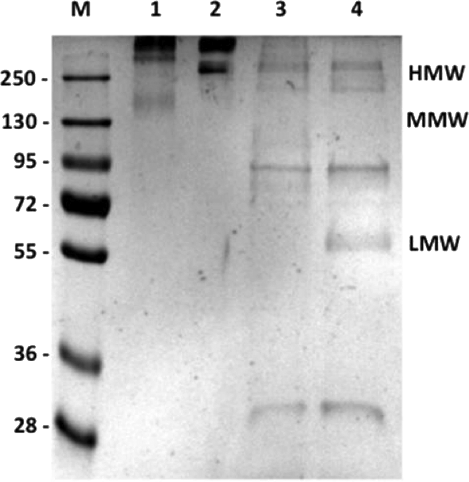
Coomassie staining of PAGE separated AdipoQ from bovine serum. Three μg AdipoQ per lane were separated by PAGE and proteins stained with Coomassie blue. Lane 1, untreated purified HMW AdipoQ; lane 2, heat-treated (95°C) HMW AdipoQ; lane 3, reduced (DTT) HMW AdipoQ; lane 4, reduced (DTT) and heat-treated (95°C) HMW AdipoQ. Size marker (Fermentas Prestained Protein Ladder, SM1811), values are given in kDa.
Preparation of MAB against AdipoQ
C57BL/6 mice were used for immunization. After six boosts given over a period of six weeks, serum samples of all immunized mice were collected and serum dilutions tested using HMW AdipoQ- and total AdipoQ-coated microtiter plates. The mouse with the highest extinction was used for fusion. Hybridomas were screened using ELISA, retested, subcloned, and recloned. Clone P10E1 reacted with AdipoQ. Thus the new MAb P10E1 is suitable for detection of AdipoQ in ELISA.
Specifying MAb P10E1 for binding to bovine AdipoQ isoforms
To test which of the different isoforms of AdipoQ are detected by the monoclonal antibody P10E1, we analyzed AdipoQ in bovine serum—native, treated with heat (95°C), and/or DTT employing SDS-PAGE and Western blot (Fig. 2). Unreduced and non-heat-treated AdipoQ (Fig. 2, lane 1) showed bands in the range from MMW to HMW AdipoQ. The heat-treated, non-reduced sample (Fig. 2, lane 2) showed a strong/broad band of hexameric MMW AdipoQ at 138 kDa, but no dimeric 56 kDa AdipoQ was observed. The reduced, non-heat-treated sample shows monomeric and some weak MMW bands but no trimeric 67 kDa AdipoQ. Reduction and heat denaturation (Fig. 2, lane 4) of AdipoQ displayed the monomeric 28 kDa isoform.
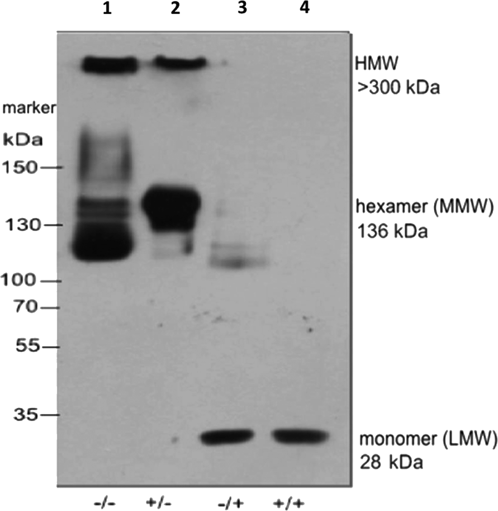
SDS-PAGE of bovine serum with monoclonal antibody (P10E1) notifying AdipoQ bands. Bovine serum (dilution 1:50) was separated by gradient SDS-PAGE (4–12%) and blotted. The marker is a pre-stained plus (Fermentas, SM1811). Lane 1 (-/-), no heat denaturation/no reduction. The novel antibody binds several isoforms of AdiponectinQ in untreated serum (above 120 kDa). Lane 2 (+/-), heat denatured (95°C, 5 min)/no reduction, the antibody binds isoforms of AdipoQ above 120 kDa mainly the 136 kDa hexameric isoform. Lane 3 (-/+), no heat denaturation/reduction with DTT. The antibody detects fragments of MMW AdipoQ and the monomeric 28 kDa isoform. Lane 4 (+/+), heat denatured (95°C, 5min)/reduction with DTT. The antibody recognizes 28 kDa monomers (LMW).
The MAb P10E1 thus detects the monomeric isoform of 28 kDa, the hexameric 136 kDa isoform, and, under native conditions, several isoforms of MMW AdipoQ and HMW AdipoQ using SDS-PAGE and Western blotting.
Detection of AdipoQ in histological sections of bovine adipose tissue by MAb P10E1
To test whether the P10E1 antibody is applicable for histological evaluations of bovine tissue, 10 μm sections from bovine retroperitoneal adipose tissue were stained with the protein-G-purified novel MAb P10E1. Sections were counterstained with Mayer's hematoxylin. Reddish P10E1-stained AdipoQ (red dye) was located in the cytoplasm of the adipocyte (Fig. 3A). Nuclei counterstained Mayer's hematoxylin (blue) are shown in Figure 3A and in Figure 3B (negative control).

Immunohistological staining of AdipoQ in bovine retroperitoneal adipose tissue. Immunostaining of bovine retroperitoneal adipose tissue with novel antibody P10E1. Immunostaining was detected using biotinylated goat-anti-mouse-Ig and 3-amino-9-ethylcarbazol as a substrate. Sections were counterstained with Mayer's hematoxylin. For negative control, retroperitoneal adipose tissue was used and PBS replaced primary antibody. (
Discussion
Here we describe the preparation and application of a MAb directed against bovine AdipoQ, able to detect AdipoQ in ELISA, on histological paraffin sections and Western blot. We showed that the MAb P10E1 detects the monomeric, LMW, MMW, and HMW isoforms of bovine AdipoQ. Thus MAb P10E1 is suitable for detection of AdipoQ in native as well as in denatured/reduced form, using SDS-PAGE and Western blotting, thus likely targeting a linear epitope. The broad reactivity against the different isoforms allows for comparing AdipoQ isoforms from different physiological and pathophysiological conditions when applying densitometry to the bands observed in Western blotting.
AdipoQ is expressed in various tissues.(13) In adipocytes it is found in the cytoplasm.(14) Our histological analysis showed expression of the AdipoQ protein located mainly around the nuclei and adjacent cytoplasm but also some staining scattered in the cytoplasm. This confirms previous observations of the subcellular distribution of AdipoQ in 3T3L1 adipocytes, with localization in the perinuclear area and punctuate staining in the cytoplasm(14) and demonstrates the suitability of MAb P10E1 for histological analysis.
Footnotes
Author Disclosure Statement
The authors have no financial interests to disclose.