
Abstract
A simple, rapid competitive immunochromatography (ICG) strip test was developed to detect morphine in urine samples using a monoclonal antibody produced in-house and conjugated to gold nanoparticles. Hybridoma cells were cultured and the Amor-HK16 monoclonal antibody against morphine was obtained from the supernatant after purification by salting out and passing through a Protein G-Agarose affinity column. Morphine was obtained from morphine sulfate and a C6-hemisuccinate derivative of morphine was prepared, conjugated to bovine serum albumin, and immobilized to a nitrocellulose membrane as the test line. Goat anti-mouse antibody was used as a binder in the control line in the detection zone of the strip. Colloidal gold particles of diameter approximately 20 nm were prepared and conjugated to the monoclonal antibody. The detection limit of the test strip was found to be 2000 ng/mL of morphine in urine samples. Reliability was determined by performing the ICG test on 103 urine samples and comparing the results with those obtained by thin-layer chromatography. The sensitivity of the test was 100%, and the analysis time for the assay was approximately 5 min. The new ICG method was adequately sensitive and accurate for the rapid screening of morphine in urine.
Introduction
Various methods, such as immunochromatography (ICG),(1,2) enzyme immunoassay (EIA),(3) radio immunoassay (RIA),(4) polarization fluorimetry,(5) gas chromatography (GC),(6,7) gas chromatography-mass spectrometry (GC-MS), and liquid chromatography-mass spectrometry (LC-MS),(8) are used to detect morphine in biological fluids. ICG, which uses colloidal gold as a label, is now one of the most widely used analytical methods for screening morphine. In addition, it is also widely used in other fields of medicine, such as detection of toxins,(9,10) infectious diseases,(11,12) drugs,(13) and hormones.(14,15)
The ICG method is used widely mainly because of its short procedure time, simple implementation, requiring no special instruments and no trained technicians, and low cost. The aim of this study was to develop a nanogold-based immunochromatographic assay for the detection of morphine in urine using the Amor-HK16 monoclonal antibody.(16)
Materials and Methods
1-ethyl 3-(3-dimethyl aminopropyl) carbodiimide (EDC), bovine serum albumin (BSA), Protein G-Agarose, succinic anhydride, tetrachloroauric acid (HAuCl4), trisodium citrate, and polyethylene glycol 8000 (PEG) were obtained from Sigma-Aldrich (St. Louis, MO). CELLine classic 1000 was purchased from Integra Bioscience (Hudson, NH). HF-75 nitrocellulose (NC) membranes, glass fiber pads, and sample and absorption pads (cellulose) were obtained from Millipore (Bedford, MA). The antibody against morphine (Amor-HK 16) had been prepared previously in this laboratory.
Preparation of 6 morphenyl hemisuccinate
Two grams of morphine sulfate was dissolved in 75 mL of distilled water, the pH of the solution was adjusted to 9 using 0.1 M ammonium hydroxide, and the solution was passed through a filter paper. The isolated morphine was then air dried over CaCl2.
A C6-hemisuccinate derivative of morphine was prepared by the method proposed by Spector and colleagues.(17,18) One gram morphine and 2 g succinic anhydride were dissolved in 20 mL of benzene and refluxed. After 2 h, another 1 g succinic anhydride was added, and the solution was refluxed for an additional hour. The mixture was cooled, the solvent was removed by nitrogen gas flushing, the residue was dissolved in 10 mL of water, and the pH was adjusted to 2. The product was filtered (separation of unreacted succinic anhydride), and the solution pH was adjusted to 9 by adding 2.5 N NaOH. The precipitate was removed by filtration. The pH of the solution was adjusted to 5 and the solution was incubated at 4°C overnight. The residue 6 morphenyl hemisuccinate (6MHS) was collected and air-dried over CaCl2.
Conjugation of 6MHS to BSA
The 6MHS (30 mg) and EDC (14 mg) were dissolved in 3 mL of distilled water and the pH of the solution was adjusted to 9. The solution was incubated at room temperature while stirring for 1 h. BSA (63 mg) was dissolved in 2 mL of distilled water and added drop-wise to the above solution while stirring, and the mixture was incubated overnight at room temperature. The final product (morphine-BSA) was dialyzed against three changes of distilled water and frozen at −70°C.
Production and purification of monoclonal antibody
Hybridoma cells were revived and cultured in RPMI 1640 supplemented with 10% fetal bovine serum (FBS). Large-scale production of monoclonal antibody (MAb) was performed in a CELLine classic 1000 system flask in RPMI containing 3–5% FBS.
The supernatant of hybridoma cells that contained MAb against morphine was separated. After precipitation with an equal volume of saturated ammonium sulfate and dialysis against phosphate buffered saline (PBS), it was applied onto a Protein G-Agarose affinity chromatography column. The column was washed with phosphate buffer (20 mM, pH 7) until the effluent liquid produced no color with Bradford solution. The bound antibody was eluted with 0.1 M glycine–HCl (pH 2.7). The eluted antibodies were neutralized with Tris base (1 M, pH 9) and dialyzed against PBS.
Preparation and characterization of colloidal gold
Colloidal gold was synthesized by the Frens method.(19) Briefly, an aqueous solution of chloroauric acid (0.01% w/v HAUCl4) and a sodium citrate solution (1% w/v) were prepared. The chloroauric acid solution (400 mL) was heated to boiling and 12 mL of the 1% sodium citrate solution was added while stirring to yield colloidal gold nanoparticles. The color of the solution changed from light yellow to purple-red. The diameter of colloidal gold nanoparticles was checked with a transmission electron microscope. The colloidal gold solution was scanned by spectrophotometer between 380 and 700 nm.
Optimal antibody–gold concentration
The optimal antibody–gold concentration was obtained by the Slot method.(20) A range of antibody amounts (0–5 μg) in a total volume of 40 μL (in distilled water) was added to 500 μL of the gold colloid made in the previous stage and incubated for 5 min at room temperature. Then, 200 μl of 10% NaCl was added to all tubes. The tube in which no change of color to blue was observed had the lowest amount of antibody that stabilized gold.
Labeling of antibody with colloidal gold
The colloidal gold prepared in the previous stage was conjugated to anti-morphine. The pH of the colloidal gold solution was adjusted to 8.5 with 1 M NaOH, and 3.2 μg/mL monoclonal antibody (Amor-HK16) were added slowly while stirring. The mixture was stirred overnight at 4°C. Ten mL 10% BSA were added to the mixture to block the residual surface of the nanogold particles, stirred for 30 min, and centrifuged at 15,000 rpm for 45 min at 4°C. The pellet was suspended in 400 mL phosphate buffer (PB, 10 mM, pH 7.2, containing 1% w/v BSA). This solution was centrifuged under the same conditions. The pellet was re-suspended in 400 mL of the same buffer and centrifuged for another 30 min in the same conditions. The pellet was re-suspended in 20 mL of PB (10 mM, pH 7.2, containing 1% w/v BSA and 0.1% sodium azide), and the optical density was adjusted to 4 at 520 nm with the dilution buffer. The anti-morphine-coated colloidal gold probe was stored at 4°C.
Assembling ICG test strip
The assemblage of the lateral flow test strip is described briefly here. The sample pad was immersed in PB (20 mM, pH 7.2, containing 0.5% [w/v] BSA, 0.5% [v/v] Tween-20, and 0.05% [w/v] sodium azide) for 30 min, and after washing with PB (20 mM, pH 7.2) was placed in a polyethylene glycol (PEG) solution (7.6% in PB) for 5 min and dried at room temperature. The absorption pad was used without pretreatment. The conjugate pad (fiberglass membrane) was treated with a 5% (w/v) glucose solution in PBS (10 mM, pH 7.2) and dried at room temperature. The HF-75 nitrocellulose (NC) membrane was treated with 0.05% BSA solution in PB (20 mM, pH 7.2) for 15 min, washed with PB (20 mM, pH 7.2), and dried at room temperature. The four parts were assembled on a polystyrene membrane with an adhesive surface so that the NC membrane, which served as a test zone, was located in the central part. The absorption pad (cellulose) was mounted on the top part of the NC membrane, the conjugate pad was mounted on the bottom part of the NC membrane, and the sample pad (cellulose) was mounted below the conjugate pad. A schematic showing the assembly of the test strip is shown in Figure 1. Strips 60 mm in length and 5 mm in width were cut. One μL of goat anti-mouse antibody (1 mg/mL) and 1 μL of morphine-BSA (58.3 ng/mL) in PBS (10 mM, pH 7.2) were coated on the NC membrane as control and test lines, respectively. Six μL of gold–Ab conjugate were coated on the conjugate pad and air-dried.
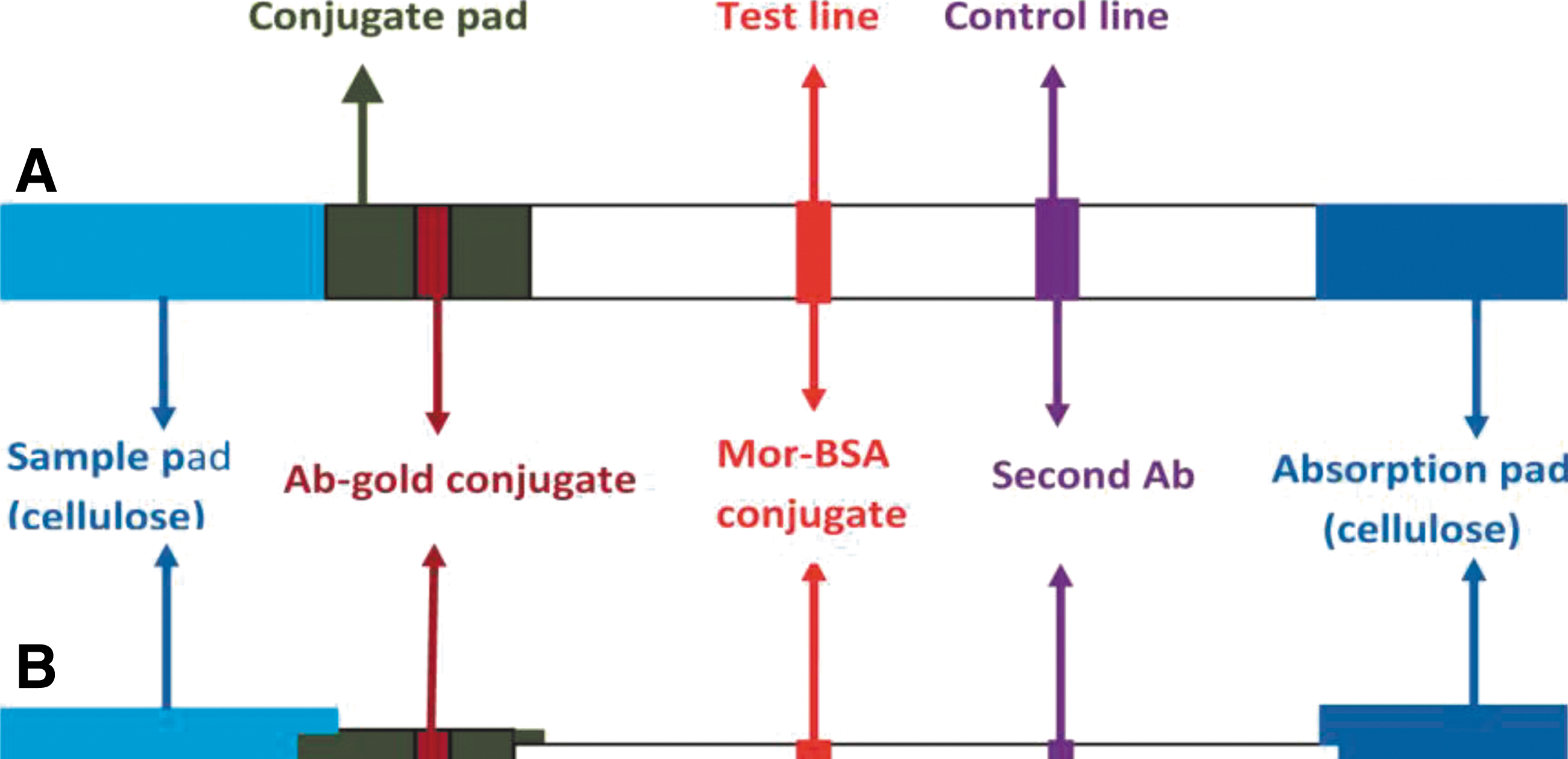
Schematic of a test strip showing its components. (
Sample collection
Forty-three morphine-positive urine samples, which were simultaneously assayed by commercial morphine test strips and thin layer chromatography (TLC), were collected from two clinical laboratories, and 50 morphine-negative urine samples, which were assayed by commercial strips, were collected. Furthermore, 103 additional urine samples, which tested positive with commercial morphine test strips with a cut-off value of 300 ng/mL but negative by TLC, were collected.
Results
Characterization of colloidal gold particles
Chloroauric acid was reduced to gold atoms by sodium citrate, and the gold atoms were nucleated into nanogold particles. Figure 2 illustrates the size of gold particles obtained by transmission electron microscopy measurement. The average diameter for the colloidal gold was approximately 20 nm.
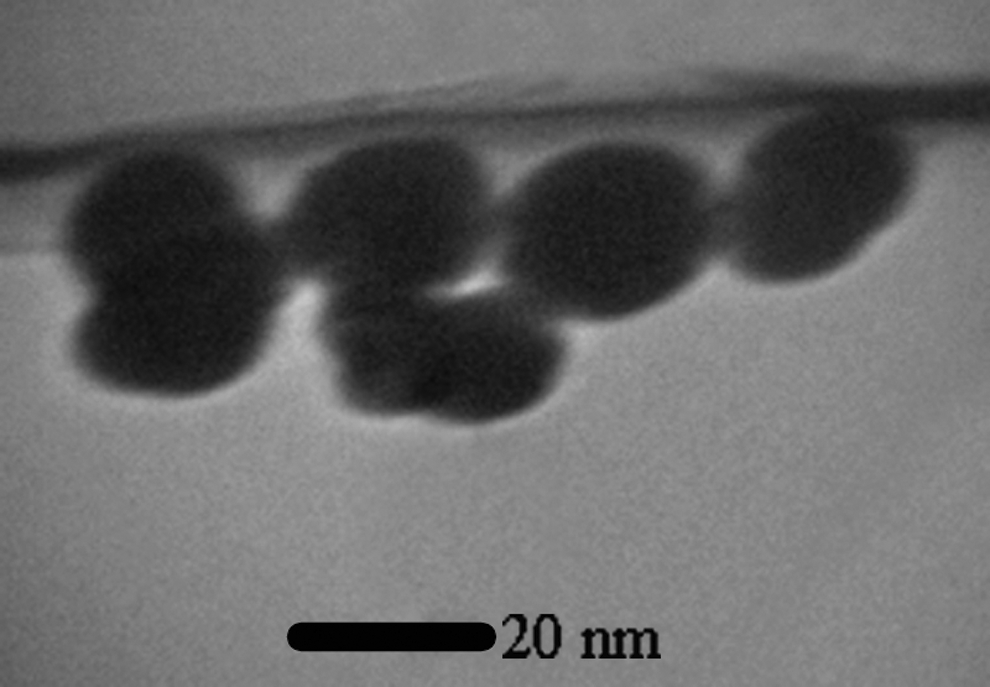
Transmission electron microscope image of colloidal gold. The average diameter for the colloidal gold was approximately 20 nm.
Optical density observed during scanning spectrophotometry performed between 380 and 700 nm showed there was only one maximum absorption wavelength at 521 nm (Fig. 3).

Spectra of the colloidal gold solution. Optical density observed during scanning spectrophotometry performed between 380 and 700 nm showed there was only one maximum absorption wavelength at 521 nm.
Optimal antibody–gold concentration
In this study, 3.2 μg/mL of morphine monoclonal antibody was determined as the minimal concentration for the stabilization of the colloidal gold.
Evaluation of detection limit
Using different concentrations of morphine–BSA on the test line, the detection limit of the test strips was adjusted to 2000 ng/mL.
Accuracy
A comparison was conducted using the assembled test strips and a commercial morphine rapid test. Presumptive positive results were confirmed by TLC. The results are shown in Table 1.
Forty-six out of 103 urine samples, which tested positive with commercial strip tests but negative with TLC, were negative with the assembled test strips in this study.
Analytical sensitivity
Morphine-free urine was spiked with morphine at the following concentrations: 0, 500, 1000, 1500, 2000, and 3000 ng/mL. A detection limit of 2000 ng/mL was obtained. The data are summarized in Table 2.
No., number of assays.
Precision
A study was conducted over 5 days, using different lots of product to demonstrate within and between runs. An identical panel of specimens containing no morphine, ±25%, ±50%, and −75% of detection limit morphine was prepared for each day. The results are given in Table 3.
No., number of assays per day.
Cross reactivity
A study was conducted to determine the cross reactivity of the test with compounds, such as apomorphine (500 μg/mL), codeine (2 μg/mL), heroin (4000 μg/mL), naloxone (1000 μg/mL), naltrexone (1000 μg/mL), and papaverine (100 μg/mL). Only codeine had a cross-reaction (100%) at the cut-off concentration of 2000 ng/mL.
Effect of urine-specific gravity
Ten urine specimens of normal, high, and low specific gravity ranges were spiked with 1000 and 3000 ng/mL of morphine. The test strips were tested and the results demonstrate that varying ranges of urine-specific gravity do not affect the test results.
Effect of urinary pH
Ten urine specimens with pH of 5, 6, 7, 8, and 9 were spiked with 1000 and 3000 ng/mL of morphine. The test strips were checked and the results demonstrate that varying ranges of pH do not interfere with the performance of the test.
Discussion
The ICG assay or simple form strip assay has been used for some time. This technique is based on an ICG method that uses an antigen-antibody in a manner that provides rapid detection of the analyte. Colloidal nanogold particles have been successfully applied for the development of a one-step strip test. In the present study, we constructed an ICG lateral flow test strip for the screening of morphine in urine using MAb and a colloidal gold label. This one-step assay is based on the principle of a competitive immunochemical reaction between the morphine in urine samples and morphine-BSA immobilized on the membrane for the limited antibody site.
Immunoglobulin is directly absorbed on the colloidal gold surface, and this process can be accomplished by non-covalent reactions, including London–van der Waals force and hydrophobic interactions.(21) The intensity of the color produced is related to the size of colloidal gold particles. In this experiment, gold particles with a diameter of approximately 20 nm were chosen to construct test strips. Particles smaller than 15 nm were found to be too small to produce a strong color, while gold particles larger than 60–70 nm self-aggregate after the particles have been stored at 4°C for several days.(22) In this study, an ICG test strip was developed with Amor-HK 16 monoclonal antibody against morphine. The cut-off value was set at 2000 ng/mL. When a test strip is immersed in a urine sample, the flow first reaches the gold conjugate pad. The mixture then reaches the test line and if the concentration of morphine in the urine sample is ≥2000 ng/mL, complete inhibition of the interaction between the bound antigen and the labeled antibody occurs and a visible band is not observed. At concentrations <2000 ng/mL, the labeled antibody interacts with morphine bound to the solid phase, which leads to the formation of a visible band. The intensity of this band is also inversely dependent on the concentration of morphine in the sample. If the test band has any intensity of rose color in the control line, this indicates that the sample contains no morphine or that its content is lower than 2000 ng/mL (Fig. 4). If only the control band is observed in the test region of the ICG test strip, this indicates that the content of morphine in the sample is ≥2000 ng/ml. If the control band is not detected, the test is not valid and should be repeated. Because small doses of codeine and foods containing poppy seeds can give a positive opiate screening result, some laboratories do not use test strips with a 300 ng/mL cut-off value. Workplace drug-testing programs in the United States raised the opiate cut-off value to 2000 ng/mL in 1998. The study of Fraser and Worth(23) on urine opiate screening and a confirmation cut-off of 2000 ng/mL indicated that increasing the cut-off value from 300 to 2000 ng/mg led to >300% reduction in the confirmed-positive rate for codeine and morphine and a 47% reduction in codeine-only confirmations. Furthermore, the cut-off value of the Synchron Systems Opiate assay (a homogenous enzyme immunoassay method) is 2000 ng/mL. In our system, setting a cut-off value of 2000 ng/mL for the developed ICG, rather than 300 ng/mL, led to a 45% reduction in false positive results compared with commercial ICG kits available in local markets.
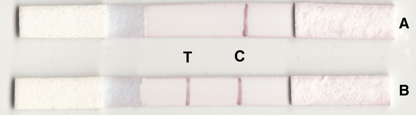
Possible results of the lateral flow test strip (competitive format). Positive (
High reproducibility, as well as high sensitivity and specificity of the rapid test, was evaluated and confirmed using various samples from addicted persons as well as normal controls (Table 1). The test strip showed no cross-reactivity with other compounds except codeine. To obtain uniform capillary flow, PEG and polyvinylpyrrolidone were tested in the treatment of a sample pad, from which PEG was selected. In addition, the use of PEG increased the intensity of color in the test band. Increasing the concentration of BSA in the treatment of the NC membrane as a blocking agent to prevent non-specific reactions can cause a decrease in color intensity.
Conclusion
In conclusion, a rapid and sensitive ICG test using Amor-HK16 with a detection limit of 2000 ng/mL to detect morphine in urine was developed. This test was not affected by specific gravity or the pH of urine. Adjusting the cut-off values for such screening tests to a concentration of 2000 ng/mL will lower the chance of false positives in the case of non-drug addicts, especially when no confirmatory tests are performed.
Footnotes
Acknowledgments
This study was financially supported by Tehran University of Medical Sciences.
Author Disclosure Statement
The authors have no financial interests to disclose.