
Abstract
Neutron scattering is a powerful technique that can be used to probe the structures and dynamics of complex systems. It can provide a fundamental understanding of the processes involved in the production of biofuels from lignocellulosic biomass. A variety of neutron scattering technologies are available to elucidate both the organization and deconstruction of this complex composite material and the associations and morphology of the component polymers and the enzymes acting on them, across multiple length scales ranging from Angstroms to micrometers and time scales from microseconds to picoseconds. Unlike most other experimental techniques, neutron scattering is uniquely sensitive to hydrogen (and its isotope deuterium), an atom abundantly present throughout biomass and a key effector in many biological, chemical, and industrial processes for producing biofuels. Sensitivity to hydrogen, the ability to replace hydrogen with deuterium to alter scattering levels, the fact that neutrons cause little or no direct radiation damage, and the ability of neutrons to exchange thermal energies with materials, provide neutron scattering technologies with unique capabilities for bioenergy research. Further, neutrons are highly penetrating, making it possible to employ sample environments that are not suitable for other techniques. The true power of neutron scattering is realized when it is combined with computer simulation and modeling and contrast variation techniques enabled through selective deuterium labeling.
Introduction
The characterization of biomass and how it evolves in response to the various processes applied to it in the production of biofuels has proven to be challenging because of the large range of length and time scales involved. Moreover, the interdispersed ordered and disordered regions present in biomass—vital to its biological function—further complicate its study. Many of the approaches used to study biomass processing, such as chemical pretreatment, are carried out by analyzing samples at sequential time points during processing. The penetrating nature of neutron scattering makes neutron scattering techniques ideally suited for in situ studies of how biomass changes over time. The ability to probe changes in biomass structure in real time during pretreatment has the potential to provide information about the system that is not accessible using other approaches.
This review discusses neutron scattering technologies being developed to provide the fundamental information needed to drive rational design of biomass, enzymes, and pretreatment approaches for cost-efficient production of biofuels. Unlike most other experimental techniques, neutron scattering is uniquely sensitive to hydrogen (and its isotope deuterium), an atom abundantly present throughout biomass, and a key effector in many biological, chemical, and industrial processes for the production of biofuels. Sensitivity to hydrogen, the ability to replace hydrogen with deuterium to alter scattering levels (called deuteration), the fact that neutrons cause little or no direct radiation damage, and the ability of neutrons to exchange thermal energies with materials provide neutron scattering technologies with unique capabilities for bioenergy research. Further, neutrons are highly penetrating, making it possible to employ sample environments that are not suitable for other techniques. Examples of current bioenergy research that involve the use of a variety of neutron techniques, including small-angle scattering, fiber diffraction, crystallography, reflectometry, and inelastic scattering, are given below. Suitable instruments available at international user facilities and the capabilities of representative instruments at the Spallation Neutron Source (SNS) and High Flux Isotope Reactor (HFIR) of Oak Ridge National Laboratory are also presented.
Small-Angle Neutron Scattering
Small-angle neutron scattering (SANS) is an ideal technique for examining complex structures in the nanometer to micron range. The technique is broadly applicable to virtually any kind of sample, such as intact plants, ground biomass, or individual proteins in dilute solution. Here, the ability to substitute deuterium for hydrogen makes it possible to study components such as lignin, hemicellulose, cellulose, and cellulases within intact structures through the application of “contrast variation” in which the use of mixtures of hydrogenated and deuterated solvents differentially highlights various components within the intact structures, particularly if one of the components has been enriched in deuterium. The length scales probed by SANS are relevant to both biological function and industrial treatments of the material. The information that can be extracted from SANS data depends on the sample. In the case of proteins in solution, the size and shape of the particle can be determined. For more complex systems, such as the highly interacting biopolymer networks that make up biomass, statistical parameters that provide insight into the bulk characteristics of the network can be extracted from the data. By leveraging the highly penetrating and non-destructive properties of neutrons, sample environments suitable for harsh processing conditions can be safely employed for in situ characterization of biomass to afford new insight into biomass processing that is uniquely accessible to SANS.
Several recent examples from the literature demonstrate the use of SANS for enabling conceptual advances in our understanding of biomass pretreatment. The structure of a cellulase, Cel7A from Trichoderma reesei, was probed in solution as a function of pH. 1 The study showed that the enzyme undergoes a structural transition at its pH of maximal catalytic activity, suggesting that it could be engineered to function more effectively in other environments. By using SANS, radiation damage to the samples during the measurement was avoided. Deuterium labeling has the potential to enable visualization of enzymes on biomass, which would afford a unique view of the enzymatic digestion of cellulose.
Biomass morphology and the changes it undergoes in response to pretreatment have also been studied. A comparison of highly purified crystalline cellulose and crystalline cellulose extracted from switchgrass showed clear structural differences at length scales corresponding to the cellulose fibril diameter. 2 The impact of ionic liquids on the morphology of biomass at mesoscopic length scales has also been studied using SANS. 3,4 Important insight into the effect of dilute acid pretreatment, an industrially relevant process for biofuel production, was gained through a combination of SANS, wide-angle x-ray diffraction, and nuclear magnetic resonance (NMR). 5 In addition to showing an increase in the crystalline cross-section of the cellulose microfibril, the growth of lignin aggregates within the network of cellulose microfibrils was clearly observable. These exciting results run counter to the prevailing wisdom regarding the effect of pretreatment on cellulose structure and suggest that the degree of cellulose crystallinity itself is a poor indicator for enzymatic digestibility, but rather removal of hemicellulose and redistribution of lignin may be the determining factors for increasing biomass conversion. The behavior of lignin in aqueous solution was studied by SANS and molecular dynamics simulations that show a highly folded and scale-invariant surface at length scales up to 100 nm. 6 Interestingly, the work also showed significant penetration of water into the hydrophobic core of lignin. The ability of neutrons to penetrate into robust sample environments raises the possibility for studying chemical pretreatment in situ, thereby providing the most complete picture of the morphological transitions taking place in biomass at nanometer length scales during pretreatment. No other experimental technique can provide such information.
There are many suitable SANS instruments available to the scientific community for performing these kinds of studies. The National Institute of Standards and Technology has two instruments (NG3 and NG7), as does the Institute Laue Langevin (D11 and D22). 7 –9 Los Alamos Neutron Scattering Center has one SANS instrument (LQD). 10 The ISIS facility is home to an established instrument and is bringing a second one on-line. 11 Oak Ridge National Laboratory (ORNL) has three SANS instruments that have recently become available: the GP-SANS and Bio-SANS instruments at the High Flux Isotope Reactor, and the EQ-SANS instrument at the Spallation Neutron Source. 12 –14 The ORNL instruments have complementary technical capabilities, as summarized in Table 1. SANS is among the most heavily used neutron techniques, and most neutron scattering facilities across the world include SANS in their suite of instruments. 15
Instruments for Small-Angle Neutron Scattering (SANS) and Neutron Reflectometry
Neutron Fiber Diffraction
While SANS provides information for length scales greater than a nanometer, higher resolution is needed to understand biomass and its conversion at an atomic level. The plant cell wall is a fibrous material. Embedded in the hydrated matrix gel of lignin and hemicellulose in the secondary cell wall are aligned bundles of crystalline elemental cellulose fibrils that provide mechanical strength. Materials such as biomass, with naturally occurring order and fiber alignment, are ideally suited to structural studies using high angle (high resolution) fiber diffraction techniques. X-ray fiber diffraction has been widely used to study the structure of biomass and its crystalline fibrous cellulose component at a molecular level for about 100 years. However, hydrogen atoms are virtually invisible to X-rays, even in highly crystalline samples that diffract to beyond atomic resolution (1 Å). This is a severe limitation because hydrogen bonding is thought to be important in determining the structure and properties of cellulose. By using neutron fiber diffraction, hydrogen atoms can be located providing crucial additional information on the hydrogen bonding and hydration properties of cellulose that are related to its recalcitrance. 16
One example of how neutron fiber diffraction has contributed to bioenergy research is in guiding the alteration of conventional ammonia fiber expansion (AFEX) pretreatment strategies. 17 Conventional AFEX under hydrous conditions mechanically disrupts biomass with a significant redistribution of lignin and hemicelluloses, but with no major decrystallization of cellulose or change in crystal structure. 18 Cellulose occurs naturally in two allomorphs—cellulose Iα and Iβ, collectively referred to as cellulose I—that have been characterized by X-ray and neutron fiber diffraction. 19,20 Neutron fiber diffraction on samples of cellulose isolated from biomass has helped to reveal that, under controlled anhydrous conditions, ammonia can penetrate into the cellulose fibers to form crystalline complexes. 21 Upon removal of ammonia, the cellulose is found in another crystal form called cellulose IIII, which has greater susceptibility to enzymatic hydrolysis. 22 These results on model systems have been used to alter AFEX pretreatment to improve overall hydrolysis yield in preliminary studies with corn-stover biomass. The important contribution from neutrons in this work has been in providing detailed structures and information on the location of hydrogen atoms and their rearrangement during the transition from cellulose I to cellulose IIII. When combined with information from molecular dynamics simulations and quantum mechanics calculations, neutron fiber diffraction has provided an explanation for the greater susceptibility of cellulose IIII to enzyme hydrolysis compared to cellulose I.
No instrument has been built specifically for performing neutron fiber diffraction studies, but most that are available to the scientific community for neutron macromolecular crystallography are suitable. Those include D19 and LADI at the Institute Laue Langevin, BIX-3 and BIX-4 at the reactor run by the Japanese Atomic Energy Agency, iBIX at J-PARC, BioDIFF at the Jülich Centre for Neutron Science, and the PCS at Los Alamos Neutron Science Center. 23 –27 Several other instruments are being built or planned at neutron scattering facilities across the world. Oak Ridge National Laboratory is building three instruments that will soon be available for neutron macromolecular crystallography: MaNDi and TOPAZ at the Spallation Neutron Source and IMAGINE at the High Flux Isotope Reactor. 28 These instruments have complementary technical capabilities (Table 2).
Instruments for Neutron Macromolecular Crystallography and Fiber Diffraction
Neutron Macromolecular Crystallography
Crystallography is a leading technique for the atomic-resolution analysis of macromolecules such as proteins. Through the use of neutrons, it is possible to locate hydrogen atoms within biological macromolecules. This provides special insights that are difficult, if not impossible, to obtain via other techniques such as X-ray crystallography and NMR. 29 Enzymes represent a significant proportion of the cost of producing lignocellulosic biofuels. Although the application of neutron macromolecular crystallography to biofuels enzymes is at an early stage, it has the potential to provide unique information on enzymatic processes that could be used to produce improved enzymes for more efficient production of biofuels.
In one of the first applications of neutron macromolecular crystallography in bioenergy research, the catalytic mechanism of the enzyme D-xylose isomerase was studied. A major problem for the efficient production of biofuels is the presence of xylose in biomass hydrolyzates. Xylose is a pentose sugar that cannot be fermented by the industrial yeast Saccharomyces cerevisiae. One approach to engineer S. cerevisiae with a metabolic pathway to produce xylulose, the fermentable keto isomer of xylose, is isomerization of xylose to xylulose by expression of xylA, the gene found in some anaerobic fungi and bacteria that encodes D-xylose isomerase. Neutron crystallography was first applied to this enzyme in order to provide new insights into its catalytic mechanism, in particular the location and movement of hydrogen atoms at different stages between enzyme, substrate, and solvent. 30 More recently it has been used to understand why activity is so sensitive to the pH value. 31 Activity is highest at pH 8, but under the often acidic (pH<6) conditions of biomass conversion, as well as in the cytosol of S. cerevisiae, the metal cofactors required for isomerization are expelled from the active site. Neutron crystallography revealed that although a hydronium ion templates the active site for metal binding at pH 8, at low pH the ion is hydrated to a proton with an accompanying collapse of active side residues, due to their interaction with this proton. These observations provide an explanation for why the required cofactor metal cations are not bound and, therefore, why there is a dramatic decrease in the activity of the enzyme at low pH values. More importantly, they provide insights that will allow rational design to improve catalytic performance and binding specificity, and which are generalizable to a broad range of biofuels enzymes with different thermal and pH stabilities.
Several more powerful instruments are becoming available for neutron macromolecular crystallography, as described above. Although it is very much a newly blossoming field of structural biology, it is likely to remain a flux-limited technique best exploited by deuterating proteins to increase their neutron scattering power. 32 It is also most productively used when combined with X-ray crystallography because of the complementary nature of the information supplied by the two techniques. Sophisticated computational tools are now available that allow joint refinement of protein structures using both X-ray and neutron data to obtain more accurate and complete (including hydrogen) structures. 33
Neutron Reflectometry
Neutron reflectometry is a non-destructive technique for investigating the structure of thin-films, interfaces, and membranes. In its first application to biofuels research, it has been used in combination with quartz crystal microbalance dissipation to provide information about the interactions of cellulases with cellulose films. 34 Neutron reflectometry can provide a 1-dimensional scattering density profile perpendicular to a film. In this application, it was combined with H2O/D2O contrast variation to provide the profile of water, and therefore swelling in the cellulose film, and also to reveal increases in surface roughness during cellulase digestion. The technique has other potential applications in bioenergy research. In particular, it could be used for studying changes in cell membranes associated with increased ethanol tolerance by microorganisms, and sugar transport from biomass hydrolyzates across those membranes during fermentation. There are numerous instruments available for neutron reflectometry, as recently reviewed by Teixeira et al. 35 The Liquids Reflectometer at the Spallation Neutron Source at Oak Ridge National Laboratory, one of the first instruments commissioned at the facility, has a high flux that enables time-resolved studies of specific structural features that have not been accessible in the past.
Inelastic Neutron Scattering
The dynamics of atomic groups or molecules that make up biological materials contain key information indispensable for understanding response to changing processing conditions. Unlike other techniques for probing structural dynamics, neutron scattering is sensitive not only to the temporal, but also to the spatial characteristics of dynamical processes through the dependence of the scattering signal on the neutron momentum transfer. The large incoherent neutron scattering cross section of hydrogen, compared with that of other elements, allows researchers to use partial sample deuteration to probe the dynamics of selected components in complex systems such as biomass. Neutron inelastic scattering can be used to characterize such phenomena as the glass transition in lignin, which is thought to be of fundamental importance to many pretreatment processes. At longer time scales, collective dynamics involved in diffusive motion of solvent molecules, membrane fluctuations, and even domain motions in active enzymes can be studied. Although neutron spectrometers are widely available at neutron facilities across the world, their application to bioenergy research is at a very early stage. 3 –5 Oak Ridge National Laboratory has three neutron spectrometers at the Spallation Neutron Source, which together cover an unprecedented dynamical range of six decades, from picoseconds to microseconds (Table 3).
Instruments for Inelastic Neutron Scattering
Computational Modeling
Computer simulation and modeling are vital for interpreting multi-scale neutron scattering data to build an atomistic and mesoscale understanding of the structure, dynamics, and degradation pathways of lignocellulosic materials. With experiment, neutron scattering can be used to test simulation models by direct comparison. Simulation provides complementary physical models of lignocellulosic biomass systems and enzymes that rationalize physical properties and enable interpretation of the neutron scattering profiles. Computer simulation has been combined with neutron scattering to address critical questions concerning biomass structure and its interaction with enzymes.
The contribution of lignin to biomass recalcitrance is closely associated with temperature-dependent changes in the polymer structure on heating and cooling. The temperature dependence of the structure and dynamics of individual softwood lignin polymers was examined using extensive molecular dynamics simulations. 36 Lignin was found to transition from glassy, compact states to mobile, extended states with increasing temperature at T>150°C, i.e., above typical pretreatment temperatures. The molecular dynamics simulations demonstrated that the low-temperature collapse of lignin is thermodynamically driven by an increase in translational entropy and density fluctuations of those water molecules removed from the hydration shell. Thus, the results distinguish lignin's collapse from enthalpy-driven coil-globule transitions observed for other polymers. 37
In other work, molecular dynamics simulation was combined with SANS to characterize the structure of aggregates of softwood lignin on multiple length scales. 36 SANS experiments were consistent with the aggregates possessing a surface fractal dimension, dexp=2.62±0.02, which corresponds to a highly folded surface. Complementary molecular dynamics simulations were performed on representative atomistic models of softwood lignin aggregates. A value of 2.65±0.01 for the surface fractal dimension was obtained from the simulations using the formal definition of a surface fractal, which is in excellent agreement with the experimental value obtained from the SANS data. The size distribution of the surface pores of the aggregates indicates that many pores are large enough to be able to accommodate the catalytic domains (radius ∼20 Å) of cellulolytic enzymes. This is consistent with evidence that cellulase binding directly to lignin contributes to the inhibitory effect of lignin on cellulose hydrolysis. 38 Extensive water penetration and heterogeneous lignin chain dynamics were also revealed by the simulations. The complementarity and synergy of SANS and molecular dynamics simulations were illustrated by the fact that, together, they demonstrated that surface fractal dimension is constant over three orders of magnitude in length, i.e., ∼1 Å (molecular dynamics), ∼10 Å (molecular dynamics and SANS), and 100 Å (SANS).
Molecular dynamics simulations and quantum mechanics calculations have also been combined with neutron fiber diffraction studies to obtain a more complete understanding of the structure and properties of cellulose in biomass. In cellulose I, the parallel cellulose chains are arranged in sheets that are held together by strong hydrogen bonds between the hydroxyl groups of neighboring chains. These sheets stack through more frequent but weaker interactions, such as van der Waals forces. Quantum mechanics calculations revealed that these stacking interactions are highly cooperative and contribute just as much to the stability of cellulose Iβ as hydrogen bonding. 39 Neutron fiber diffraction has shown that the hydrogen bonding arrangement in naturally occurring cellulose is disordered, which is important because the nature of hydrogen bonding in cellulose has long been thought to play a key role in determining its properties, in particular its recalcitrance to hydrolysis. 19,20 A statistical mechanical approach was developed to investigate this disorder, and it was found that the hydrogen bonding has an adaptive nature that serves to stabilize the cellulose fibers and make them more resistant to degradation. 40 A combination of neutron fiber diffraction, quantum mechanics calculations, and molecular dynamics simulations revealed that most chains (70–80%) in cellulose have the same hydrogen bonding arrangement, but that disorder exists at defects in the fibers and also at their surfaces. 41 Further molecular dynamics simulations in the presence of water revealed that the central core of the fiber maintained an average structure that is essentially the same as the crystallographic structure, but that chains at the surfaces have significantly different structures and hydrogen bonding arrangements and are more disordered. 17 The surface chains interact strongly with water, forming a dense layer near the surfaces that will affect the way hydrolyzing enzymes interact with cellulose fibers.
Simulating complex biomolecular systems is computationally intensive, particularly for materials such as lignocellulose in which characteristic length and time scales extend over several orders of magnitude. The computer power available at the National Center for Computational Science at Oak Ridge National Laboratory is therefore a valuable resource for simulating phenomena from the Angstrom and femtosecond levels of atomic detail to microsecond and micrometer mesoscale phenomena, enabling detailed simulation of complex heterogeneous lignocellulosic biomass systems. However, in addition to computer power, development of coarse-grained mesoscale and experimental data-driven techniques will be important for allowing even larger ensembles of heterogeneous biomass systems to be simulated.
Deuteration
Deuteration of biomolecules is valuable for structural studies of biological systems using neutron scattering techniques because neutrons scatter through their interaction with atomic nuclei and are sensitive to isotopic substitutions. This property of neutrons is exploited by the contrast variation technique, which makes it possible to alter the scattering from an individual component in a multicomponent system by varying the amount of hydrogen and deuterium in the solvent. For example, the scattering contribution from a membrane protein in a detergent solution can be selectively observed in 18% D2O in a H2O solution that attenuates the scattering contribution of the detergent. 42 Figure 1 shows how the calculated scattering length density of lignin and cellulose with various levels of deuterium incorporation vary with the ratio of H2O/D2O ratio in the solvent. There is very little contrast between hydrogenated cellulose and lignin, both of which have a contrast match point of ∼40% D2O in H2O buffer. However, as the level of deuterium incorporation is increased in the biomolecules there is a corresponding increase in the separation of the contrast match points of cellulose and lignin, assuming uniform deuterium incorporation into these molecules. The use of deuterated proteins also improves the sensitivity of crystallography experiments and makes it possible to work with smaller crystals.
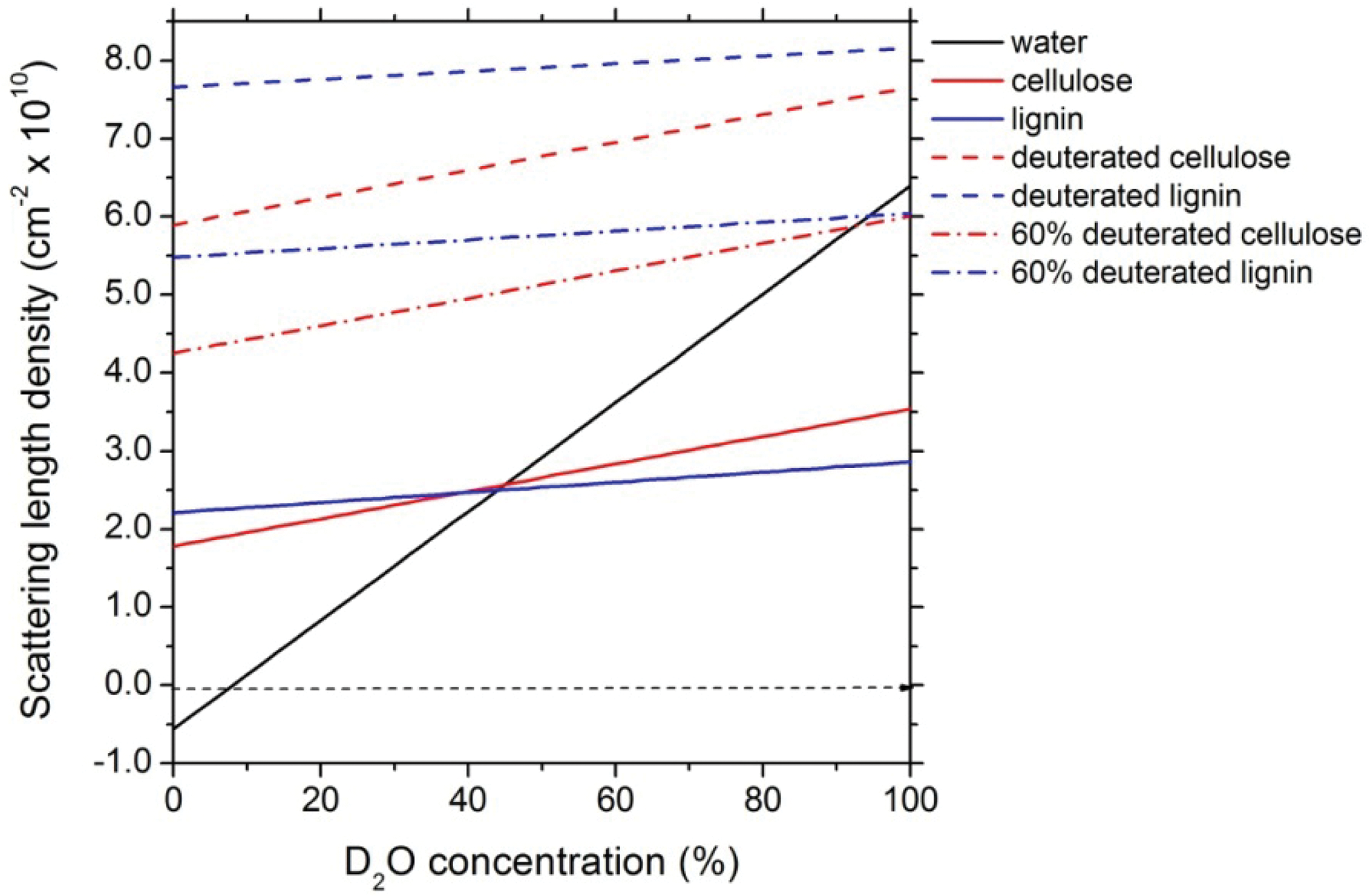
Variation in the scattering length density of water and lignocellulose components in different ratios of H2O/D2O solvent mixtures
The types of deuterated biomolecules available for structural studies are constrained by the requirement to cultivate the source organisms in D2O. Although methods for production of highly deuterated biomolecules in bacteria, particularly Escherichia coli, and algae have been described and utilized for decades, deuteration of plant biomass to the levels desirable for neutron studies is hampered by the deleterious effects of D2O on germination, root formation, and growth. 43 Many species of unicellular algae can tolerate up to 98% D2O, but higher plants generally exhibit growth inhibition at levels of D2O above 50%. Several species of higher plants have been grown to obtain deuterium incorporation of 30%. 44 Winter rye was reported to have exceptional D2O tolerance compared to other plants. 45
The amount and location of deuterium in deuterated biomolecules are typically determined by Fourier transform infrared spectroscopy (FTIR), mass spectroscopy, and NMR. 46 –48 The complexity of the signals obtained from whole biomass samples complicate quantification by these methods. Isolation of cellulose followed by acetylation or nitration and subsequent NMR analysis only provides information on non-exchangeable hydrogen in the cellulose component. 49 –51 By measurement of the loss of proton signals compared to non-deuterated controls, 1 H-NMR has been used to estimate deuterium incorporation in cellular extracts. Recently, two complementary NMR methods, 1 H 2 H solid state and ionic liquid dissolution, were used to analyze deuterium incorporation in kale plants grown hydroponically to 31% deuterium incorporation. The solid-phase technique provided a rapid estimate of total deuterium content in dried kale, while liquid-phase analysis of the dissolved kale provided information on incorporation sites, with deuterium preferentially incorporated in carbohydrates compared to lignin. 52 The differential incorporation of deuterium into the carbohydrate and lignin components in plants suggests that it will be possible in future studies to separate the scattering contributions of these components in plants grown in partially deuterated media to a greater extent than is predicted by the calculated scattering length densities shown in Figure 1, allowing the structure of individual components in intact biomass samples to be studied.
Several laboratories have been established to develop deuteration methods and to provide a deuteration service in support of neutron scattering facilities. The longest established include the D-Lab at the Partnership for Structural Biology in Grenoble, the Biological Deuteration Laboratory at Los Alamos Neutron Scattering Center, and the Biological Deuteration Laboratory operated by the Center for Structural Molecular Biology at Oak Ridge National Laboratory.
Conclusions
Neutron scattering encompasses a suite of powerful materials characterization tools that can be used to probe the structures and dynamics of complex systems. It can provide a fundamental understanding of the processes involved in the production of biofuels from lignocellulosic biomass. The true power of neutron scattering is realized when it is combined with computer simulation and modeling and contrast variation techniques enabled through selective deuterium labeling. In contrast variation, it is possible to study isotopically labeled components of a large, complex system within the intact superstructure by varying the scattering length density of the background through the use of hydrogenated and deuterated solvents. The creation of deuterated biomaterials, either the substrate or the enzymes, can also allow masking to provide truly unique insight into the deconstruction processes by making it possible to visualize individual components within a complex system. Further, neutrons are highly penetrating, making it possible to use novel sample environments for in situ materials processing that cannot be used in other experimental methods for structural determination. Data interpretation through advanced computational modeling methods makes it possible to visualize structure and dynamics across a broad range of length and time scales.
Footnotes
Acknowledgments
This work was funded by the Genomic Science Program of the Office of Biological and Environmental Research, US Department of Energy, under FWP ERKP752. This research at Oak Ridge National Laboratory's High Flux Isotope Reactor and Spallation Neutron Source was sponsored by the Scientific User Facilities Division, Office of Basic Energy Sciences, US Department of Energy.
Author Disclosure Statement
No competing financial interests exist.