
Abstract
We present a new format of apta-sensing composite particles for Surface-Enhanced Raman Spectroscopy (SERS) detection of malathion. These apta-sensing microspheres acquire their extraction capability through aptamer-target analyte interaction and Raman signal enhancement for SERS detection of the pesticide. Micron-sized polymer particles were synthesized by precipitation polymerization using methacrylic acid and ethylene glycol dimethacrylate as co-monomers in acetonitrile. Conjugation with colloidal gold nanoparticles (AuNPs) via modification with 2-aminoethanethiol led to polymer-AuNP composites with controlled aggregation of AuNPs onto the polymer surface. The thiolated aptamer targeting malathion was attached to the metal surface by thiol-gold interaction, resulting in polymer-AuNP-aptamer composite microspheres. The new material was characterized using two techniques: scanning electron microscopy imaging and dynamic light scattering measurements. The polymer-AuNP-aptamer particles were incubated with a phosphate buffer solution containing malathion at a suitable concentration level for 30 min, washed with water, and then dried on a microscope glass slide prior to spectra acquisition. The proposed apta-sensing SERS substrate successfully allows the direct detection of the target molecule at 3.3 μg mL−1. Only basic equipment is required for analyte separation, making the apta-sensing microspheres well-suited for potential industrial applications that require on-site analyte detection.
Introduction
In the 1970s, it was first reported that the Raman signal intensity of molecules could be dramatically increased by chemisorption onto a roughened noble metal surface. 1,2 Since then, the researchers' interest in Surface-Enhanced Raman Spectroscopy (SERS) and SERS-based technologies has grown. In recent years, SERS has been used to detect small molecules, peptides, proteins, and DNA, resulting in an increasing number of publications. Some features of SERS, such as its high sensitivity, utility for fingerprinting and multiplexing, minimal background signal from water, and the elimination of long and expensive procedures, make it a particularly versatile and effective technique for bioanalytical and homeland security applications. 3 –5 Moreover, the electromagnetic enhancement mechanism of SERS can create localized surface plasmons from the matching of the resonance frequency of the valance electrons of a noble metal with the frequency of the incident light, which can further enhance the Raman signal of the target. 6
Malathion, the most common organophosphate insecticide used in the United States, is applied primarily in agriculture and domestic settings for eradication and control of pests. 7 Its structure is shown in Fig. 1. Presently, there are no definitive data indicating chronic toxicity associated with malathion exposure and, due to its low toxicity in humans, it is considered safe to use. 7,8 However, because of its widespread use, it is necessary to develop detection methods to monitor the presence of malathion in different environmental compartments (e.g., surface waters, soil, etc.). Analytical methods for measuring malathion levels, along with those of other organophosphorous pesticides, have recently been developed using gas chromatography-mass spectrometry, gas chromatography-nitrogen phosphorus detectors, or high performance liquid chromatography (HPLC)-diode array detectors, among others. 9 –16 These methods are capable of measuring the target analyte within satisfactory limits of detection; however, they often involve time- and labor-intensive sample preparation and separation procedures prior to the final detection. These can take up to 80% of the total time spent on analysis. 17,18
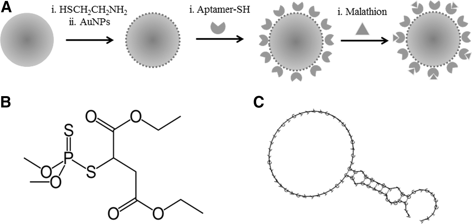
Illustration demonstrating preparation of polymer-AuNP-aptamer substrates for SERS detection of malathion
SERS-based sensors can be classified into two categories: intrinsic and extrinsic sensors. With intrinsic (or direct) sensing, the molecular fingerprint of the target analyte is acquired directly. This method has been applied to a variety of small biological molecules and used in microfluidic devices for detection of dyes. 3,19 Extrinsic sensors employ SERS-active nanotags that consist of distinguishable SERS-active reporters or molecules that have unique spectral fingerprints for indirect measurements of the analyte of interest. Extrinsic SERS sensors for small molecule detection are limited by the difficulty in obtaining reliable SERS spectra of complex structures such as DNA or proteins in comparison to direct measurement of the small molecule itself. However, some systems of this kind have been developed for small molecule detection, including colloidal gold nanoparticles (AuNPs) functionalized with a 16-amino acid antibody segment coupled to the Raman reporter molecule 5,5′-dithiobis(succinimidyl-2-nitrobenzoate) to detect protective antigens for anthrax screening or displacement of rhodamine 6G by glutathione on nanoparticles for reverse detection in aqueous solution. 20,21
To provide the molecular affinity of SERS sensors and improve their reliability, the use of cyclodextrin inclusion complexes (CICs) or molecularly imprinted polymers for direct detection of analytes has been reported. 22,23 An alternative strategy to chemical modifications for molecular recognition is to use DNA aptamers, which can bind to selected targets with a high level of affinity. Aptamers present unique advantages over chemical modifications, including well-established screening protocols and versatility in modifications for labeling and surface chemistry. Use of aptamers in SERS sensors has been reported for extrinsic detection of cocaine by analyzing the conformational changes to the aptamers upon binding of the target analyte. 24,25 For this purpose, it is necessary to obtain a reliable and highly reproducible DNA signal, which can be a limitation of using such methodologies. In this study we exploit the specificity of DNA aptamer recognition, combined with the simplicity of intrinsic analyte detection, and demonstrate a method for direct detection of malathion using DNA apta-SERS substrates.
Materials and Methods
Materials
Ethylene glycol dimethacrylate (EGDMA), methacrylic acid (MAA), 2-aminoethanethiol, N-(3-dimethylaminopropyl)-N′-ethylcarbodiimide hydrochloride (EDC), azobisisobutyronitrile (AIBN), tetrachloroauric (III) acid, sodium citrate, and 2-(N-morpholino) ethanesulfonic acid (MES) were obtained from Sigma-Aldrich (St. Louis, MO), and (3-aminopropyl)triethoxisilane (APTES) and analytical-grade malathion were purchased from Fluka (Buchs, Switzerland). EGDMA and MAA were freed of inhibitors prior to use via a disposable, pre-packed column (22.5×2.0 cm) obtained from Sigma Aldrich. AIBN was recrystallized from methanol prior to use. All organic solvents, also purchased from Sigma Aldrich, were at least HPLC-grade. All other chemicals were used as received. Ag-coated SERS Diagnosis Membranes (SER-DM™) were supplied by iFyber (Ithaca, NY).
Preparation of SERS Substrates
Synthesis of gold nanoparticles
AuNPs were prepared by reduction of chloroauric acid with sodium citrate, according to a methodology adapted from Frens. 26 In brief, 50 mL of a HAuCl4 (1 mM) aqueous solution was heated to boil, and a fixed amount of sodium citrate solution (1%) was then added. The reaction mixture was cooled after 20 min of boiling. The concentration of AuNPs in the resulting solution was determined by absorbance measurements (λ=520 nm) using a Nanodrop ND-1000 spectrophotometer (Thermo Scientific, Rockford, IL) and molar extinction coeffiecients for AuNPs derived from Yguerabide et al. 27
Preparation of glass-gold nanoparticle substrates
Glass bottom microwell dishes with an uncoated coverslip (P35G-14-C, MatTek Corporation, Ashland, MA) were cleaned with nitric acid, thoroughly rinsed with deionized (DI) water and dried under nitrogen flow. Then, 100 μL of aqueous 5 mM APTES was deposited on the glass surface and left there for 15 min. The substrates were again rinsed with DI water and dried under nitrogen. Subsequently, 100 μL of colloidal gold solution was deposited on the modified glass and allowed to dry. The glass-gold substrates were finally rinsed with DI water and dried prior to use.
Synthesis of polymer microspheres
Cross-linked polymer microspheres were prepared by precipitation polymerization. Purified MAA (0.46 mmoles, 39.4 μL), EGDMA (2.32 mmoles, 453.2 μL), and initiator AIBN (0.2 mmoles, 33 mg) were mixed with 20 mL of acetonitrile and placed in a glass vial fitted with a screw-cap, which served as a reaction vessel. The mixture was degassed by a gentle nitrogen stream for 10 min. The bottle was sealed and introduced into a temperature-controllable incubator equipped with a Spindrive orbital shaker platform (Bel-Art, Wayne, NJ) powered by a standard magnetic stirrer that allowed for slow agitation of the solution during the course of the polymerization. The temperature was maintained at 60°C for 20 h. The polymer microspheres that formed were separated from the reaction medium by vacuum filtration on a Magna nylon membrane filter with 0.45-μm pore size (Osmonics, Minnentoka, MN) and then sequentially washed with acetonitrile.
Preparation of polymer-AuNP composite microspheres
Polymer microspheres were functionalized, taking advantage of the presence of available carboxyl groups from the MAA. Polymer microspheres (0.2 g) were dissolved in 15 mL of MES buffer at pH 5.5 and mixed with 200 mg of EDC previously dissolved in 5 mL of water. The mixture was stirred for 30 min, and 75 mg of 2-aminoethanethiol (cysteamine) in 2 mL of water were added. The suspension was maintained by stirring for 3 h, after which the polymer-cys functionalized particles were filtered, washed with water, and resuspended in 5 mL of water. A 1-mL aliquot of this suspension was added to 10 mL of AuNP solution under stirring, leading to the formation of polymer-AuNP composite microspheres.
DNA aptamer development
The aptamer targeting malathion was developed using the modified Systematic Evolution of Ligands by EXponential enrichment (SELEX) method of Bruno et al. 28 Briefly, analytical-grade malathion was immobilized on a PharmaLink affinity column (Thermo Scientific) per instructions detailed by the manufacturer. The column was incubated with the SELEX DNA library, and the bound DNA was eluted and amplified by polymerase chain reaction (PCR). The PCR product was used for six subsequent rounds of SELEX. The aptamer designated as M17 Forward (M17-F), purchased from Integrated DNA Technologies, Inc. (Coralville, IA), had the following sequence: 5′-ThioMC6-ATCCGTCACACCTGCTCT-TATACACAATTGTTTTTCTCTTAACTTCTTGACTGC-TG GTGTTGGCTCCCGTAT-3′. 29 The aptamer structure is schematically depicted in Fig. 1C. The aptamer contained a thiol C-6 modifier on the 5′-end for conjugation to gold nanoparticles attached to the surface of the polymer microspheres. A stock solution was made by reconstituting the aptamer to 100 μM in nuclease free water. The stock was stored at −20°C for further use.
Preparation of polymer-AuNP-aptamer conjugates
Polymer-AuNP particles functionalized with DNA-aptamers on the surface were prepared according to the methodology developed by Taton et al.
30
The following equation was used to calculate the amount of aptamer to attach to the polymer-AuNP particle, where An is the surface area of the microsphere; Cn is the concentration of the original AuNP solution (particles L−1); Do is the aptamer density on the particle (35 pmol cm2); and V is the volume of the reaction (L):
A mole excess (1.5 times) of the amount of aptamer calculated with equation 1 (∼10 μL) was then added to a 500-μL suspension of polymer-AuNP particles and rotated on a rocking table at 1 Hz in the exclusion of light for 16 h. Next, 0.250 vol of 1M NaCl/0.1 M sodium phosphate buffer (buffer I) was added, and the particles were rotated for an additional 4 hours. The conjugated particles were centrifuged at 3,000 rpm for 4 min to remove excess aptamer and resuspended in 500 μL of 0.1 M NaCl/10 mM sodium phosphate buffer (buffer II). The particles were centrifuged and resuspended two more times and finally resuspended in 125 μL of buffer II.
Characterization Techniques
Dynamic light scattering
The sizes of the AuNPs, the polymer microspheres, and the polymer-AuNP-aptamer conjugates were analyzed using dynamic light scattering (DLS). Suspensions of the three different types of particles were prepared using water as dispersant and transferred to 400-μL disposable sizing cuvettes. The diffusion of the particles moving under Brownian motion was measured using a Zetasizer Nano-ZS (Malvern Instruments Ltd, Worcestershire, UK).
Scanning electron microscopy
Scanning electron microscopy (SEM) was used to characterize the polymer-AuNPs. The images were obtained using a LEO 1550 FESEM (Keck SEM). The dried particles were first coated with an electrically conductive gold-palladium coating approximately 10-nm thick and then placed on a silicon wafer for imaging. The resolution at 5 KeV was 2.5 nm.
SERS Measurements
SERS spectra were obtained using a Renishaw InVia Confocal Microscope system (Renishaw Inc; Hoffman Estates, IL) equipped with a 785-nm edge laser and Renishaw CCD Camera fitted to a Leica microscope. The spectra for polymer-AuNP-aptamer and glass-Au-aptamer substrates were taken with a 50× objective at an exposure time of 10 s and a laser power of 1%. The ∼1.0-μm laser spot was focused on a single particle on the surface. All spectra were obtained using 785 nm excitation. The spectra were analyzed using GRAMS AI spectroscopy suite (Thermo Scientific).
Glass-gold substrates
To the glass-gold substrates a 10 μL-aliquot of 1-μM thiolated aptamer solution in buffer I was added, and incubation was carried out overnight at 4°C. The substrates were rinsed with DI water and dried before SERS measurements. In the same manner, glass-gold substrates were incubated with a 1-mM (330 μg mL−1) solution of malathion in ethanol for 24 h.
SERS diagnosis membrane
The membranes were cut into small squares (0.5×0.5 cm size) and incubated with a 1mM (330 μg mL−1) solution of malathion in ethanol for 24 h. The membranes were then rinsed with DI water and placed onto a glass slide for SERS measurements.
Glass-gold-aptamer-malathion experiments
For the glass-gold-aptamer-malathion experiments, a 100-μL aliquot of 3-nM thiolated aptamer solution in buffer I was incubated on the glass-gold substrate at 4°C for 24 h. After this incubation period, a 100-μL aliquot of malathion in buffer II was added. After 15 min, the substrates were thoroughly rinsed with DI water, allowed to dry, and SERS spectra were obtained.
Polymer-AuNP-aptamer-malathion binding experiments
Aliquots (125 μL in buffer II) of the polymer-AuNP-aptamer particles were incubated with the same volume of a solution of buffer II containing malathion at a suitable concentration level for 30 min. The particles were then centrifuged, separated from the supernatant, and washed with an equal volume of DI water three times. Prior to spectra acquisition, a volume of 100 μL was allowed to dry on a microscope glass slide that was previously cleaned with nitric acid. The same procedure was followed for experiments with spiked tap water.
Results and Discussion
We propose a new composite material based on polymer-AuNP-aptamer sensing particles capable of acting as capture and signal-enhancer for SERS detection of malathion (Fig. 1A). Fabrication of multimeric SERS substrates is better suited than methods based on aggregation of gold nanoparticles through changes in the electrostatic charge, which can affect reproducibility. To obtain reference SERS spectra of the analyte malathion, we used Ag-coated SERS diagnosis membranes as a positive control substrate. For the same purpose, SERS substrates of AuNPs aggregated on the surface of APTES-coated glass slides were prepared to obtain a reference SERS signal of each component of our multimeric system.
Preparation and Characterization of Polymer-AuNP-Aptamer Substrates
Polymer-AuNP microspheres
When designing our composite SERS substrate, different parameters, such as the morphology of the polymer support as well as the type, size, and organization of the AuNPs, were taken into account. In this work, we have adapted the methodology proposed by Prasad et al., to form polymer-AuNP composites with controlled aggregation of AuNPs onto the polymer surface. 31 DLS analysis of the synthesized AuNPs indicated a size of ∼40 nm with a Gaussian distribution shown in Fig. 2C.
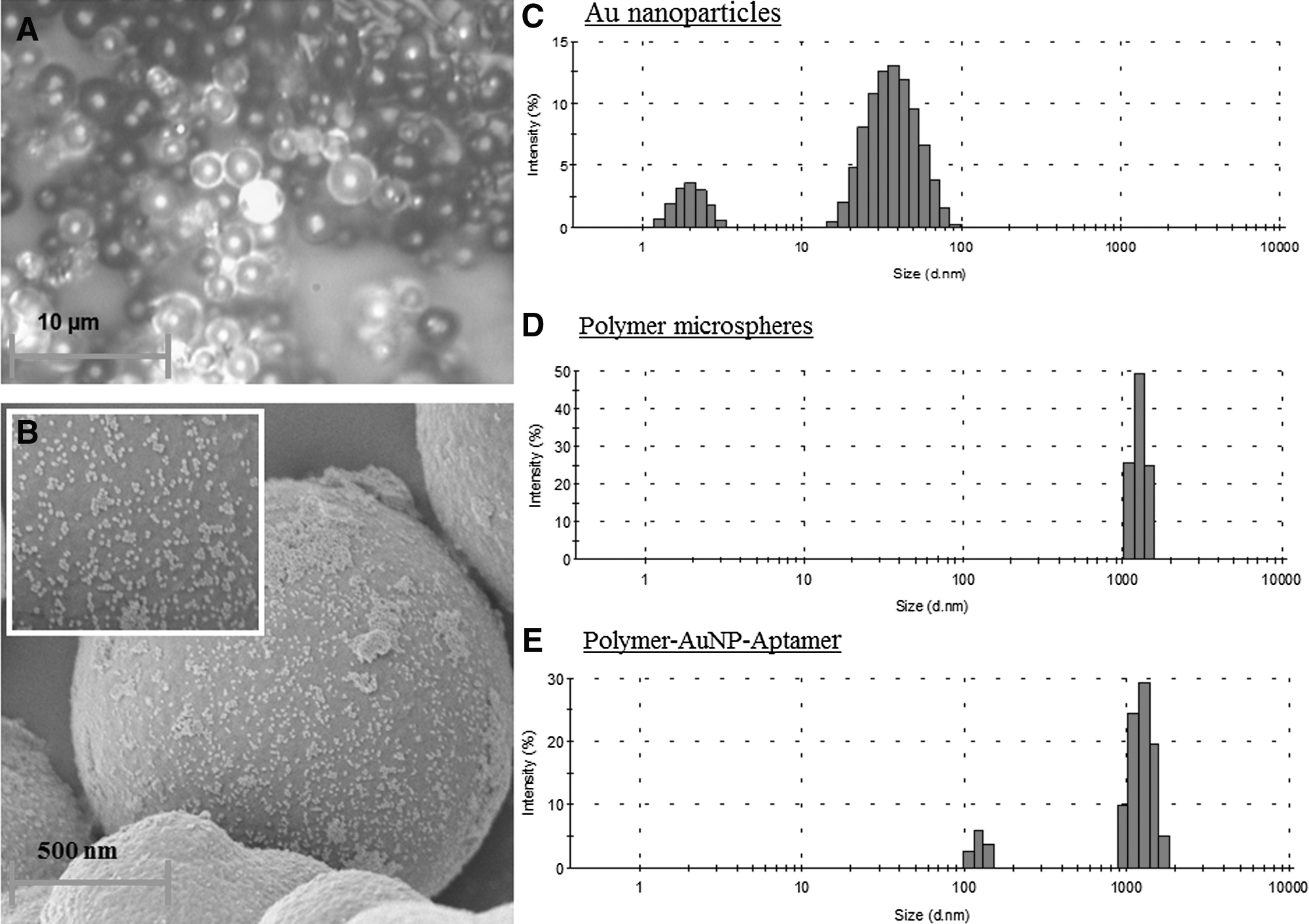
Light microscope images of polymer-AuNP-aptamer substrates immobilized on glass surface
Studies have shown that whereas the signal resulting from measurements obtained using colloidal nanoparticles is highly dependent on the number of nanoparticles in the laser beam, the variation in signal using microsphere-AuNP composite substrates is independent of the number of microspheres in the sample. 32 We decided to use as support polymer microspheres with a diameter size comparable with the focal volume of the laser so that each Raman measurement is restricted to a single microsphere surface. Fig. 2A shows an image taken with the microscope coupled to the Raman spectrometer with a single bead (center) illuminated by the laser. For practical considerations, particles in the range of μm size are also easier to handle and can be subjected to routine processes such as centrifugation or filtration without affecting their performance. Additionally, it was important to minimize the porosity of the polymer beads in order to restrict the distribution of AuNPs onto the surface of the microspheres. The polymer microspheres should show a suitable chemical functionality to allow for subsequent preparation of polymer-AuNP composite particles. Polymer microspheres were prepared by polymerization via precipitation using MAA and EGDMA as co-monomers and acetonitrile as porogen. Acetonitrile permits formation of particles with a low level of porosity, and MAA provides carboxyl groups on the surface for conjugation with AuNP via modification with 2-aminoethanethiol. Because size distribution can affect the quality of the Raman signal, especially in terms of reproducibility of the signal, it was important to obtain a narrow size distribution. In this case, based on prior knowledge obtained from our laboratory, the polymerization was conducted directly with 2.5% monomer concentration and produced monodispersed polymer particles of an appropriate size (diameter of 1.6 μm), with no obvious signs of aggregation and a narrow size distribution determined by DLS measurements ( Fig. 2D).
Fig. 2B is an SEM image of the surface of a composite particle prepared as described in the Materials and Methods section, based on the methodology proposed by Prasad et al. 31 AuNPs can be seen distributed throughout the polymer surface either in a monodispersed manner or forming small aggregates. Although beyond the scope of this work, it was found that the addition of smaller amounts of AuNPs produced poorer coverings, whereas the addition of a greater amount of gold caused the formation of independent gold clusters. It is of note that there were no signs of irreversible aggregation of polymer-AuNP composite particles over several months. Although the particles sedimented after approximately 2 hours in suspension, it was possible to resuspend them easily by vortexing. This provides an additional advantage of the proposed composite material compared to the instability of the AuNPs themselves, which is one of their main limitations. 33
Polymer-AuNP-aptamer conjugates
Conjugation of the aptamers to the polymer-AuNP particles was carried out following a methodology adapted from Taton et al. 30 The maximum number of units of oligonucleotide per nanoparticle is limited and depends on the area of the nanoparticles and the sequence and length of the aptamer. 34 The amount of aptamer used in the conjugation was calculated according to equation 1 considering the entire polymer microsphere as a gold particle. After functionalization with aptamer, the particles remained in suspension without settling to the bottom of the vial for longer than polymer-AuNP particles, suggesting effective surface coverage with aptamer. This phenomenon can be attributed to a different charge on the gold surface. Fig. 2E shows the size distribution of the polymer-AuNP-aptamer particles obtained by DLS. The composite microspheres retained their morphology after conjugation with the AuNPs and aptamers, and only a small excess of gold appears in the histogram.
To distinguish between the background signal of DNA and the signal characteristic of the target analyte, SERS measurements were conducted using previously reported substrates. Fig. 3 shows the SERS spectra obtained using glass-gold substrates prepared as described in the Materials and Methods section after incubation with different amounts of the aptamer thiol-C6-M17-F. Although the signal obtained appears in a region of the spectrum where the signal of citrate has been reported, the increase in the intensity of the signal acquired when greater amounts of DNA were immobilized on the substrate indicates that the observed signal between ∼800 cm−1 and ∼1800 cm−1 corresponds to the DNA. 32,35 Similarly, bands corresponding to DNA can be observed in the same region of the spectra for our polymer-AuNP-aptamer system, as shown in Fig. 4 . This result is consistent with previous studies reporting SERS detection of DNA. 36 According to that work, it is possible to obtain a highly reproducible signal of DNA by applying an appropriate thermal treatment. This kind of process is required for indirect measurements, in which the molecule bound to the aptamer is detected via a target-induced conformational change. 24,37,38 However, direct sensing of malathion can be carried out using our polymer-AuNPs-aptamer system in a suitable solution without any previous thermal treatment by directly measuring characteristic peaks of the target analyte.
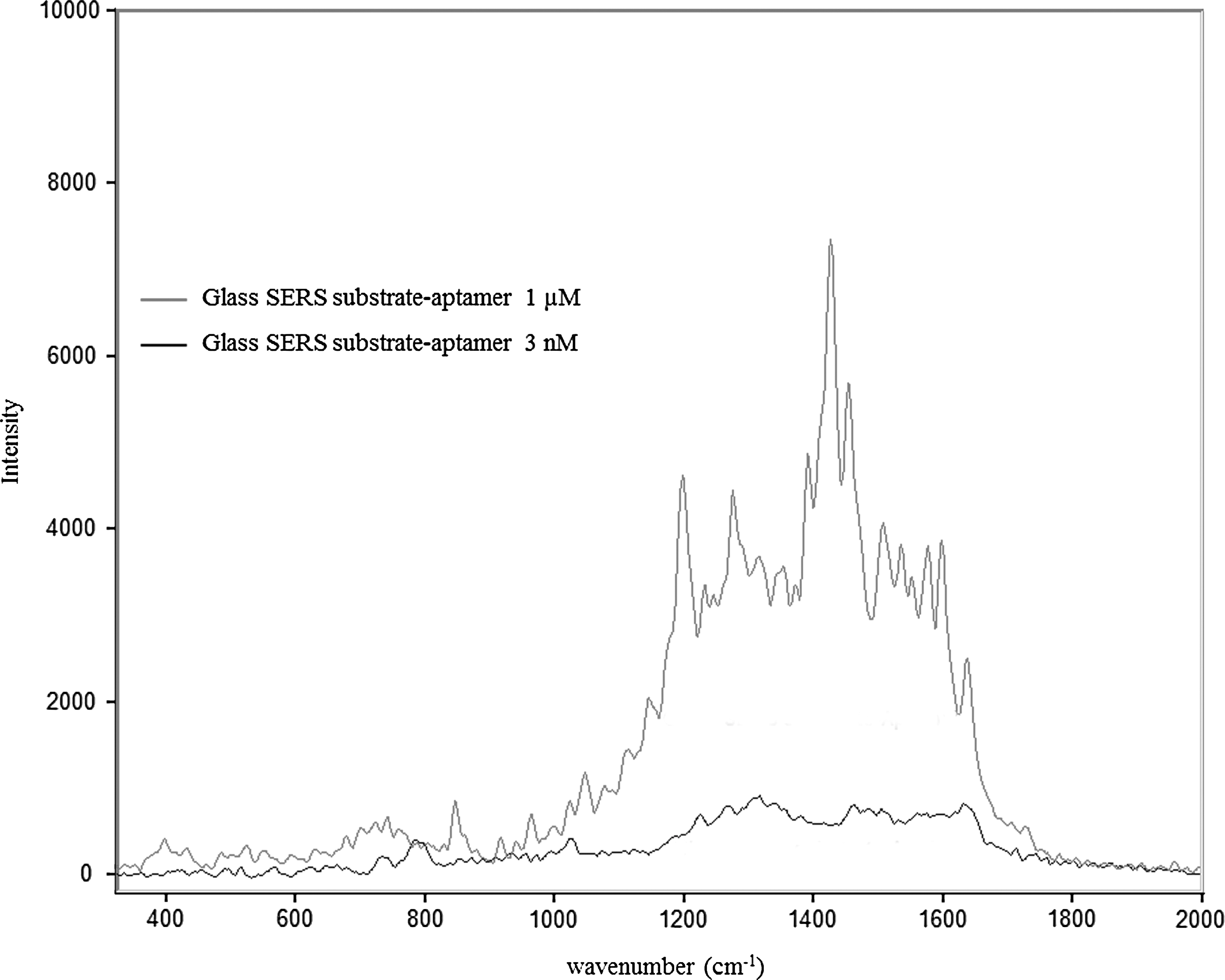
SERS spectra taken on glass SERS substrate conjugated with 1 μM or 3 nM concentration of aptamer M17-F modified with a 5′ thiol group containing a six-carbon spacer.
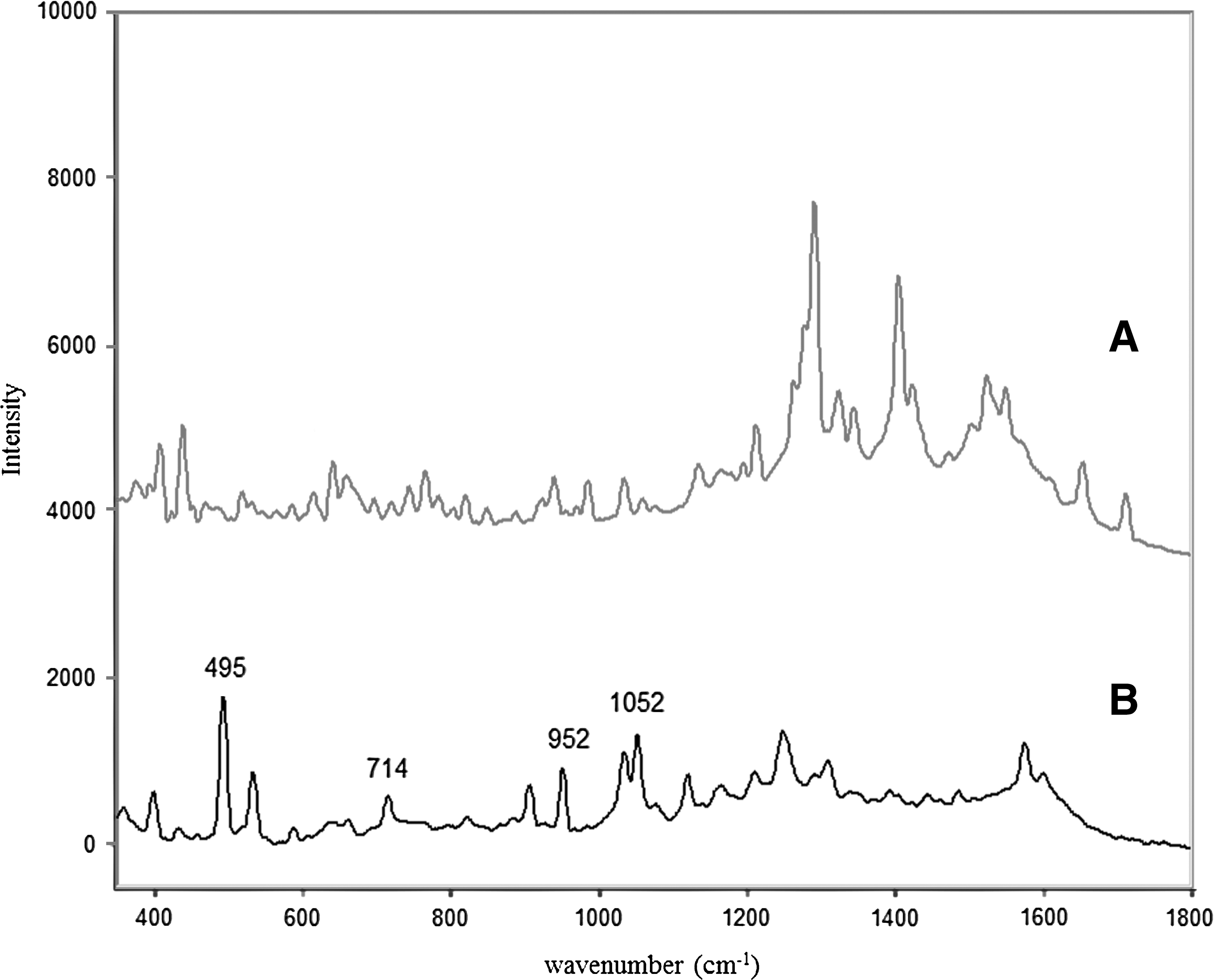
SERS spectra of polymer-AuNPs-aptamer substrate incubated with a blank buffer solution
Sensing Experiments for Malathion
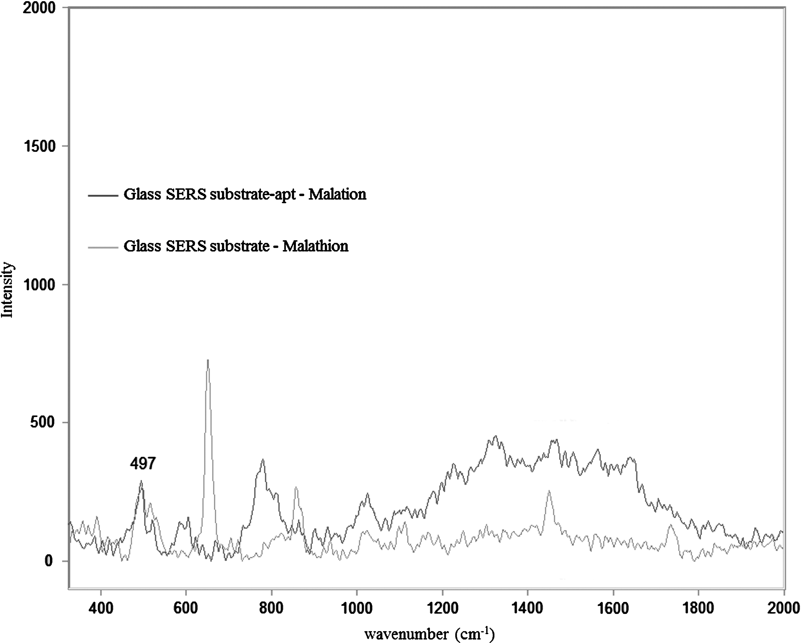
Although the electromagnetic enhancement mechanism does not require the analyte to be in direct contact with the SERS substrate, for best enhancement the distance between the analyte and the metal surface should be in the range of a 0.01–10 nm. 6 To perform direct measurements of the analyte, the polymer-AuNPs-aptamer system can operate as an extractant of the pesticide from the solution by specific interaction between the aptamers and the analyte for which they were designed. Conformational changes produced by the rearrangement in the presence of target analyte allow the molecule to be in close proximity to the metal surface. Given that Raman measurements were made with the samples dried on a glass slide, the aptamers exhibited different conformations on the surface of the particles, resulting in variability in the DNA signal. However, operating in a suitable concentration range, characteristic peaks of malathion were clearly observed in every measurement, indicating that the aptamer-analyte interaction was preserved in the vicinity of the surface. Fig. 5B shows the SERS spectra of polymer-AuNP-aptamer particles incubated as described in the experimental section with a 16.5 μg mL−1 (20 μM) solution of pesticide, together with the spectra obtained using the SERS Diagnosis Membrane as a reference substrate ( Fig. 5A ). Multiple matches can be observed in the wave numbers of the peaks assigned to malathion, (e.g., 500 cm−1, 527 cm−1, 714 cm−1) in a region of the spectra free of DNA signal, enabling direct detection of the pesticide. These peaks assigned to malathion were not present in the spectra obtained from blank samples ( Fig. 4A ). Finally, we selected the peak at ∼495 cm−1, which was better defined and provided greater sensitivity. As shown in Fig. 6 , the same peak (∼497 cm−1) is also present in the spectra obtained after incubation of the pesticide with the reference glass-gold substrates functionalized with thiolated aptamer on the surface. According to previous data, 495 cm−1 can be assigned to the P-S stretching mode of malathion. 39
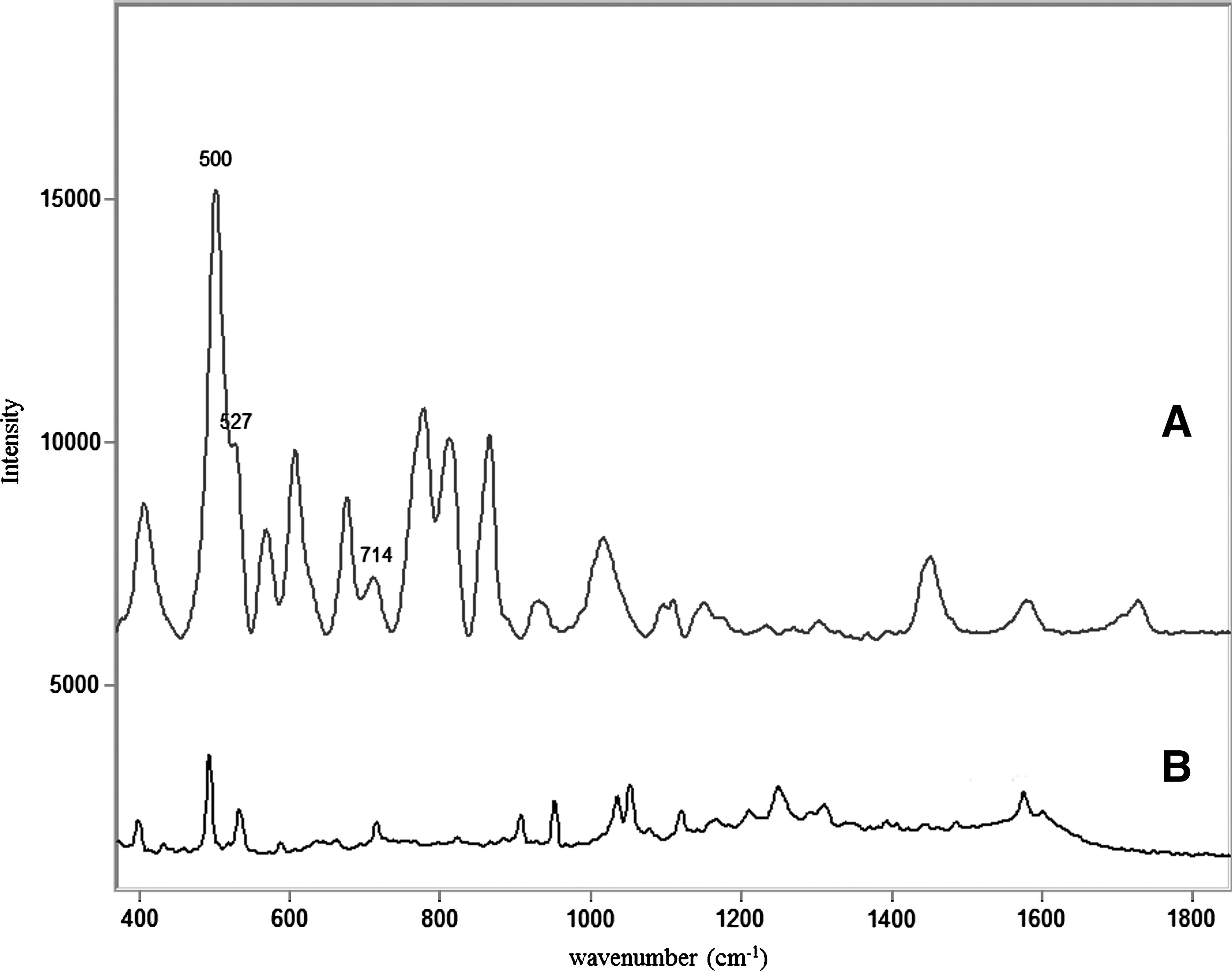
Comparison of SERS spectra of 330 μg mL−1 malathion in ethanol incubated 24 h on iFyber™ SERS Diagnostic Membrane
SERS spectra of glass SERS substrate incubated with 330 μg mL−1 malathion in ethanol for 24 h
It is noteworthy that malathion could not be detected with the polymer-AuNP microspheres lacking modification with the aptamer following washing with DI water. The washing step was able to remove all material nonspecifically adsorbed onto the surface during the 30-min incubation period. This suggests that modification of the polymer-AuNP beads with the aptamer promotes binding of the pesticide by specific interaction with the aptamer.
Analytical Performance and Application
The apta-sensing system for malathion was applied to pesticide solutions at different concentration levels. Selectivity for the target molecule in a sample, which would also likely contain potential interfering molecules, may be an issue. In this sense, experiments with tap water spiked with malathion have also been carried out. The ionic content usually present in tap water is also a challenge for the operation of our system.
Limits of detection
The theoretical limits of detection (LOD) calculated as three times the average signal of the background noise obtained in the analysis of six blank samples were 0.3 and 0.75 μg mL−1 for standard solutions and spiked tap water solutions, respectively. 40 However, in practice, such a value is in a concentration range at which the variability in the signal is very high. Consequently, an experimental limit of detection of 3.3 μg mL−1 was estimated considering the smallest concentration of analyte in the test sample that was reliably distinguished from zero according to the judgment of the researchers (Fig. 7). This experimental LOD is comparable with the value provided by other studies that address the direct SERS detection of malathion. 41
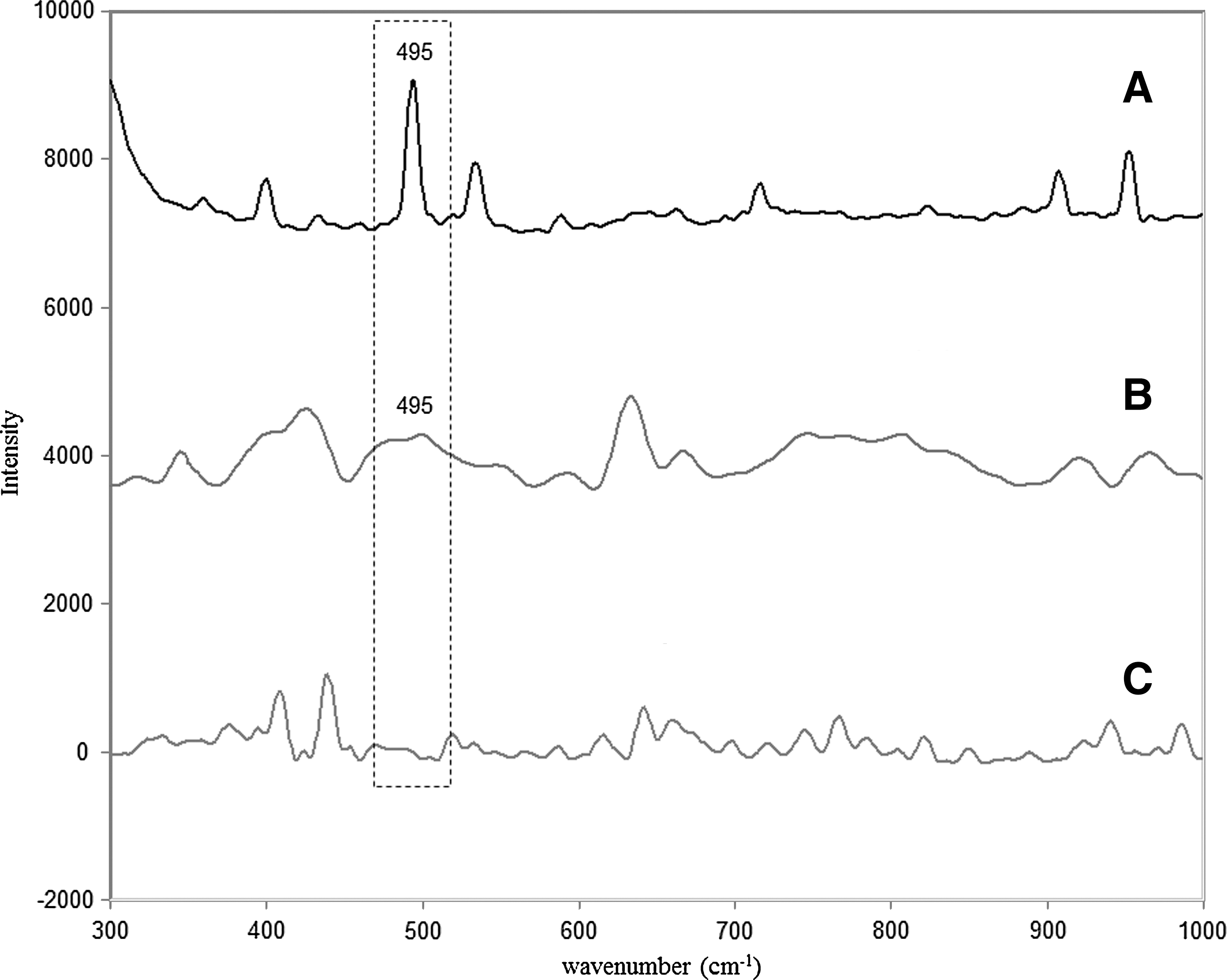
Comparison of SERS detection of malathion using 495 cm−1 peak with polymer-AuNPs-apt substrates at: 16.5 μg mL−1 of malathion
Linearity
It is important to stress that the poor solubility of malathion in aqueous solution (145 μg mL−1, log Pow 2.75) is the primary limiting fact of the apta-sensor for this specific pesticide. 42 Above a concentration level of 33.3 μg mL−1 it is possible to observe the formation of a film of malathion that prevents focus of the laser on the sample, resulting in no Raman signal. Taking these facts into account, the range of application can be reliably restricted to one order of magnitude, and thus, the polymer-AuNP-aptamer successfully allows the detection of the target molecule in a concentration range of 3.3-33.3 μg mL−1. Linearity was checked with fortified tap water samples, and good correlation was observed in the range of concentration studied, with r=0.998.
Precision
Repeatability was studied in terms of the relative standard deviation (RSD) of the values of concentration found at different spiking levels in tap water samples. Table 1 shows the expected and calculated concentrations of malathion with their respective RSD values. Although the value of RSD for concentration levels close to the estimated limit of detection is 25%, for a higher level of concentration (16.5 μg mL−1) the RSD drops to acceptable levels of 14%.
Theoretical and Calculated Concentration of Malathion Present in Tap Water Samples Spiked with the Analyte
Number of replicates, n=6
Conclusions
In this paper, we present a new format of apta-sensing composite particles for SERS detection of malathion. The developed polymer-AuNP-aptamer microspheres combine extraction capability by aptamer-target analyte interaction and Raman signal enhancer for SERS detection of the pesticide. Working under described experimental conditions, the polymer-AuNP-aptamer successfully allows the direct detection of malathion at 3.3 μg mL−1, which compares well with other reported SERS substrates. 41 The apta-sensing microspheres are a system well-suited for industrial and agricultural applications, as only basic equipment is required for analyte separation. As hand-held and bench-scale Raman spectroscopy systems become more powerful, apta-sensing microspheres can become a reliable method for on-site pesticide detection.
Footnotes
Acknowledgments
The authors thank Dr. Kit Umbach and the Cornell Center for Materials Research for assistance and access to the facilities. The authors gratefully acknowledge financial support received from the United States Department of Agriculture (Grant 2009-35603-05066).
Author Disclosure Statement
No competing financial interests exist.