
Abstract
Conventional detection methods for Listeria monocytogenes contamination in food are time-consuming and expensive, creating a need for a more inexpensive and efficient detection method. The objective of this study was to develop an inexpensive biosensor to detect L. monocytogenes in food samples. Screen-printed carbon electrode (SPCE) strips previously used by diabetic patients for glucose monitoring were modified with gold nanoparticles (AuNPs) and specific antibodies to L. monocytogenes. To facilitate detection L. monocytogenes was amplified by conjugating secondary enzyme-labeled antibodies with AuNPs. This assay has the ability to detect L. monocytogenes at 2 log CFU/g in wild blueberry samples and exhibits significant specificity over other enteric pathogens such as Escherichia coli O157:H7 and Salmonella Typhimurium . These results indicate that modifying the electrodes with AuNPs is crucial in the development and production of SPCE biosensors. The use of this optimized amperometric immuno-biosensing strip is an inexpensive, rapid method for detecting foodborne pathogens and has commercial potential.
Introduction
Listeria monocytogenes is an opportunistic, intracellular pathogen and an important cause of foodborne infection worldwide as it is tolerant to extreme pH, temperature, and salt conditions. 1 Unlike other foodborne pathogens such as Escherichia coli O157:H7 and Salmonella species, L. monocytogenes has caused sporadic outbreaks of listeriosis, which has fatality rates up to 30%. 2 Listeriosis is a clinical diagnosis made when the organism can be isolated from usually sterile body sites such as blood, central nervous tissue, or a placenta/fetus. In affected patients, it septicemia, meningitis, or encephalitis can occur, and intrauterine or cervical infections may develop in pregnant women, possibly resulting in spontaneous abortion (in the second or third trimester) or stillbirth. 3 Because L. monocytogenes is very tolerant to extreme conditions and can grow at temperatures as low as 3°C (permitting multiplication in refrigeration), it can be isolated from many foods, including raw milk, pasteurized fluid milk, cheeses, ice cream, raw vegetables, fermented raw-meat sausages, raw and cooked poultry, raw meats, and raw and smoked fish. 3 In the United States, it is estimated that 2,500 people become seriously ill with listeriosis each year, and 500 of these people die. 4
Conventional detection methods for L. monocytogenes are labor-intensive, time-consuming, and expensive. The process can take days to complete, as the culture needs to grow on Oxford agar specific for L. monocytogenes for about 24–48 hours before the experiment can take place. 5 As the food industry grows and food is being increasingly mass produced and consumed, the need for a more efficient, simpler, and lessexpensive method to detect foodborne pathogens such as L. monocytogenes is becoming more urgent. Biosensors that can detect pathogens in a short period of time, from minutes to a few hours, are in high demand and have been developed as an alternative to traditional methods. 6 This technology relies on biological receptor compounds (antibodies, enzymes, nucleic acids, etc.) and physicochemical or electrochemical transducers in biosensor systems to direct observations of specific biological events and provide high specificity and sensitivity. 7 Although these types of methods can greatly reduce detection times, they are too expensive and impractical for commercial use. 8
The use of screen-printed carbon electrode (SPCE) strips as a disposable amperometric biosensor that is inexpensive, portable, and easy to handle has great potential for the detection of L. monocytogenes. 9 The detection of bacteria using SPCE involves a transducer that translates a biochemical reaction into a measurable response, such as the oxidation of a substrate leading to the emission of electrons. 10 Carbon is ideal for these screen-printed electrodes because it provides a wide range of working potentials and is chemically inert. Carbon also has a very pure crystal structure that provides a high signal-to-noise ratio and low residual current. For the production of SPCEs, carbon paste is printed on a matrix through a mask net with a defined pattern. 11
SPCE strips are most commonly used in amperometric immunoassays in which antibodies or immunoreagents are immobilized on the surface of electrodes. 8 Electrochemical immunosensors use enzyme labels for either antigen or antibody. The peroxidases, phosphatases, urease, and glucose oxidases have been the most extensively used labels. 9 The antigen-antibody interaction is then measured through a specific enzyme substrate. 10
Use of SPCE strips has been improved through the modification of the electrode surface; in particular, gold (Au), silver (Ag), and platinum (Pt) are of great interest because of their versatility and beneficial properties. 9,12 Interest in gold nanoparticles (AuNPs) has been especially high due to their good biological compatibility, excellent conducting capability, and high surface-to-volume ratio. 13 On electrodes, AuNPs have the ability to amplify the detection signal, improve the electron transducer, and reduce the detection limit in electrochemical biosensors. 8,14 Metallic nanoparticles are useful in immunoassays because when they are immobilized on the electrode surface, they provide more surface area on which antibodies can immobilize and capture target bacteria. While the gold film aids in conductivity, it is the NPs that increase the surface area and provide higher antibody immobilization. Additionally, AuNPs are more commonly used in electrochemical applications as they have the ability to increase sensitivity cost effectively by enhancing electrode conductivity and facilitating electron transfer by transducing the binding reaction of antigens at antibody-immobilized surfaces. 12,15,16
The objective of this study was to develop an enzyme-linked immunosorbent assay (ELISA)–based biosensing strip to use for quick and inexpensive detection of L. monocytogenes through AuNP-modified SPCE. The electrode surfaces were modified by AuNPs to create a larger electrode reaction surface, assist in electron transfer, and increase conductivity, thereby lowering the detection limit. The biosensor was further examined for its sensitivity in detecting L. monocytogenes inoculated on wild blueberries as well as its specificity for L. monocytogenes over other enteric pathogens such as E. coli O157:H7 and S. Typhimurium.
Materials and Methods
Apparatus and Construction of Sensor
SPCE strips made from carbon/graphite and silver resin inks and containing working and reference/counter electrodes were obtained from Apex Biotechnology (Hsinchu, Taiwan). Each SPCE strip was attached to a strip holder, which was then wired to a PalmSense potentiostat (Palm Instrument, BZ Houten, Netherlands) and an HP (Palo Alto, CA) IPAQ personal digital assistant (PDA) (Fig. 1A). When the switch of the strip holder was pushed down, the emission of electrons from the reaction site was collected and transferred to the PalmSense, therefore generating an amperometric graph on the PDA. Two tests measuring the emission of electrons were conducted—cyclic voltammogram (CV) and amperometric (AMP) testing. For the oxidation of hydrogen peroxide (H2O2) to occur as a result of its reaction with horseradish peroxidase (HRP), which is bound to the secondary antibody, a potential must be applied to the working electrode. The CV tests consist of four scans run at 100 mV/s. In a graph of a CV test, there is an oxidation peak and a reduction peak. The peaks indicate the potential at which the reaction has reached the greatest rate of electron transfer. In contrast, the amperometric test, measures the current at a set potential over a period of 50 seconds at 300 mV. The response currents from the strips with and without bacteria were compared using Δcurrent, which is calculated by subtracting the response current of the blank strips from the response current of the L. monocytogenes target strips. Positive Δcurrent indicates that there was a stronger reaction from the target bacteria-containing strips than from the strips without bacteria due to the lack of antibody-antigen interaction on the blank strips. 8 The HRP-labeled antibody is responsible for the oxidation of H2O2. 10
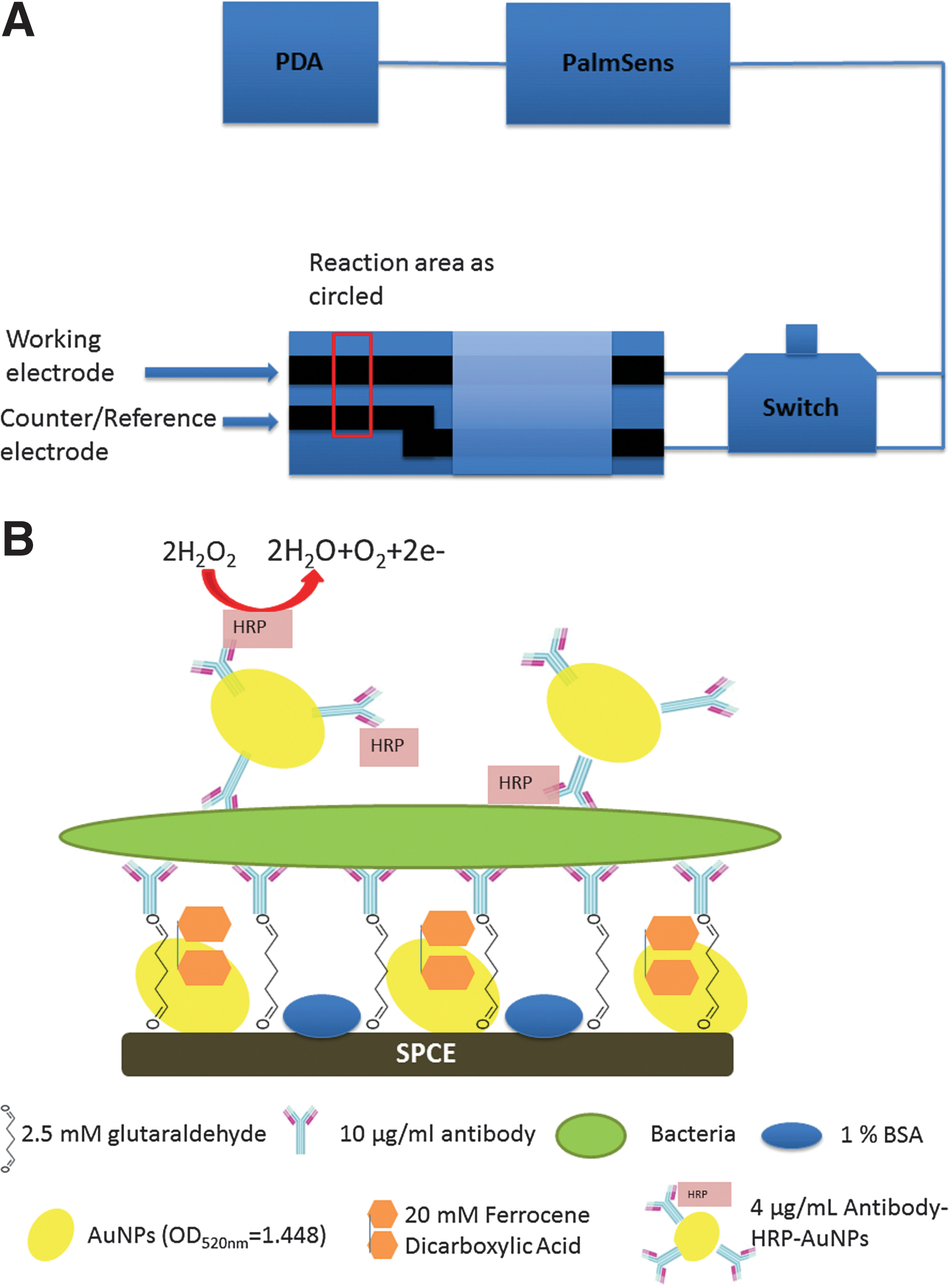
Diagram of apparatus and schematic of optimized procedure. The sensor apparatus includes SPCE strip, strip holder, switch, PalmSense, and PDA. The SPCE strip was attached to a chip holder, which was wired to a PalmSense, and a PDA. When the switch of the strip holder was pushed down, the emission of electrons from the reaction site was collected and transferred to the PalmSense, thereby generating an amperometric graph on the PDA. Once the substrate is added to the strip surface and incubated for 40 seconds, the switch on the strip holder is depressed and the emission of electrons is detected and analyzed by the PalmSense. Analysis of the data yields a graph on the PDA depicting the emission of electrons over a period of 50 seconds that can then be downloaded for further analysis
Reagents and Solutions
Ethanol (100%) was obtained from Pharmco-Aaper (Brookfield, CT); anti-L. monocytogenes, anti-L. monocytogenes+HRP, anti-E coli O157:H7, and anti-E. coli O157:H7+HRP were purchased from Meridian Life Science (Saco, ME). Phosphate buffered saline (PBS; 0.1 M, pH 7.2) and PBS with 0.05% Tween 20 (PBST) were used in the experiments. The anti-L. monocytogenes antibody was diluted to the desired concentration from a stock solution of 2 mg/mL of antibody into PBS buffer while the secondary antibody, labeled with HRP, was diluted from a stock solution of 1mg/mL into PBS buffer. Sodium citrate (C6H5Na3O7), glycerol, H2O2, potassium chloride (KCl), sodium chloride (NaCl), disodium phosphate (Na2HPO4), and monopotassium phosphate (KH2PO4) were purchased from Fisher Scientific (Fair Lawn, NJ). The mediator 1,1′-ferrocene-dicarboxylic acid (FeDC) was obtained from Sigma Aldrich (St. Louis, MO). Gold chloride trihydrate (HAuCl4·3H2O), which was used to make AuNPs, was purchased from RICCA Chemical (Arlington, TX).
Preparation of AuNPs
AuNPs (13 nm) were prepared by reduction of HAuCl4·3H2O solution with sodium citrate using a procedure modified from Lin et al. 8 The peak absorbance of the AuNPs was read at 520 nm using a DU Series 500 spectrophotometer (Beckman Coulter, Brea, CA). The ideal OD520nm value of AuNPs ranges from about 1.8–2.5.
Culturing Samples
The target bacteria, L. monocytogenes (ATCC 19115, 7644, and 15313) were purchased from the American Type Culture Collection (ATCC, Manassas, VA), grown in brain heart infusion broth (BHI; Acumedia Manufacturers, Lansing, MI) for 24 hours at 37°C, and enumerated on Oxford agar plates (Acumedia Manufacturers). The concentration of bacteria used for optimization (with the exception of sensitivity experiments) was 5 log CFU/mL. Decreasing concentrations from 105 CFU/mL to 101 CFU/mL were prepared for the sensitivity experiments by performing serial dilutions of the bacterial culture in 0.1% peptone water. E. coli O157:H7 (ATCC 35150 and 700599) and S. Typhimurium (ATCC 6962 and 14028) were also purchased from the ATCC and grown in BHI broth. E. coli O157:H7 and S. Typhimurium were isolated on MacConkey sorbitol agar and xylose lysine deoxycholate agar (MSA, XLD; Acumedia Manufacturers), respectively, for 24 hours at 37°C prior to the experiments. The standard concentration for E. coli O157:H7 and S. Typhimurium was also 5 log CFU/mL.
Optimization of Protocol
SPCE strips were washed with 100% ethanol to remove the protective membrane, rinsed with double-distilled H2O, and air dried before substances were added to the surfaces of the electrodes. An aliquote (4 μL) of each substance was placed on the working electrode only, and the strips were incubated for 30 minutes at 2°C. Substances were added in the following order, as depicted in Fig. 1B: 2.5 mM gluteraldehyde (cross-linking agent), AuNPs, 10 μg/mL anti-Listeria antibodies, 20 mM FeDC (mediator), 1% bovine serum albumin (BSA) in PBS (blocking solution), 5 log CFU/mL of L. monocytogenes or BHI/peptone water dilution for blank strips, and 4μg/mL anti-Listeria antibodies+HRP/AuNPs. After incubation, the strips were washed four times with 100 μL of PBS buffer, except after the addition of the secondary antibody when PBST was used. After the final incubation, 50 μL of the substrate composed of 3% H2O2 with 4.5 mM FeDC was added to both the working and reference electrodes for 40 seconds, and amperometric testing was conducted.
AuNPs were adhered to the strip surface via the gluteraldehyde, which acts as a cross-linking agent. The adhered AuNPs create a larger surface area to increase antibody immobilization for greater target capture. The mediator FeDC was applied as part of the surface modification and in the substrate to facilitate the shuttle of electrons from the reaction site back to the electrode for optimal electron detection. In this study, 1% BSA in PBS was used as a blocking agent to diminish nonspecific binding of the antibodies and to decrease false positives. After the addition of target bacteria, the enzyme-linked secondary antibody was applied. HRP was chosen as the conjugation enzyme because of how well it oxidizes, and especially with H2O2 as the oxidation agent, to ensure a reliable emission of electrons for detection.
Conjugation of Secondary Antibody with AuNPs
Strips prepared with 4 μg/mL anti-Listeria antibodies+HRP conjugated with AuNPs were compared with strips made using 4 μg/mL anti- Listeria antibodies+HRP according to the optimized protocol described herein.
Assay Sensitivity
Decreasing concentrations of bacteria from 5 log CFU/mL to 1 log CFU/mL were tested using the optimized protocol described. Each concentration of bacteria had a corresponding blank that contained the same dilution factor of BHI in peptone water.
Assay Specificity
Mixtures of L. monocytogenes (ATCC 19115, 7644, and 15313), E. coli O157:H7 (ATCC 35150 and 700599), and S. Typhimurium (ATCC 6962 and 14028) at 5 log CFU/mL were compared against a blank strip (containing a BHI/peptone water dilution) using the optimized protocol to determine the specificity of the assay against other enteric pathogens.
Testing of L. Monocytogenes-Inoculated Blueberry Samples
A 25-g sample of wild blueberries was weighed for both the control and L. monocytogenes trials. Blueberries were rinsed in ethanol and sterile water and dried under ultraviolet light for 2 hours. After the blueberries were dried, they were inoculated with a mixture of L. monocytogenes strains 19115, 7644, and 15313 by pouring the culture over the blueberry sample and mixing for 3 minutes. After inoculation, the blueberries were dried for 2 hours. After the blueberries were completely dry, each 25-g samplewas added to a stomacher bag with 25 mL of 0.1% peptone water and stomached for 2 minutes. Once the samples were stomached, the liquid portion was then diluted and plated onto trypticase soy agar and modified Oxford agar plates to determine viable cell counts. Samples were filtered with a syringe and a 5-μm filter before application to the SPCE strips.
Statistical Analysis
The experiments were repeated three times. Bacterial populations were reported as log CFU/mL (or /g). Analysis of variance (ANOVA) was performed using SYSTAT 12 software (Systat Software Inc, Chicago, IL). Significance of difference was defined as p<0.01.
Results
Optimized Protocol
The optimized protocol is depicted in Fig. 1B. Results show that the average Δcurrent for a 5 log/CFU sample was 147 μA (Fig. 2). After optimization of the protocol, it was determined that the following was the order and concentration of substances that resulted in the highest Δcurrent and most reliable response: 2.5 mM gluteraldehyde, AuNPs (OD520=1.448), 10 μg/mL the first anti-L. monocytogenes antibodies, 20 mM FeDC, 1% BSA in PBS, L. monocytogenes sample (5 μL), and 4 μg/mL of the secondary anti-L. monocytogenes antibodies with HRP/AuNPs. The substrate was composed of 4.5 mM FeDC and 3% H2O2, which reacts with the HRP-labeled antibody and is reduced to water and emitting electrons (Fig. 1B). This emission of electrons shapes the amperometric curve and determines the presence of L. monocytogenes contamination.
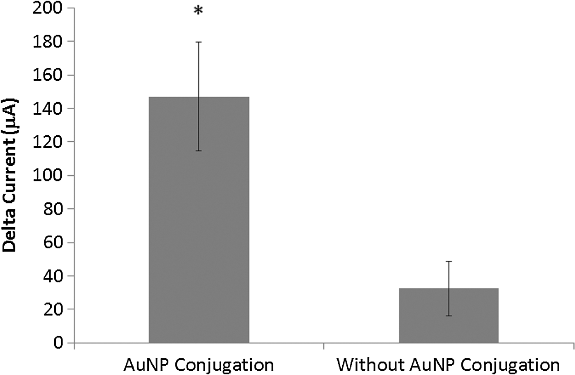
Enhancement of the sensitivity of the biosensor was achieved using conjugation of secondary antibodies with AuNPs. Comparison of both conjugated and non-conjugated antibody shows that adding the second layer of AuNPs with the secondary antibody increases Δcurrent by adding extra conductance and improving assay response. * indicates that the conjugation of secondary antibody significantly increases Δcurrent (p<0.01) and therefore increases the sensitivity of the assay.
Conjugation of Secondary Antibody with AuNPs
Blank and L. monocytogenes strips containing the secondary antibodies with and without AuNPs conjugation were compared. Results indicate that the Δcurrent of strips containing AuNP-conjugated secondary antibodies was statistically higher than the SPCE strips without the conjugated antibody (p<0.01) (Fig. 2 ).
Sensitivity and Specificity Studies
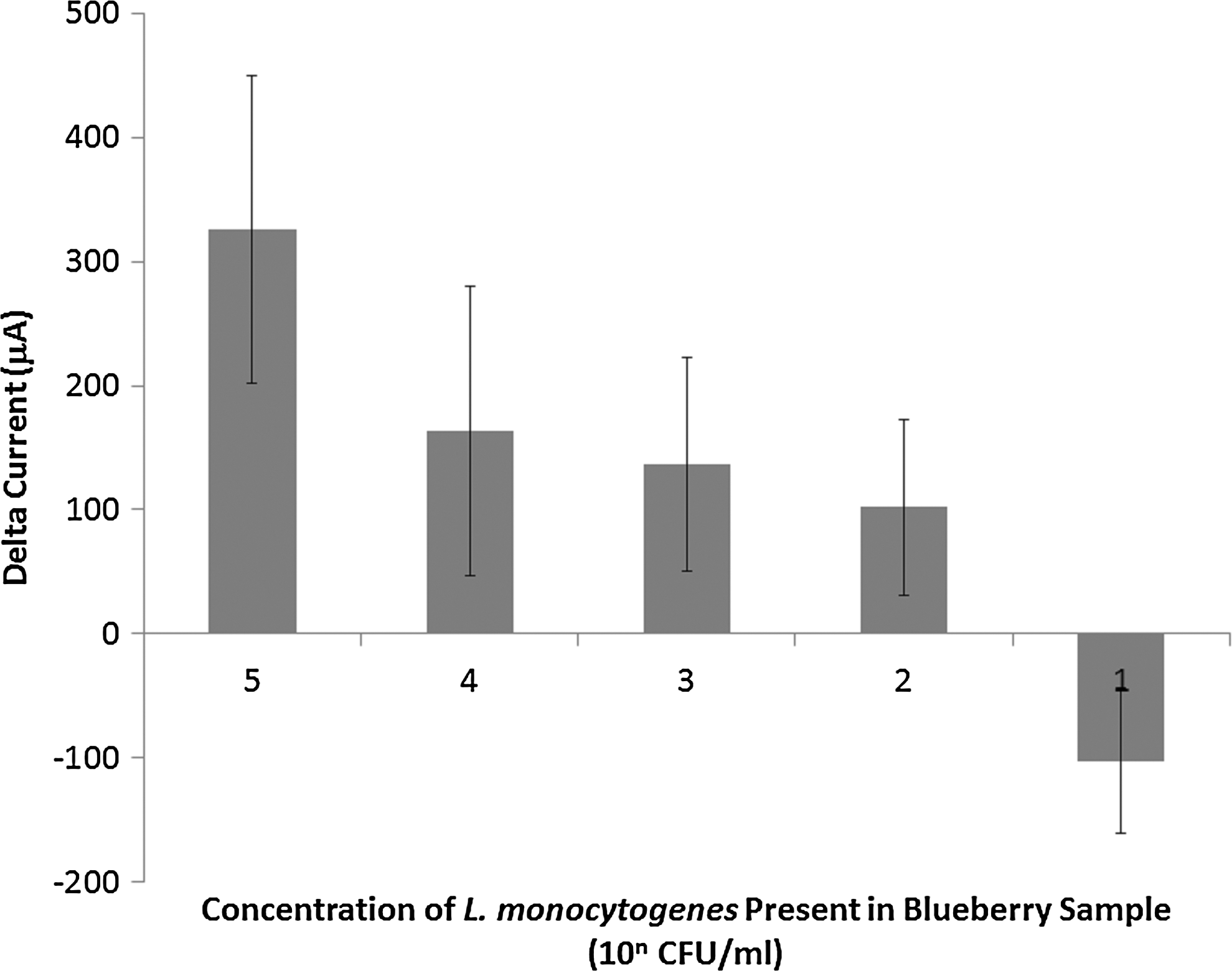
To determine the sensitivity of the assay, decreasing concentrations were tested from 5 log CFU/mL to 1 log CFU/mL. The results shown in Fig. 3 indicate that there was a positive Δcurrent in concentrations of L. monocytogenes as low as 1 log CFU/mL and a linear correlation that indicates this is a possible detection limit. However, considering the average Δcurrent±standard deviations obtained with other enteric pathogens included in the specificity study, such as E. coli O157:H7 and S. typhimirium (Fig. 4), the detection limit for L. monocytogenes was defined as 2 log CFU/mL.

Sensitivity study in which decreasing concentrations of L. monocytogenes from 101 CFU/mL to 101 CFU/mL were tested against blanks containing the same dilution of BHI/peptone water as the concentration of bacteria that was applied. Detection limit of the assay was determined to be 2.25×10² CFU/mL, as there was a linear correlation of positive detection and the response of other enteric pathogens was less than the response for 102 CFU/mL of Listeria. This is the lowest concentration of bacteria per mL that can be confirmed to be Listeria and not other enteric pathogens.
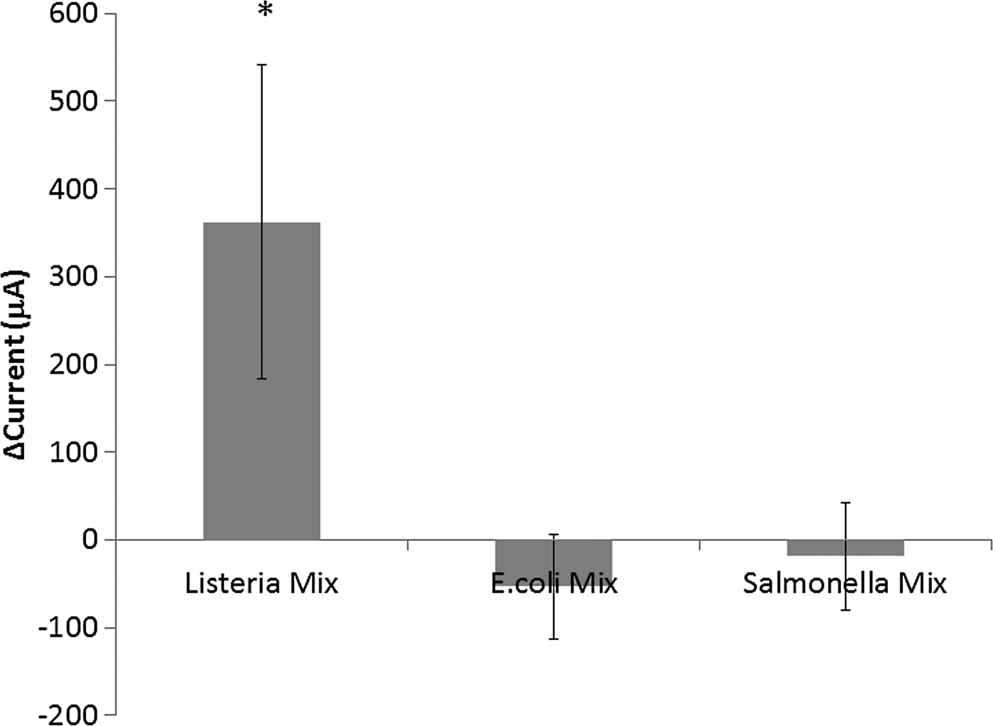
Specificity study; mixtures of L. monocytogenes (ATCC 19115, 7644, and 15313), E. coli O157:H7 ( ATCC 35150 and 700599), and S. Typhimurium (ATCC 6962 and 14028) were all applied to the SPCE strips at a concentration of 105 CFU/mL and compared against a blank strip using the optimized protocol to determine the specificity of the assay against other enteric pathogens. *p<0.01 vs. the E. coli O157:H7 and Salmonella mixes.
Application in Real Samples
Upon testing of blueberries inoculated with L. monocytogenes, there was a positive Δcurrent as low as 2 log CFU/g, but a negative Δcurrent at 1 log CFU/g (Fig. 5). These results indicate that in food samples the detection limit is 2 log CFU/g, and the assay was unable to detect bacteria at lower concentrations due to background noise.
Food study of L. monocytogenes-inoculated blueberry samples. Results indicated a positive Δcurrent and detection of Listeria samples in blueberries as low as 2 log CFU/g. Concentration of 1 log CFU/g led to a negative Δcurrent and, therefore, the assay was not able to detect bacteria at concentrations lower than 2 log CFU/g. However, since only 4 log of sample was applied to the strips this would mean that the biosensor was still able to detect as low as one bacterial cell per strip.
Discussion
Recently, it has become increasingly common to apply nanotechnology to enhance the sensitivity and reliability of biosensors. AuNPs have been of interest due to their versatile and efficient properties. They not only increase the surface area for antibody immobilization on the electrode surface, but also improve electron transmission capacity in solution. 9 It has been shown that the conductive qualities of gold also enhance the electrochemical signal by transducing the binding reaction of antigens at antibody-imobilized surfaces. 9,12
Research to determine the optimized protocol employed in this study used a first layer of AuNPs to modify the SPCE surface, as demonstrated by Lin et al, who determined that the use of the AuNP layer in conjunction with the FeDC mediator improved the electrochemical capacity to enhance the oxidation and reduction of the HRP/H2O2 reaction and increased the transmission of electrons between the FeDC mediator and the SPCE. 8 The AuNPs on the SPCE immunosensing strips increased the effective area of the working electrode and decreased the distances for the reductive-form mediator to diffuse to the stereo-electrode structures. The attached AuNPs could help overcome the long-range barriers to the formation of complexes of antibodies and bacterial cells that deliver an electron via peroxidase to the electrode. 8
The present study added to this research by conjugating the secondary antibody with the AuNPs, providing a route for shuttling electrons from the HRP/H2O2 reaction site back to the SPCE via the increased conductance and surface area provided by the AuNPs and the FeDC present in the 4.5 mM FeDC+3% H2O2 substrate. The presence of the AuNPs in the antibody solution also provides for a larger surface area on which the antibody-antigen interaction can take place, thus allowing more HRP to be oxidized if more secondary antibody is present. 12 The results indicate that conjugation with AuNP significantly increased the Δcurrent, with an average of 147 μA for the AuNP-conjugated antibodies and 32 μA for the non-conjugated antibodies. This shows that the conjugation of the secondary antibody significantly increases the Δcurrent and aids in the increased sensitivity of the assay.
In the present study, the results indicate that there was a positive Δcurrent with concentrations of L. monocytogenes as low as 2 log CFU/mL, as well as a linear correlation. This implies a detection limit of only one bacterial cell per SPCE strip used in our study. Detection of the other enteric pathogens included in the study is most likely attributable to the assay's use of polyclonal antibodies and their ability to cross-react with certain strains of E. coli and Salmonella. This assay could potentially be more specific via the use of monoclonal antibodies or even polyclonal antibodies with less cross-reactive properties.
In the food industry, it is imperative to be able to detect live bacteria to prevent the production and distribution of contaminated food. Since dead bacterial cells would give a false positive if detected, the food industry is focused on detecting lives cells that would cause infection. Although the sensor described here is not specific to live cells, a quick and routine enrichment procedure prior to the detection assay could be conducted to select for live cells and minimize false positive results due to the presence of dead bacteria.
Finally, the real-world applicability of the assay was determined by testing L. monocytogenes-inoculated wild blueberries. The results are in agreement with the sensitivity study performed with the BHI/peptone dilutions of bacteria and indicated that the biosensing strip is reliable, both in theory and in real application and can detect L. monocytogenes down to 2 log CFU/g. The detection limit in food is 102 CFU/g, which indicates that in food samples this biosensor has the ability to detect as low as one bacterial cell on the electrode surface of SPCE strips.
Conclusions
These results demonstrate that an optimized reliable biosensing strip utilizing AuNP-modified carbon electrodes has been developed to detect L. monocytogenes contamination in food samples with the desired specificity. The detection limit of this assay is 2 log CFU/mL (or CFU/g), and an SCPE strip can detect as little as one bacterial cell. This is a rapid and efficient biosensor as it only takes approximately 1 hour for the accurate detection of contamination. This sensor has the potential to be commercialized and it would be extremely beneficial in the food industry as it is inexpensive and portable.
Footnotes
Acknowledgments
This research was supported by the Maine Agricultural and Forest Experiment Station at the University of Maine with external publication number 3302. This work was also supported in part by grant NSC-100-2911-I-009-101, Taiwan. The authors thank Apex Biotechnology Corp. for providing the screen-printed carbon electrode strips.
Author Disclosure Statement
No competing financial interests exist.