




Abstract
Reproductive performance of livestock animals, including dairy and beef cattle, influences the productivity of agriculture in the US and worldwide. Artificial insemination (AI) is one technology with great potential to improve livestock reproductive efficiency. Particularly, gains in reproductive performance of AI sires can be made by identifying and eliminating bulls with inferior fertility/semen quality by testing and discarding individual semen collections/ejaculates with inferior fertilization potential and by eliminating defective spermatozoa from semen collected for AI. Both sire fertility testing and semen purification can potentially be improved through the application of nanotechnology. The necessary first step in this process is the identification and validation of sperm quality biomarkers; they are the differentially expressed sperm-borne proteins/ligands that can be objectively quantitated to measure sperm quality and to estimate future fertility. Proteins expressed on the sperm surface can be targeted using magnetic nanoparticles to allow for rapid, efficient removal of defective spermatozoa from semen. This article reviews recent progress in the identification of such biomarkers (e.g., ligands of lectins from Arachis hypogaea and Lens culinaris, sperm proteins ubiquitin and post-acrosomal, WW domain-binding protein [PAWP]), and also describes recent trials of nanoparticle-based technologies for fertility testing and the nanopurification of bull semen for commercial AI.
Introduction
Though the term nanotechnology is a fairly recent addition to our vocabulary, cell and reproductive biologists have been using nanoparticles for several decades. Notably, colloidal gold particles coupled to lectins or antibodies have been used for immunolabeling various sperm ligands and imaging by electron microscopy, providing superior resolution of subcellular antigen detection and localization. 1 More recently, fluorescent nanoparticles called quantum dots have been developed to improve the resolution and specificity of fluorescence-based immunolabeling at light microscopic levels. 2 Another recent application is the use of nanoparticles as vectors for DNA and protein transfection into various cell types, including spermatozoa. Using this technology, transgenic animals could be created by sperm-mediated gene transfer coupled with in vitro fertilization (IVF). 3 Finally, DNA-binding nanoparticles have been coupled with DNA probes designed to hybridize to genes unique to the male Y chromosome, with the goal of improving semen sexing, a cell-sorting method by which the sex ratio of calves born after artificial insemination (AI) can be altered to favor one gender over another. 4
Various nanotechnology-based devices have been developed to test for animal diseases, as well as for testing of reproductive function and fertility. Of particular interest to this review is the development of nanoparticle-based lateral flow devices (so called “dip stick tests”) for fertility testing in male livestock animals and for male infertility diagnostics in humans. Such test devices can also be adapted for forensic sperm detection in crime scene investigation. 5 One such device, the fertility-associated antigen (FAA) test has been tested on bulls used for AI, but did not appear to be informative of their fertility. 6
Large animal breeding by AI is on the rise in the US and worldwide. 7 However, only about 50% of inseminations result in a full-term pregnancy, in part due to lack of a thorough understanding of the molecular mechanisms determining the fertilizing potential of a semen sample. 8 The success rate of AI can be improved via better management of fertility and reproductive performance in both male and female livestock animals. 9 –11 Gains in reproductive performance of bulls can be made at the level of individual sires (i.e., by eliminating bulls with inferior fertility) and at the level of individual semen collection (i.e., by discarding semen collections/ejaculates with inferior fertilizing potential, and/or by eliminating defective sperm cells/spermatozoa from collected semen). Young sires are admitted into an AI breeding program based on a breeding soundness evaluation and extensive, costly, and time-consuming progeny testing. This approach could be greatly streamlined if reproductive traits and semen parameters could be reliably predicted from genomic analysis and semen evaluation of young sires. This review will focus on new, cost-efficient ways of improving semen analysis and increasing the fertilizing potential of AI doses, with particular focus on nanotechnology-based approaches.
Biomarker-based Flow Cytometric Semen Analysis
Bovine semen analysis is still performed using mainly subjective, light microscopy-based methods; however, new biomarkers and instrumentation are now being introduced for automated, high-throughput semen analysis reflective of fertility in AI services. 12,13 Semen evaluation via traditional light microscopy determines the number of sperm cells in a semen collection (sperm concentration per mL of semen), percentage of spermatozoa with visible structural defects (sperm appearance/morphology, presence of abnormal and immature sperm forms), sperm motility (percentage of spermatozoa with certain pattern and velocity of movement), and the presence of semen contaminants such as sperm fragments, white blood cells, and bacteria. Such an analysis provides useful information about the semen sample, yet new methods for estimating future fertility of a sire are still sought. 14 Furthermore, not all sperm abnormalities are detectable with standard light microscopy. Semen samples with cryptic (i.e., non-obvious) sperm defects and reduced fertilizing ability may appear normal by conventional semen analysis standards and be deemed suitable for insemination. In-depth analysis carried out quickly and with repeatable precision on a large number of spermatozoa is of paramount importance to farm animal biotechnology. As a result, flow cytometric evaluation using biomarkers to detect specific spermatozoan characteristics is growing in popularity in both andrology laboratories and agricultural studs. At the same time, new methods are being sought to remove defective spermatozoa from semen collected from sires with high genetic value. The goal is to produce as many AI doses as possible per sire per collection.
To develop new biomarkers to assess the quality of a semen sample, or to purify it, candidate biomarkers can be identified using a variety of approaches, including sperm fractionation and proteomic analysis. Proteomics refers to the qualitative and quantitative comparisons of the cells' protein make-up, or “proteome,” aimed at identifying cellular mechanisms involved in biological processes. Comparison of fertile and sub/infertile bull sperm proteomes as well as proteomic characterization of bovine seminal plasma and normal/defective sperm fractions have given some insight into which proteins—and the level at which they are present in the sample—are indicative of fertility or infertility. 8,15 –17
Fluorescently labeled biomarker probes can be used to assess a variety of structural and functional properties of spermatozoa: proper packaging of DNA in the sperm head (sperm chromatin integrity); presence and integrity of the sperm head cap indispensable for sperms' ability to penetrate the egg coat during fertilization (sperm acrosomal status); the ability of the spermatozoa to produce and regenerate energy necessary for sperm movement (mitochondrial membrane potential); and sperm cell viability (percentage of live/dead spermatozoa in collected semen). Antibodies are often used to detect and quantify proteins that are up- or down-regulated in defective spermatozoa. 13 The goal of our ongoing research is to validate the candidate biomarker proteins or ligands that are uniquely associated with defective spermatozoa, even if defects are subtle or unnoticed during evaluation using standard light microscopy. The central hypothesis of this research is that these biomarkers, unique to defective spermatozoa, are indicative of poor semen quality and decreased fertility. Thus, such biomarkers are described as negative biomarkers of fertility. In addition to semen analysis, biomarkers present on the sperm surface, such as sperm protein ubiquitin and binding partners (ligands) of several plant lectins, are potential targets for nanoparticle-based semen purification, as will be discussed below. 13
Flow cytometry is a method in which fluorescently labeled cells (in this instance, spermatozoa) travel individually at high speed (hundreds or thousands of spermatozoa per second) through the flow cell of a flow cytometer, where they are illuminated by one or more lasers. This causes light scattering and fluorescence excitation of biomarker-recognizing fluorescent probes bound to a specific site on the spermatozoa; the signals are detected and quantitated by photo-detectors, and the data are routed to a computer program. The program presents the information in the form of relative fluorescent intensity units, which are typically displayed as either scatter plots or histograms. 18 Analysis of the scatter plots and histograms allows for specific sperm populations to be gated off, yielding information regarding fluorescence intensity, percentage of sperm population with certain fluorescence characteristics within a total sample, median fluorescence intensity, etc.
Flow cytometric semen analysis has been increasingly adopted by the AI industry. Sub-fertile bulls or their AI doses can cost producers a significant amount of money because of reduced conception rates of the inseminated cows. The lack of precision in conventional semen analysis, coupled with the subjective nature of such an assessment, implies that some acceptable semen may be erroneously rejected, and at the same time, semen of unacceptable quality may be used for inseminations. 14 In contrast, flow cytometry is fast, accurate, highly repeatable, and can analyze significantly more spermatozoa per sample (e.g., 10,000 cells measured in a few seconds in a single sample) than standard semen analysis. 19 Sperm characteristics commonly measured by flow cytometry include sperm viability, mitochondrial function, membrane potential, chromatin structure, and acrosomal status. 20 –28
Sperm acrosome is the cap-like structure covering the front part of the sperm head. It harbors proteolytic enzymes—molecules that, upon sperm binding to the egg coat, digest a slit through which the fertilizing spermatozoon penetrates the egg. Acrosome can be inherently compromised in otherwise normal spermatozoa; it can also be damaged or prematurely activated (so-called premature “acrosome reaction”) during semen processing for cryopreservation or during freeze-thawing of the cryopreserved AI semen. Sperm acrosomal status can be probed by flow cytometry using fluorescently labeled lectins—plant proteins that recognize and bind glucosidic residues in different parts of the acrosomal membrane. 29 The binding partners/ligands of lectins are concealed within the intact acrosome and, therefore, are not recognizable to lectins. In a compromised acrosome, lectin ligands become exposed on the sperm head surface and are available for lectin binding. This makes lectins suitable for nanodepletion-based semen purification. Metallic nanoparticles coated with a lectin will readily bind to the surface of spermatozoa with damaged acrosomes upon nanoparticle mixing with semen. A potent magnet is then used to concentrate the defective, nanoparticle-coated spermatozoa on the bottom of the test tube used for nanopurification. Normal, fertile spermatozoa can be skimmed off the top, while the defective spermatozoa are held on the bottom of the tube by magnetic force.
Two widely available plant lectins are known for their affinity to damaged sperm acrosomes, and both can be coupled to fluorescent dyes for flow cytometric analysis and to metallic nanoparticles for semen purification. Pisum sativum agglutinin (PSA) derived from the pea plant, and Arachis hypogaea agglutinin (PNA, or peanut agglutinin) derived from the peanut plant, are the most commonly used lectins because of their specificity. 30 Spermatozoa with reacted, damaged, or abnormally formed acrosomes acquire green fluorescence after labeling with PNA lectin coupled to the green fluorescent dye fluorescein isothiocyanate (Fig. 1A); as shown in Fig. 1, spermatozoa with intact, normal acrosomes have no fluorescence. 27 Of significance for semen analysis and purification, the percentage of the acrosome-damaged, PNA-binding spermatozoa in an ejaculate correlates with semen quality and reproductive performance of AI bulls. PNA values correlate with the conventionally established parameters of sperm morphology and sperm concentration. 28,29 A third lectin, Lens culinaris agglutinin (LCA) from the lentil plant, has also been used to evaluate bull sperm quality. 31 Lectin LCA binds to the entire surface of defective spermatozoa, but only to the acrosomal surface in normal spermatozoa (Fig. 1B ). Consequently, distinct patterns of LCA-induced fluorescence are seen in normal vs. defective spermatozoa labeled with fluorescently tagged LCA. In our preliminary unpublished data, we found a positive correlation between LCA and ubiquitin staining and a negative correlation between LCA and percent normal sperm morphology.
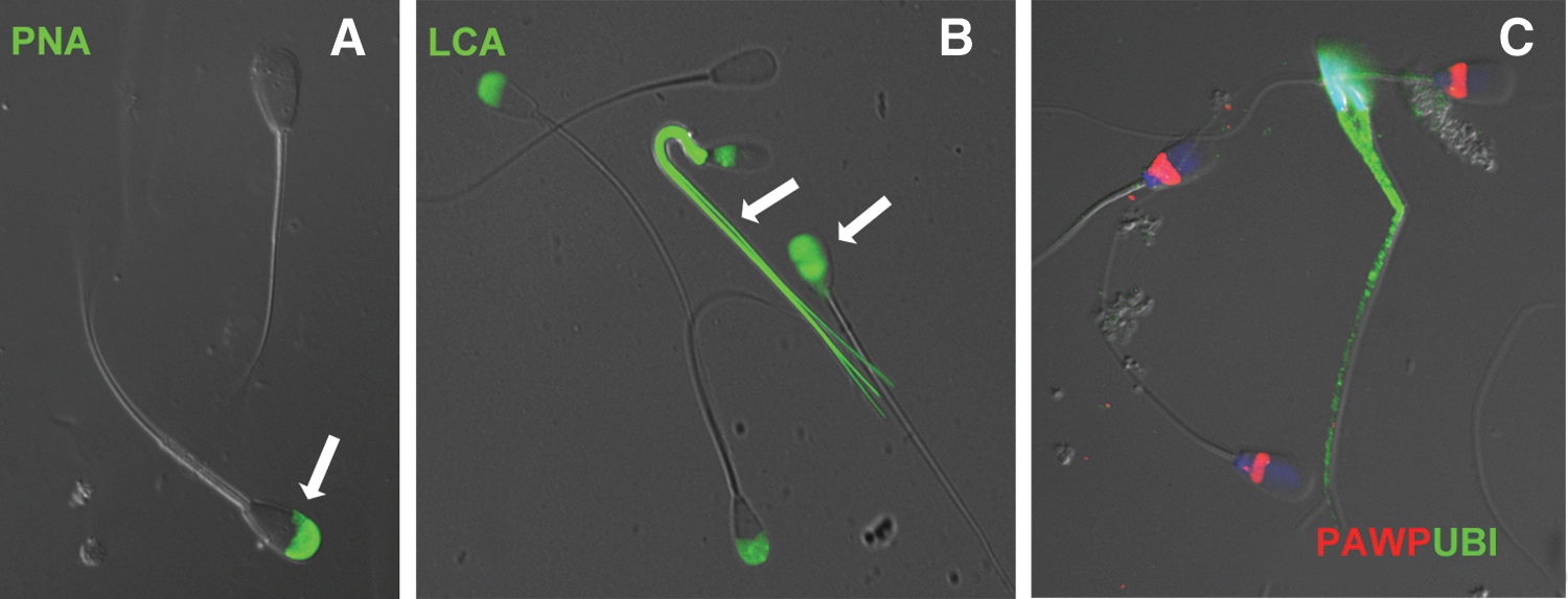
Patterns of biomarker labeling in normal and defective bull spermatozoa. Lectin PNA (green) binds to the caps of the sperm heads in spermatozoa with compromised acrosomes (arrow), but shows no labeling of intact spermatozoa
Candidate sperm quality/fertility biomarkers include proteins that are exclusively or predominantly associated with morphological or molecular sperm defects (“negative” fertility biomarkers) and proteins more abundant in morphologically and functionally normal spermatozoa (“positive” biomarkers of sperm quality/fertility). One of the protein biomarkers of bull sperm quality that has been studied in depth is ubiquitin. Similar to lectin ligands, ubiquitin is present on the surface of defective spermatozoa and can be easily targeted by nanoparticles with specific affinity to it (particles coated with ubiquitin-binding antibodies or with recombinant, genetically engineered proteins that specifically interact with ubiquitin). Contrary to lectins, and perhaps as an added advantage for semen nanopurification, ubiquitin recognizes not only spermatozoa with damaged acrosomes, but also those with other types of sperm head and tail defects, including cells with compromised DNA. 32 During sperm maturation in the epididymis–a sperm storage gland attached to the testicular surface–abnormal spermatozoa are tagged on their surfaces by ubiquitin through the process of protein ubiquitination. 31,33 Though some of these ubiquitin tagged spermatozoa may disintegrate and be removed in the epididymis, many appear in the ejaculate, and their increased content is indicative of poor semen quality or even infertility. 32,34 Increased binding of fluorescently labeled anti-ubiquitin antibodies to the sperm surface reflects the occurrence of sperm abnormalities (Fig. 1C). This can be detected by flow cytometry as an increase in the relative fluorescence induced due to the presence of ubiquitin on the sperm surface.
Ubiquitin has been used as a sperm biomarker in numerous species, including men, stallions, bulls, and boars. 34 –37 In humans, sperm ubiquitin also correlates negatively with pre-embryo development after assisted fertilization. 38 Ubiquitin has also been used for validation of other candidate biomarkers of sperm quality, including platelet-activating factor receptor (PAFR), and more recently, post-acrosomal, WW domain-binding protein (PAWP). 39,40 Proper integration of PAWP in the sperm head structures appears to be reflective of bulls' sperm quality and of sperm head morphology in particular. Defective spermatozoa displayed various anomalies of PAWP labeling and often show the presence of ubiquitin on their surface (Fig. 1). 41 Consequently, correlations are being explored between flow cytometric PAWP levels and the parameters of bull sperm quality and fertility.
Nanoparticle-based Fertility Test
Heparin-binding proteins (HBP) secreted by the sex accessory glands are present in bull spermatozoa and seminal plasma. One particular protein from this family, HBP-30, is more abundant in semen of bulls with high fertility and has come to be known as fertility-associated antigen (FAA) in the AI industry. 42 High semen content of FAA was predictive of high fertility in bulls of several breeds in trials grouping bulls based on the presence or absence of FAA. 42 –44 Groups of FAA-positive bulls were consistently more fertile (by 9–40%) than groups of FAA-negative bulls. 42 Cows covered by FAA-positive bulls were impregnated earlier in the breeding season, resulting in increased numbers of older and heavier calves at weaning, and AI trials on beef cows inseminated using semen from FAA-positive or FAA-negative bulls established FAA as a biomarker of sperm quality. 45 The ReproTest (Midland Bioproducts Corporation, Boone, IA) a lateral flow device (i.e., a dipstick fertility test similar to over-the-counter human pregnancy tests), based on the colloidal gold nanoparticle design, has been developed and marketed. In this device, a semen sample is loaded into a sample well and flows through a series of pads soaked with specific antibodies. The first pad releases primary antibodies conjugated to nanogold particles that bind specifically to the target protein, in this case FAA. The nanogold-tagged sample solution flows toward a test line region in which the secondary antibody, which recognizes a different region of the target protein, is immobilized on several pads in a concentration gradient-like fashion. Target protein molecules tagged with nanogold are captured on these lines, which turn red (the color of colloidal gold in visible light spectrum), indicating a positive test. Extra primary antibodies not bound to target protein flow farther and are captured by anti-immunoglobulin G (IgG) antibodies at the far end of the strip (positive control).
Semen Nanopurification
Removal of defective spermatozoa from bull semen can be achieved by a variety of techniques. Spermatozoa can be overlaid by culture medium in a test tube, and the most motile cells allowed to swim to the upper layer of the medium where they can be collected. This so-called swim-up technique is also used for human-assisted fertilization, but is too lengthy and too low volume to be used for bulk processing of bull semen in an AI stud. 46 A gradient separation method commonly used in test tube, in vitro fertilization (IVF), involves centrifugation at high speed/g-force through a gradient of colloid that retards defective spermatozoa but allows normal, motile spermatozoa to pass through and settle in the pellet, which is then recovered and used for IVF. 47 A discontinuous 45%/90% Percoll gradient is commonly used for bovine IVF. While highly efficient, this method of sperm separation is time-, reagent-, and equipment-intensive, and therefore not suitable for bulk processing of semen for AI. Consequently, magnetic separation methods have been explored by andrologists. 48,49
Based on the discovery of the mechanism for surface ubiquitination of defective spermatozoa in the bull epididymis, we have developed a method for nanodepletion of defective spermatozoa from bull semen during semen preparation for AI doses/straws. 17,33 Nanoparticles composed of magnetite and mixed iron oxides were prepared using a proprietary method for conjugation of antibodies and lectins. Custom conjugation was performed by Clemente Associates (Madison, CT). Different batches of particles were coated with commercially available anti-ubiquitin antibodies or lectin PNA. The ubiquitin-binding particles were designed to bind to ubiquitin protein, found exclusively on the surface of defective bull spermatozoa. 32,39 Particles coated with lectin PNA bind to glycans exposed by the damage to or premature remodeling of the sperm head acrosome. 29 Preliminary laboratory tests were conducted to assess the effect of nanopurification on sperm viability and semen content of defective spermatozoa and to optimize the nanopurification protocol (Fig. 2). Based on these tests, IVF trails were conducted, followed by two field insemination trials.
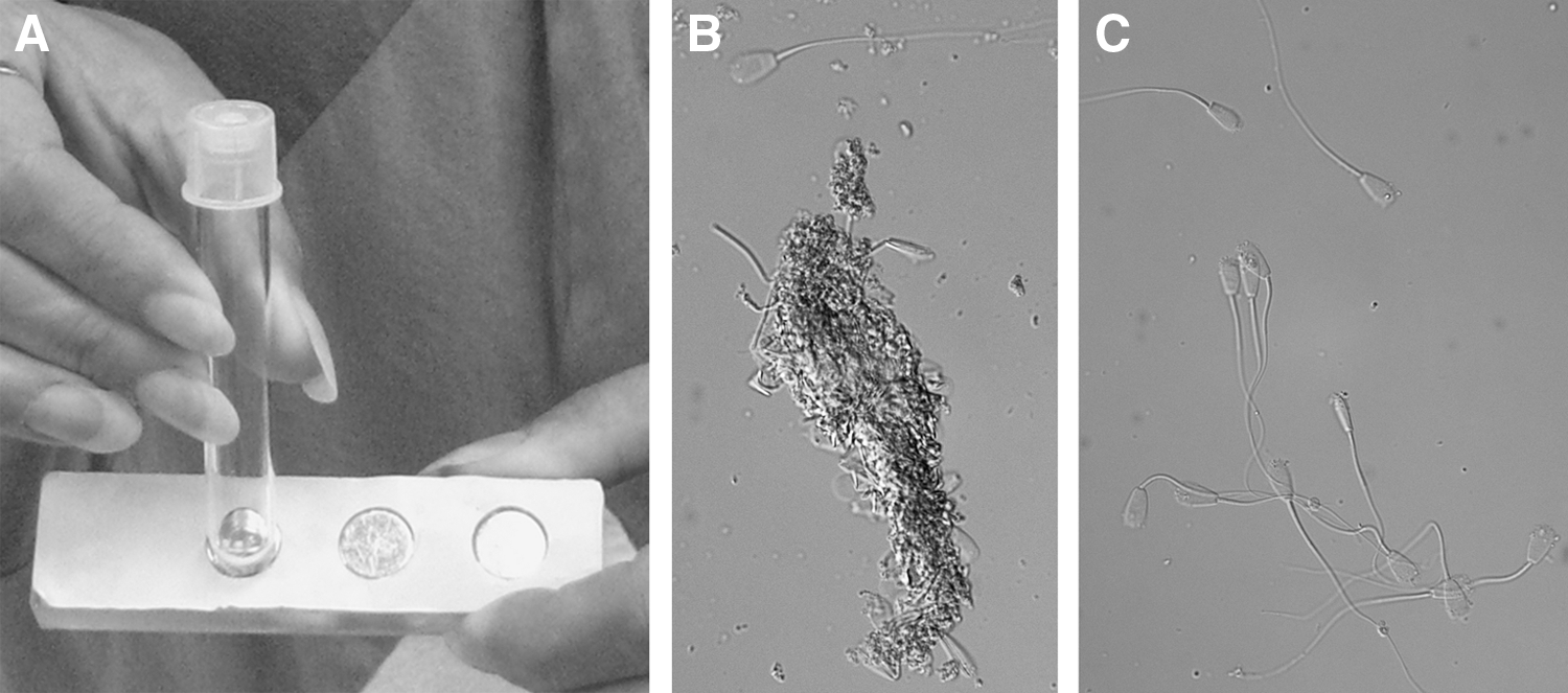
Nanopurification of bull semen for artificial insemination. A magnet is used to attract the nanoparticle-coated defective spermatozoa to the bottom of the collection tube
Data from both trials showed improvement in conception rates after insemination for some treatments and animal groups. In the first trial, a total of 499 cows and heifers were inseminated by semen from three bulls. In the second trial, 422 cows were inseminated by the same bull. In both cases, each bull's ejaculate was used at a full dose of 20 million non-purified spermatozoa, a half dose (10 million) of non-purified spermatozoa, a half dose of spermatozoa nanodepleted with ubiquitin-binding nanoparticles, or a half dose of spermatozoa nanopurified with PNA-coated nanoparticles. In both trials, conception rates achieved with a half dose of PNA-particle purified spermatozoa matched the conception rate of a non-purified full dose and were significantly higher than that of a non-purified half-dose. Differences in conception rates with nanopurified semen were observed between sires and also comparing cows to heifers in the first trial, suggesting that heifers and some sires may benefit from nanopurification more than others. Importantly, no adverse effects on inseminated animals were observed. In these trials, each type of particle was tested separately. In the future, combinations of two or more nanoparticle types will be tested to develop a multiplex nanopurification procedure. Preliminary findings were presented at the meeting of the Association for Applied Animal Andrology, in Vancouver, BC, July 28–29, 2012.
These results will encourage further exploration of nanodepletion protocols for routine use by the AI industry. Semen collections with inferior sperm quality are routinely discarded in the AI industry if they come from bulls with limited data on semen quality and field fertility. However, there is a concerted effort, guided by the US National Association of Animal Breeders (NAAB), to maximize semen extension and to utilize as many ejaculates as possible from elite bulls with high field fertility and valuable genomes. 50 It is this elite group of progeny-tested sires that would most likely be targeted for nanopurification of ejaculates with high content of defective spermatozoa. According to the NAAB, semen samples that fail to achieve quality control minimums can be released for AI use based on superior genetic value, imminent demise of the sire, or at the request of a bull's owner. 50
Two main types of sperm defects may be found in the semen of bulls used for AI: the compensable defects such as sperm tail defects and cytoplasmic droplets; and the non-compensable defects including but not limited to nuclear craters/diadems and knobbed acrosomes. The detrimental influence of compensable defects on AI outcome can be alleviated by increasing the total number of spermatozoa per AI dose. On the contrary, adding more spermatozoa per dose does not improve conception rates in bulls with a high percentage of spermatozoa with non-compensable defects. 51 Semen nanopurification will most likely benefit the former category. Even so, it may still be useful to explore whether nanopurification would increase conception rates of some bulls in the non-compensable category, particularly the ones with a low percentage of affected spermatozoa.
In future development of semen nanopurification protocols, attention will be paid to possible reprotoxic effects of nanoparticles that come into contact with spermatozoa. As the use of nanomaterials becomes more widespread, concerns increase with regard to nanotoxicity conveyed by air and food. In particular, nanoreprotoxicity studies in animal models have shown that nanoparticles administered orally or by injection can cross protective barriers in male and female reproductive systems, potentially harming fertility and causing serious birth defects, particularly in the central nervous system. 52 Such detrimental effects are being addressed by the development of biocompatible and biodegradable nanomaterials. Complete removal of unbound nanoparticles from purified semen will have to be assured to prevent nanoparticle contamination of the AI doses, from which the particles could enter the female reproductive system after insemination. Thus far, we have not observed any detrimental effects on cow health and fertility after AI with nanodepleted particles, most likely because there was minimal carryover of unbound particles in the AI doses.
Conclusions and Future Directions
Semen nanopurification trials will provide proof-of-concept of semen nanopurification in the setting of a commercial bull stud. Importantly, no side effects related to residual nanoparticles present in nanopurified semen have been observed thus far. Field trials with PNA and ubiquitin nanoparticles were preceded by extensive laboratory research, validating this approach by flow cytometry and test tube, in vitro fertilization. Depending on the observed pregnancy rates in cows vs. heifers, it is possible that nanopurification treatments could be tailored specifically to boost fertility in replacement females entering the breeding programs. The main focus of the ongoing and future AI trials will be on optimization of nanoparticle doses and nanopurification protocols, with the goal of maximizing the number of AI doses per semen collection from sires with high genetic value. Research will also be pursued to identify additional nanopurification targets—the negative fertility biomarkers expressed on the surface of defective sperm cells. Further testing will also be needed to assure the safety of nanodepleted semen; residual nanoparticles that could remain in semen after nanodepletion could enter the female body through the reproductive system and have adverse effects on reproduction and other bodily functions (nanoreprotoxicity). 52 A lateral flow device similar to that used for the FAA test could be developed for routine chute-side fertility testing of bulls.
Footnotes
Acknowledgments
The authors would like to thank their collaborators including Richard Oko, PhD, of Queen's University, Kingston, ON, Canada; Tom Geary, PhD, of United States Department of Agriculture (USDA), Fort Keogh, MT; Mel De Jarnette, PhD, of Select Sires; Eric Schmitt, DVM, PhD, of IMV Technologies; and Kari Beth Krieger, PhD, of Genex Cooperative, for their support of original research cited and discussed in this article. Collegial support from Miriam Sutovsky, Shawn Zimmerman, Young-Joo Yi, PhD, and Edward (TJ) Miles is much appreciated. Original research reviewed in this article was in part supported by National Research Initiative Competitive Grant no. 2007-35203-18274 and grant no. 2011-67015-20025 from the USDA National Institute of Food and Agriculture, to Peter Sutovsky.
Author Disclosure Statement
No competing financial interests exist.