




Abstract
Cellulose nanocrystals (CNCs) are a biobased nanomaterial attracting increasing interest for a range of potential applications. This article reviews the current literature on the pulmonary, oral, dermal, and cytotoxicity of CNCs. Current studies of the oral and dermal toxicity of CNCs have shown a lack of adverse health effects, whereas studies of the pulmonary and cytotoxicity have yielded discordant results. Additional studies are needed to support the general conclusion that CNCs are nontoxic on ingestion or contact with the skin and to determine whether CNCs have adverse health effects on inhalation or elicit inflammatory or oxidative stress responses at the cellular level. This review underscores the importance of careful sample characterization and exclusion of interfering factors, such as the presence of endotoxins or toxic chemical impurities, for a detailed understanding of the potential adverse health effects of CNCs by various exposure routes.
Introduction
Cellulosic nanomaterials are an emerging class of nanomaterials with several desirable properties: they are produced from a renewable starting material at relatively low cost, are biodegradable, biocompatible, and have high water absorption capacity, mechanical strength, and stiffness. Consequently, cellulosic nanomaterials are being studied for a number of potential applications, including polymer nanocomposites, transparent or chiral films, rheology modifiers and hydrogels, drug-delivery vehicles, artificial blood vessels, and wound dressings. 1 –7 However, before a material or technology can be commercialized, its impact on the environment and human health needs to be thoroughly assessed. The literature on cellulosic nanomaterial toxicity has recently been summarized in two review articles covering the use of cellulosic nanomaterials in biomedicine. 8,9 The present review provides a more in-depth look at the effects of cellulose nanocrystals (CNCs) on human health.
CNCs can be obtained from different starting materials, including tunicin, bacterial cellulose, algal cellulose, wood pulp, bast fibers, cotton linters, and microcrystalline cellulose. 10,11 Current studies of CNC toxicity have focused on plant fiber-derived CNCs, which are shorter and have smaller cross-sectional dimensions than those derived from animal, bacterial, or algal cellulose. Wood-derived CNCs, for example, have average lengths of 100–200 nm and average cross-sectional dimensions of 3–5 nm (Fig. 1). 11,12
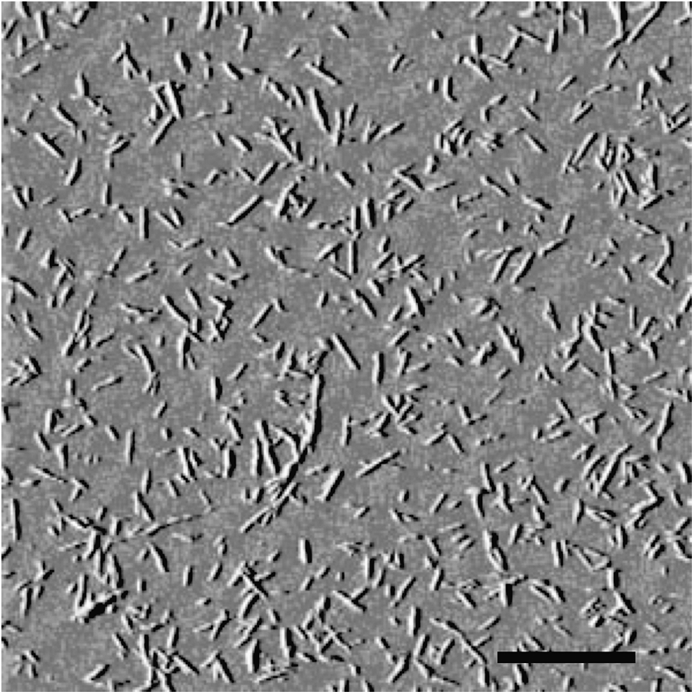
Wood-derived cellulose nanocrystals (scale bar: 1 μm). Adapted with permission from Roman M. Cellulose nanocrystals.jpg (Wikimedia Commons)
CNCs are typically prepared by acid hydrolysis of the cellulose starting material. When sulfuric acid is used, the hydroxyl groups on the CNC surface become partially esterified; the resulting sulfate half-esters impart acidic properties to CNCs. With respect to CNCs' effects on human health, their acidic properties might be of minor concern because of the buffering capacity of the human body. Their pH-lowering effect should, however, be considered in cytotoxicity assessments and can altogether be prevented by using the sodium salt form. The pKa of the sulfate half-esters on CNCs has been reported as 2.46, which means that CNCs are fully ionized–i.e., have a degree of ionization of 1.00–at a pH of 4.76 and above. 13 Consequently, sulfate group-bearing CNCs have a negative surface charge at physiological pH levels, diminished only in the low pH environment of the stomach. The negative surface charge gives rise to repulsive Coulomb interactions between the CNCs, preventing aggregation due to attractive forces, such as hydrogen bonding. However, because of the abundance of sodium and other cations in body fluids and their charge-shielding effect, aggregation of CNCs in these fluids might nevertheless occur.
Many nanomaterials have been shown to have adverse health effects upon entering the body. 14,15 Unintentional or coincidental uptake of nanoparticles into the body generally occurs by inhalation, ingestion, or transdermal absorption. In addition, nanoparticles may be present in medications or vaccines administered by injection. Because no studies have yet been published on the parenteral toxicity of CNCs, this literature review focuses on their potential and demonstrated pulmonary, oral, dermal, and cytotoxicity.
Pulmonary Toxicity
Pulmonary toxicity is the medical term for any adverse health effects that occur when a foreign substance enters the respiratory tract. The respiratory tract has three regions: the nasal–pharyngeal–laryngeal (NPL) region, the tracheobronchial region, and the alveolar (gas-exchange) region (Fig. 2). 16,17 The tracheobronchial region consists of the trachea, which bifurcates into two primary bronchi and further subdivides into secondary bronchi, tertiary bronchi, and bronchioles of progressively smaller diameter. The bronchi and bronchioles are lined by a columnar epithelium (cell lining) of 0.5–5-mm thickness that is covered with a negatively charged mucus layer (isoelectric point [pI]=2.72) of approximately neutral pH. 15,18,19
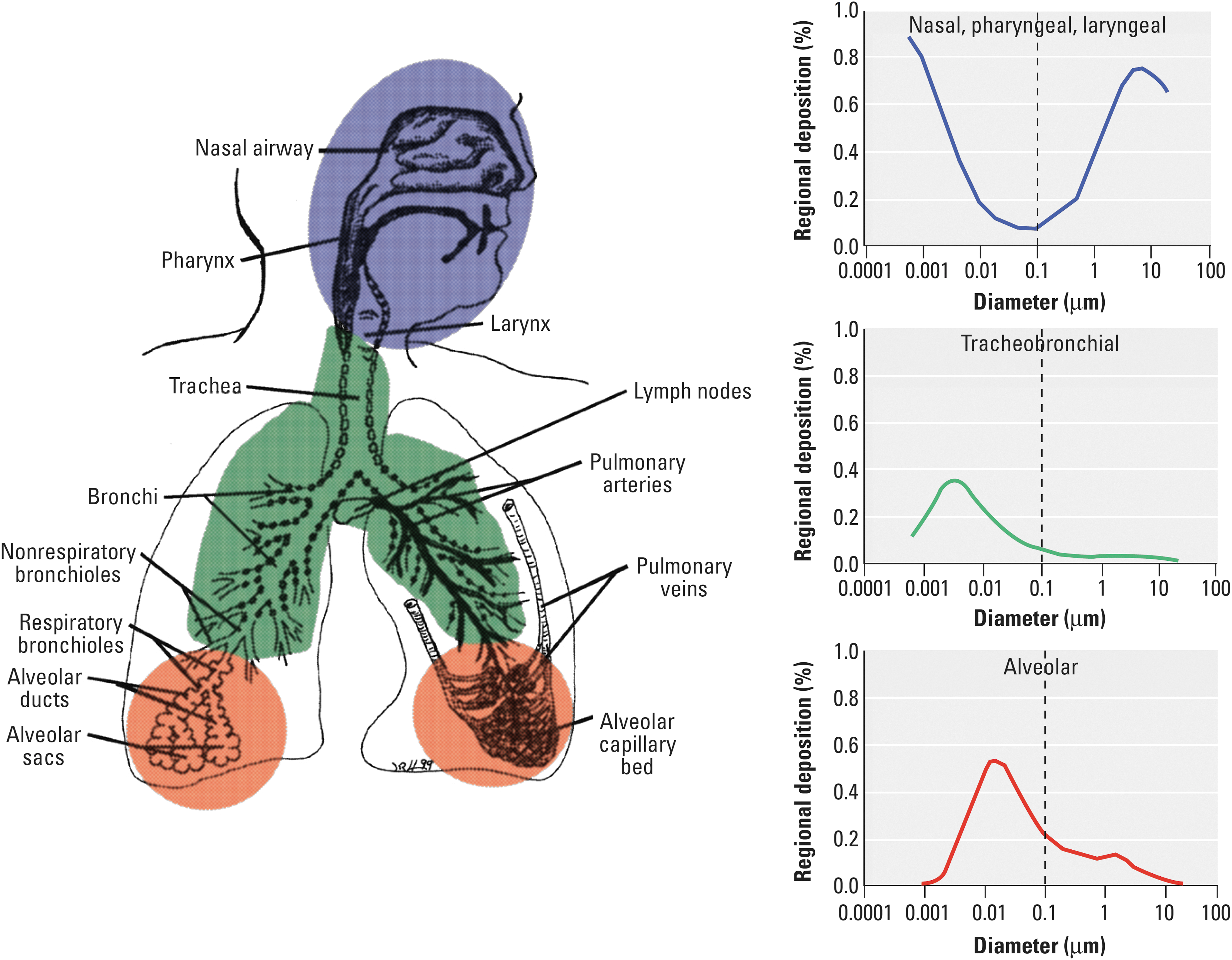
Predicted fractional deposition of inhaled particles in the nasopharyngeal, tracheobronchial, and alveolar region of the human respiratory tract during nose breathing. Based on data from the International Commission on Radiological Protection. Reproduced with permission from Environmental Health Perspectives. 17
In contrast, the walls of the alveoli (microscopic sacs responsible for gas exchange) consist of a single cell layer covered only by a thin (∼0.1 μm) liquid layer. 20 A mathematical model by the International Commission on Radiological Protection predicts that very small (∼1 nm) and very large (∼10 μm) inhaled particles are deposited primarily in the NPL region, whereas particles of about 5 nm in size are deposited approximately equally in all three regions, and particles of about 20 nm in size are deposited primarily (∼50%) in the alveolar region (Fig. 2). 16, 17 Particles that are deposited in the NPL and tracheobronchial regions are primarily cleared from the respiratory tract toward the mouth via the mucociliary escalator (through the movement of microscopic hair-like structures, termed cilia), whereas particles deposited in the alveolar region are cleared primarily by alveolar macrophages through phagocytosis (engulfment) followed by intracellular degradation or transport to the mucociliary escalator. Besides these classical clearance mechanisms, a few other mechanisms of nanoparticle clearance from the lung have been identified, including translocation through the epithelium into the central nervous system by neuronal uptake or into the interstitial space (between the alveoli and lung capillaries), potentially followed by uptake into the lymphatic system or translocation through the vascular endothelium into the blood circulation. 17 The effects of CNCs upon entering the respiratory tract and their mechanism of clearance from it will depend largely on their degree of aggregation, which determines particle size (and therefore the location of deposition in the respiratory tract) and their surface charge, which governs their interactions with respiratory mucus and cells.
To date, only a few studies have investigated the pulmonary toxicity of CNCs. Yanamala et al. assessed the adverse effects of CNCs produced by the US Forest Service's Cellulose Nano-Materials Pilot Plant at the Forest Products Laboratory (Madison, WI) in adult female C57BL/6 mice upon pharyngeal aspiration. 21 The plant produces CNCs from machine-dried prehydrolysis kraft rayon-grade dissolving wood pulp by hydrolysis with 64% sulfuric acid at 45°C for 90 min, followed by dilution, neutralization of the acid with NaOH, and membrane filtration. 22 It should be noted that the plant's purification process involves the addition of hypochlorite for color removal, which is generally not used in lab-scale methods and might affect the product's toxicity. Two starting materials were tested, a 10 wt% suspension and a freeze-dried powder. Sterile stock suspensions in USP-grade water of 5-mg/mL concentration and pH 7 were prepared from the starting materials by dilution, sonication, and autoclaving. The stock suspensions were diluted further, and mice were administered 50, 100, or 200 μg in a volume of approximately 40 μL. Pharyngeal aspiration is an administration method that involves placement of a liquid sample onto the base of the tongue of the animal and extension of the tongue, resulting in a reflex gasp and aspiration of the liquid. 23 A recent study comparing pharyngeal aspiration to inhalation of single-walled carbon nanotubes found similar outcomes for the two exposure methods. 24 Yanamala et al. found that both CNC materials elicited dose-dependent oxidative stress, tissue damage, and inflammatory responses. At a dose of 200 μg, levels of protein carbonyl and 4-hydroxynonenal, two oxidative stress markers, were on average double that of the control, and at least six of the 23 measured markers of inflammation exhibited a more than 10-fold increase on CNC administration. Moreover, the extent of the response depended on the starting material. CNCs from the 10 wt% suspension, having a mean length and width of 90.2±3.0 and 7.2±2.1 nm, respectively (determined by transmission electron microscopy) caused greater increases in oxidative stress markers and inflammatory mediators than freeze-dried CNCs, which have a mean length and width of 207.9±49.0 and 8.2±2.3 nm, respectively, whereas the latter caused a greater increase in biomarkers for tissue damage. The results of Yanamala et al. are in agreement with those of an earlier in vitro study by Clift et al., who assessed the pulmonary toxicity of cotton filter paper-derived CNCs with a three-dimensional triple cell coculture model of the human epithelial airway barrier. 25 Like Yanamala et al., Clift et al. observed a dose-dependent cytotoxicity and (pro-) inflammatory response. At a dose of 0.03 mg/mL, release of the pro-inflammatory chemokine interleukin-8 by the human bronchial epithelial cell-line 16HBE14o- in the triple cell coculture model was about double that of the control.
In a more recent study, O'Connor et al. assessed the acute inhalation toxicity of NCC™, a commercial CNC material manufactured by CelluForce (Montréal, Canada) through hydrolysis of bleached softwood kraft pulp with 64% sulfuric acid at 45°C for 60 min. 26 The assessment was performed according to test guideline 403 of the Organisation for Economic Co-operation and Development (OECD). Sprague-Dawley stock-derived albino rats were exposed by inhalation for a period of 4 h to aerosolized CNCs at a maximum concentration of 0.26 mg/L in the exposure chamber and monitored for mortality, gross toxicity, and behavioral changes for a period of 14 d. At the end of the test, all animals were subjected to gross necropsy (animal autopsy). No adverse effects of the aerosolized CNCs on the animals were observed. It should be noted, however, that the study did not involve characterization of the aerosolized CNCs; the properties of the particles inhaled by the rats—the size, shape, and surface charge, in particular—are therefore unknown.
Oral Toxicity
Oral toxicity is measured in terms of any adverse health effects of a substance entering the orogastrointestinal tract through the mouth. The orogastrointestinal tract comprises the oral cavity, the esophagus, the stomach, and the small and large intestines. Bypassing the oral cavity, an alternative route for substances into the gastrointestinal tract is via clearance from the respiratory tract by the mucociliary escalator. The orogastrointestinal tract is lined by an epithelium with varying properties along the tract (Table 1). 27 The orogastrointestinal epithelium is covered by a mucus layer, which contains various proteins, including mucin and antiseptic proteins, such as lysozymes. The thickness of the mucus layer varies from 70–100 μm in the oral cavity to over 1000 μm in the stomach, where it is the thickest. 27 Mucus in the oral cavity has a pH of about 6.6, whereas the pH of stomach mucus ranges from 1–2 at the luminal surface to about 7 at the epithelial surface. The pH of the intestine changes from 6 in the duodenum, to about 7.4 in the terminal ileum, to 5.7 in the cecum, and to 6.7 in the rectum. 28
Properties of the Orogastrointestinal Epithelium 27
Most studies of nanoparticle uptake by the gastrointestinal tract have shown that nanoparticles pass through the tract and are eliminated from the body in the feces. 17 However, some studies have demonstrated permeation of the gastrointestinal barrier by micro- and nanoparticles. 27 In addition, penetration of the buccal mucosa by nanoparticles has recently been shown. 29 Particle translocation through a mucous membrane has four components: diffusion through mucus, initial contact with the epithelium, cellular trafficking, and post-translocation events. 30 A particle's ability to diffuse through mucus depends primarily on its size, surface charge, and hydrophilicity. The main structural component of mucus is a three-dimensional network of mucin, a high molecular weight, highly glycosylated glycoprotein. Smaller particles diffuse more readily through the mucin network than larger particles. Based on a simple cubic-lattice model of cylindrical mucin fibers of 3.5-nm radius, the mesh spacing within human cervical mucus has been predicted to be 100 nm. 31 The electrostatic properties of mucin are governed by glutamic and aspartic acid residues (pK a≈4) in its polypeptide backbone and sialic acid residues (pK a≈2.6) and sulfate groups (pK a≈4) in its oligosaccharide side chains. The isoelectric point of porcine gastric mucin, which is similar in composition to human mucin, has been determined to lie between pH 2 and 3. 32,33 In other words, mucin is negatively charged in most sections of the orogastrointestinal tract. As a result of its negative charge, positively charged nanoparticles become entrapped in and diffuse much more slowly through mucus than do negatively charged ones. 34 For nanoparticles to reach the underlying epithelium, however, they have to penetrate the mucus layer quickly because of its rapid turnover. 27 In the stomach, mucus is secreted at a rate that makes it unlikely for even the smallest non-mucoadhesive nanoparticles to reach the gastric epithelium. 35
In the intestine, nanoparticles that penetrate the mucus layer have four possible exit routes through the intestinal epithelium, potentially followed by entry into the lymphatic system or blood circulation (Fig. 3): 1) through direct uptake by M-cells in the Peyer's patches of the gut-associated lymphoid tissue; 2) by passive diffusion through the enterocytes; 3) by active transcellular transport; and 4) by paracellular translocation through the tight junctions between the cells. 36,37 Route 1 has primarily been observed for uncharged hydrophobic particles, whereas Route 4 is restricted to particles with dimensions smaller than the physical dimensions of the paracellular space, estimated to lie between 1 and 3–5 nm. 36,38 Neither of these routes is therefore likely to enable significant CNC translocation through the intestinal epithelium. The passive transcellular transport mechanism (Route 2) involves partitioning into and diffusion across the cell plasma membrane and therefore requires a certain level of lipid solubility. 39 Hence, CNCs might not be expected to exit the intestine by this mechanism. Active transcellular passage (Route 3) occurs either through receptor-mediated or adsorptive-mediated transcytosis, i.e., endocytosis (active cellular uptake) at the apical plasma membrane and exocytosis (expulsion out of the cell) at the basolateral plasma membrane of enterocytes. Adsorptive-mediated transcytosis is facilitated by a positive particle surface charge, giving rise to attractive interactions with anionic sites of the plasma membrane. 40 Active transcellular transport mechanisms through the intestinal epithelium, however, will play a minor role in the clearance of nanoparticles from the intestinal lumen because of the low endocytic activity of enterocytes. 40 Consequently, significant permeation of the orogastrointestinal barrier by sulfate group-bearing CNCs is not to be expected.
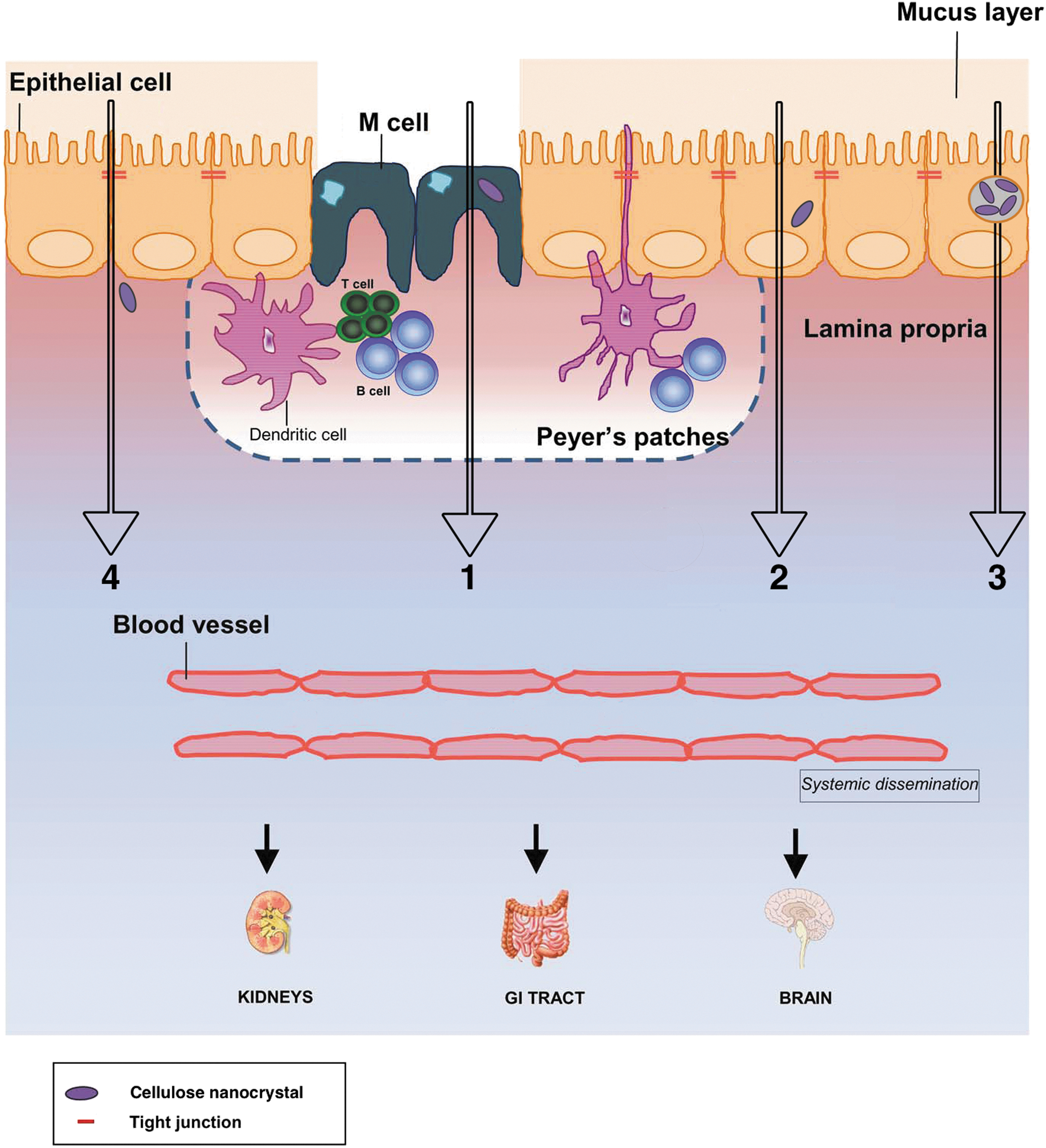
Possible mechanisms of nanoparticle translocation through the intestinal epithelium:
Only two studies of the oral toxicity of CNCs, both reported by O'Connor et al., have been published to date. 26 The studies, conducted according to OECD test guidelines 425 and 407, determined acute oral toxicity as well as oral toxicity upon repeated daily administration of NCC, respectively. Acute oral toxicity was assessed by administration of one-time doses of up to 2,000 mg/kg in aqueous suspension form directly into the stomach of Crl:CD(SD)BR rats by oral gavage (force feeding) and monitoring of the health of the rats for a period of 14 d followed by gross necropsy. Using the same rat strain and administration method, the repeated-dose test was performed by daily administration of doses of 500, 1,000, and 2,000 mg/kg for a period of 28 d. During this period, the animals were closely observed for signs of toxicity. At the end of the test, all animals were subjected to gross necropsy. No adverse effects of CNCs on rats were observed and the median lethal dose was established to be above 2,000 mg/kg.
Dermal Toxicity
Dermal toxicity is measured in terms of any adverse health effects of a substance contacting the skin. Human skin has three layers—the epidermis, dermis, and hypodermis (Fig. 4). Among other functions, the hypodermis thermally insulates the body with adipose tissue, and the dermis provides blood circulation to the epidermis. The main function of the epidermis is to provide a barrier and prevent pathogens from entering the body. The epidermis is a stratified (layered) squamous epithelium, consisting of five strata (Fig. 4). Besides keratinocytes—the main cell type, producing the structural protein keratin—the epidermis contains melanocytes, which produce the skin pigment melanin; Merkel cells, a component of the somatosensory system; and Langerhans cells, which are antigen-presenting immune cells. 41
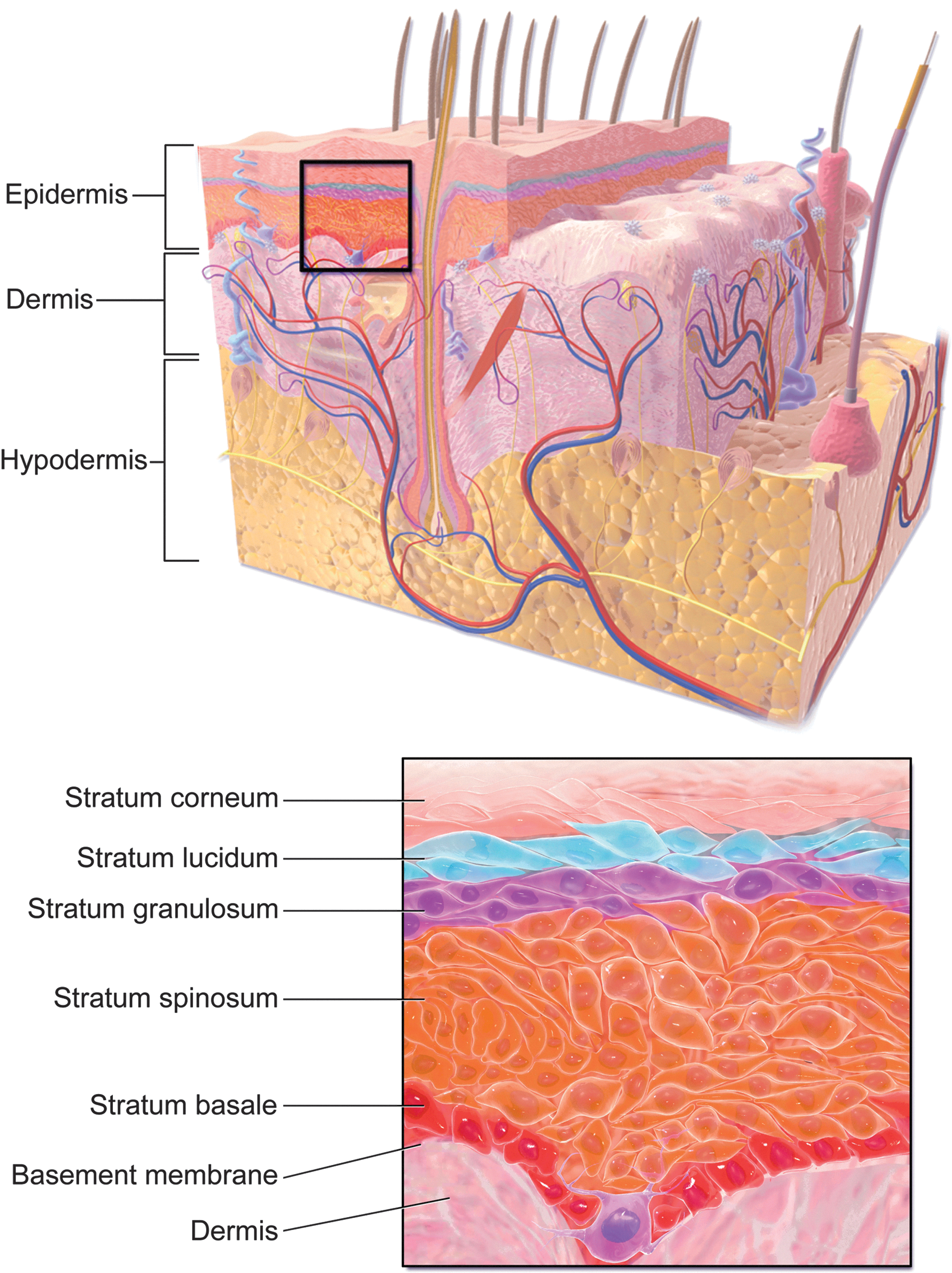
Structure of the epidermis. Adapted with permission from Blausen gallery 2014, Wikiversity Journal of Medicine.
For a skin-contacting substance to have an effect on human health, it must first penetrate the stratum corneum, the top-most layer of the epidermis. The stratum corneum is composed of clusters of corneocytes, which are terminally differentiated keratinocytes, embedded in a lipidic matrix. Penetration of the stratum corneum occurs solely by passive diffusion because corneocytes do not possess the ability for active internalization of materials. 42 Apolar regions in the lipidic matrix potentially enable intercellular diffusion through the stratum corneum of apolar, lipophilic permeates smaller than 5–7 nm, whereas polar regions in the lipidic matrix, termed aqueous pores, potentially enable intercellular diffusion through the stratum corneum of polar, hydrophilic permeates smaller than 36 nm. 43 The latter pathway, however, may be purely hypothetical because the permeability of the stratum corneum to water molecules has been shown to be very low. 44 In addition to intercellular diffusion, transcellular diffusion of substances through the corneocytes is possible. This pathway, however, requires repeated partitioning into and out of corneocytes and intracellular and paracellular diffusion through hydrated keratin and the lipidic matrix, respectively. 45 Another potential skin-penetration route, the transfollicular route, is through epidermal invaginations, such as sweat glands and pilosebaceous units, comprising the hair shaft, hair follicle, sebaceous gland, and arrector pili muscle. 43 The transfollicular route potentially accommodates permeates up to 210 μm in size but requires that they are dispersible in sweat, a dilute aqueous mixture of organic acids, carbohydrates, amino acids, nitrogenous substances, vitamins, and electrolytes; or sebum, a mixture of squalene, waxes, cholesterol derivatives, triglycerides, fatty acids, and cell debris. 43 As of yet, however, no penetration of skin-contacting substances into the sweat glands has been reported. 42 Furthermore, because of their low density and outward excretions, epidermal invaginations are thought to play a minor role in the dermal absorption of substances. The majority of studies assessing the dermal absorption of nanoparticles reported no unintentional permeation of nanoparticles through the skin. 15 Accordingly, because of their relatively large size compared to transdermal drug molecules and considerable polar and hydrophilic properties, significant permeation of the dermal barrier by CNCs is not to be expected.
The most common adverse health effect of substances that penetrate the stratum corneum is skin sensitization. 46 Skin sensitization occurs when a substance that has reached the viable layers of the epidermis, encompassing the stratum granulosum, stratum spinosum, and stratum basale, forms a stable association with skin proteins, triggering dendritic cells to migrate to the lymph nodes and activate T lymphocytes. 47 O'Connor et al. have assessed the skin-sensitizing potency of CNCs in vivo with the guinea pig maximization test (OECD test guideline 406) and the murine local lymph node assay (OECD test guideline 429). 26 In the guinea pig maximization test, Crl:(HA)BR guinea pigs were exposed to CNCs by intradermal injection of 0.1 mL of a CNC suspension having a concentration of 1.1 mg/mL. A week later, a CNC gel with a concentration of 103 mg/mL was applied topically for 48 h with a filter paper and occlusive dressing. The topical application was repeated two weeks later for 24 h. CNCs were found to be nonsensitizing at the concentrations tested. In the murine local lymph node assay, CBA/J mice were treated for three consecutive days with 25 μL of CNC suspension, applied topically to the dorsum of each ear. Three CNC concentrations, ranging from 2.5 to 10.7%, were tested. Three days after the last treatment, proliferation of lymphocytes in the draining auricular lymph nodes, as a measure of dose and potency of the test substance, was determined following euthanasia of the mice injected with a radioactive lymphocyte label and excision and processing of the relevant lymph nodes. On the basis of the test results, CNCs were not considered to be a contact dermal sensitizer at concentrations≤10.7%. In addition to their skin-sensitizing potency, O'Connor et al. determined the ability of CNCs to cause reversible and irreversible skin tissue damage, known as dermal irritation and corrosion, respectively. 26 In this study, performed according to OECD test guidelines 404 with Crl:KBL(NZW)BR albino rabbits, a CNC gel containing 0.5 g of CNCs was applied topically for 4 h with a gauze patch and semi-occlusive dressing. Following exposure, animals were observed for 14 days and examined for signs of erythema and edema. Skin response was scored on a scale of 0 to 4 at 60 min, 24 h, 48 h, and 72 h after removal of the patch, and the individual scores at each time point were used to calculate the Primary Irritation Index. No corrosive effects were observed and the material (NCC) scored a Primary Irritation Index of 0 (nonirritating).
Cytotoxicity
Cytotoxicity refers to adverse effects of a substance on cell viability. Evaluation methods of cytotoxicity can be grouped into four categories: 1) methods that assess cell damage on the basis of cell morphology; 2) methods that measure cell damage; 3) methods that measure cell growth; and 4) methods that measure specific metabolic activities. A number of research teams have conducted cytotoxicity studies with CNCs. The reports available in the literature to date are summarized in Table 2. 25,48 –57 Most studies showed CNCs to have no cytotoxic effects, as defined by ISO standard 10993-5, i.e., not to reduce cell viability by more than 30%. However, several studies found a dose dependency of CNC cytotoxicity. 25,49,53,56,57 The most pronounced decrease in cell viability was reported by Kovacs et al., who reported a decrease in rainbow trout hepatocyte viability of 20% at an estimated CNC concentration of 0.034 mg/mL and a decrease of 50%, i.e., cytotoxic effects at an estimated concentration of 0.245 mg/mL. 49 The results of Kovacs et al. are in stark contrast to the findings of a more recent study by Zoppe et al., who reported human corneal epithelial cell viabilities in excess of 90% at a CNC concentration of 0.1% (∼1 mg/mL). 56 The discrepancies in the results of current CNC cytotoxicity studies are not surprising, however, considering that they all used different cell lines, cellulose sources, CNC preparation procedures, and post-processing or sample preparation methods (Table 2).
Current CNC Cytotoxicity Studies
Reagent, reaction temperature, reaction time, reagent-to-cellulose ratio.
For the experimental conditions used.
NA, not available.
9 M H2SO4, 3:1 (v/v) mixture of 18 M H2SO4 and 12 M HCl, or 12 M HCl.
Different cellulose sources, preparation procedures, and post-processing methods, such as sonication, will obviously yield CNCs with different physicochemical properties, and CNCs with different particle dimensions and surface chemistries will likely have different effects on cell viability. Most studies, therefore, include CNC characterization to some extent. It is important to note, however, that post-processing methods such as autoclaving may alter the physicochemical properties of CNCs, making it critical to determine these properties in the final samples. A frequently overlooked factor is that cellulose sources sometimes contain endotoxins or cytotoxic chemicals from prior processing, such as pulping, that could affect cell viability. In addition, the CNC preparation or post processing procedures themselves might introduce cytotoxic impurities into the material. For example, heating of sulfate group-bearing CNCs, as done during autoclaving, is known to cause desulfation and the release of sulfuric acid from the CNC surface. 58 In ISO standard 10993-12, residues from the manufacturing process are considered integral to the medical device under evaluation. However, if one is interested in the cytotoxicity of CNCs per se, as opposed to a commercial product, the interfering effects of endotoxins and cytotoxic contaminants need to be eliminated through the use of suitable blanks. In addition, as mentioned in the introduction, the acidic or pH-lowering properties of CNCs need to be considered. A significant decrease in the culture medium's pH on CNC addition could negatively affect cell viability. If the pH-lowering effect of CNCs is to be excluded from the study, their sodium salt form should be used. Last but not least, the degree of aggregation or dispersion of the CNCs in the cell culture medium needs to be assessed for accurate information on the particle size to which the cells are being exposed. Needless to say, all of these factors (presence of cytotoxic impurities and design of test blanks, effect of CNCs on medium pH or use of the sodium salt form, and aggregation of CNCs in the culture medium) need to be carefully addressed in the report of the study. Of the reports listed in Table 2, only two considered the effects of CNC acidity and aggregation, and several of the reports failed to describe the methods of cell exposure or sample sterilization in sufficient detail to allow evaluation of their potential effects on the test results. 52,54 Future CNC cytotoxicity studies should address all of the above-mentioned factors to allow correlation of their results.
Conclusions
Current studies of the oral and dermal toxicity of CNCs have shown a lack of adverse health effects. The available studies, however, are still very limited in number (two oral toxicity studies and three dermal toxicity studies) and in the variety of tested CNC materials (CelluForce's NCC). Additional oral and dermal toxicity studies are needed to support the general conclusion that CNCs are nontoxic upon ingestion or contact with the skin. Studies of pulmonary and cytotoxicity, on the other hand, have yielded discordant results. The questions of whether CNCs have adverse health effects on inhalation and whether they elicit inflammatory or oxidative stress responses at the cellular level therefore warrant further investigation. The toxicity of CNCs will depend strongly on their physicochemical properties—in particular, surface chemistry, including particle charge, and degree of aggregation, which determines particle shape and dimensions. Therefore, these properties—which in turn depend strongly on the cellulose source, CNC preparation procedure, and post-processing or sample preparation methods, such as lyophilization, aerosolization, sonication, or sterilization—need to be carefully measured in the final samples.
Another factor that might affect the outcomes of toxicity studies are sample contaminants, such as endotoxins or toxic chemical impurities. Samples for exposure tests should therefore be carefully analyzed for such contaminants prior to testing. Ideally, because detection of toxic chemical contaminants may be difficult, control experiments should be carried out with suitable blanks from which the CNCs have been removed, for example by membrane filtration. Moreover, especially in cytotoxicity assessments, the effect of CNCs on pH and their aggregation in the cell culture medium need to be monitored. Only by careful particle characterization and exclusion of interfering factors will we be able to develop a detailed understanding of the potential adverse health effects of CNCs.
Footnotes
Acknowledgments
Funding for this work was provided in part by the Virginia Agricultural Experiment Station (Blacksburg, VA) and the Hatch Multistate Program of the National Institute of Food and Agriculture, US Department of Agriculture.
Author Disclosure Statement
No competing financial interests exist.