
Abstract
Spherical nanoparticles are attracting increasing interest for diverse commercial uses due to their many useful attributes such as applicability as controlled-release systems and their super-hydrophobicity. In this review, methods of particle formation for cellulose, its derivatives, and related species are discussed with reference to numerous specific examples. Techniques for characterization of particle size, size-distribution, and related properties under both dispersed and solid particle conditions are summarized and contrasted. Finally, we note potential applications for these surprisingly reproducible nanospheres.
Introduction
The idea of science and technology at the nanoscale, without actually using any form of the prefix “nano” was introduced by Richard Feynman in his 1959 lecture “There's plenty of room at the bottom”. 1 Typically, nanoscale refers to features with at least one dimension in the range of 1–100 nm (1 nm=10−9 m), a scale in which materials often exhibit unique physical properties relative to those associated with comparable micro- or macro-scale particles. Every year thousands of reports are published describing physical, chemical, and biological properties of nanoscale particles, new nanotechnologies, and diverse applications in areas ranging from medicine, to effective drug-delivery systems, nanoscale materials, advanced functional thin films, and faster electronics. 2 –4 In Europe, the nanoparticle system description is slightly modified by specifying that at least 50% of the individual particles must have one or more dimensions in the range 1–100 nm. 5
This review focuses only on particles incorporating cellulose, cellulose derivatives, or related materials, and on particles of those materials that can be described further as nanospheres, as opposed to whiskers or nanofibers. Such particles are of greatest interest with respect to sustained/controlled release of drugs, fragrances, and other small molecules, topics that have been widely reviewed elsewhere. 3,4,6 This review discusses methods of preparation that influence both particle size and distribution, as well as the density and composition of the particles. For example, particles ground from larger, crystalline particles are likely to be semicrystalline solids with little or no internal liquid component. Such materials are often compounded with polymer matrixes forming nanocomposites, where the particles act as reinforcing fillers. 5 However, particles made by any of the several emulsion processes discussed below are likely to contain a considerable fraction of the fluids in which they were formed. These spaces may, in turn, provide convenient internal surfaces in which smaller molecules, including drug molecules, fragrances, and other small molecules, are likely to be bound. In a manner not unlike the binding of small molecules within size-exclusion chromatography matrices, the release of these smaller particles is governed by the distribution of pore sizes in the particles. While many of the papers noted here deal with the specifics of drug release, this review focuses on the preparation methods and effect on the particles. Due to space limitations, the important role of surface modification on particle uptake will not be discussed in detail.
The diversity of nanoparticle size, shapes, and origins was reviewed by Buzea, Pacheco, and Robbie. 6 Besides dimensions, other important considerations include both the external and internal surface areas of the particles. Modifications in particle surface chemistry may alter hydrophobicity and hydrogen-bonding characteristics. 4,7 Such changes may influence particle uptake, water repellency, and other surface-dependent applications, as well as the formation of advanced functional nanostructures and materials. 8,9 The environmental effects of these nanoparticles are also an important concern, as topochemical modifications are likely to alter the way in which they bind to tissues in a living organism. 10,11
Polymeric nanoparticles, which may occur naturally or be of synthetic origin, have received significant attention due to their diversity, stability, and potential for chemical modification. Cellulose, a 1-4 linked polymer of β-D-glucose and the most abundant organic polymer in the world, has attracted interest due to its nanofibrillar, in vivo structures in diverse sources, including higher plants, fungi, algae, bacteria, and even some animals, such as tunicates. 12 Fibrous cellulose nanoparticles have been described in a recent monograph. 13 Native cellulose is hydrophilic, and the abundant surface hydroxyl groups on its nanoparticles favor adhesion with biological tissues and biocompatibility. 14 Since cellulose derivatives such as esters, ethers, and uronides are used commercially, extensive patent and original research data describing derivative formation and processing into films, textiles, paper, etc., are available and may be adapted in developing techniques for nanoparticle formulations. 15 The chemical properties of such material can be tuned easily by partial modification of hydroxyl groups usually expressed in terms of a degree of substitution (DS).
Cellulose nanoparticles include two shape-defined types—the widely reported microfibrillated cellulose, whiskers, and nanocrystals—and the less common cellulose nanospheres. 12 Cellulose nanocrystals are rod- or lath-shaped, often highly crystalline particles with lateral dimensions of 5–100 nm or greater and lengths that may exceed 1 μm. They are derived from natural sources by either acid hydrolysis or enzymatic methods followed by mechanical disruption using ultrasonics or other high-shear methods. 12,13,16 Cellulosic nanospheres with diameters greater than 50 nm may be made from cellulose using a modified acid hydrolysis method or any of several methods using derivatives. 17 For the purposes of this review we shall consider naturally occurring derivatives such as chitin/chitosan. Unlike cellulose particles, nanospheres formed from cellulosic derivatives exist in amorphous, colloidal states that arise from formation of nanospheres dispersed in two-component liquid phases through various dispersive and self-assembly processes that take advantage of the limited solubility of cellulosics in most organic solvents and their insolubility in water. Figure 1 depicts several examples of nanospherical cellulosic particles made by different processes. 18 –22 In general, solid particles, as in Fig. 1a, exhibit appreciable crystallinity and are likely to be impermeable. 18 These particles have limited permeability, and hence most modifications alter functionality on the particle surface. Particles formed by emulsion processes, nanoprecipitation, microfluidics, or self-assembly approaches tend to provide opportunities for incorporation and controlled release of drugs, fragrances, or other small “guest” molecules. 19 –21 The fraction of particle volume occupied by liquids and guest molecules is likely to vary with time and environmental parameters such as temperature, pressure, humidity, pH, etc. For example, the same particle will exhibit diameter changes of 50% or more when data obtained from “wet’ samples by techniques like dynamic light scattering (DLS) are compared with data obtained under “dry” conditions, as in transmission electron microscopy (TEM); in terms of volume, the data show an 8-fold change. Thus, application-driven particle design choices will focus on particle size and distribution, reproducibility, characteristics of guest-particle and particle-host affinities, and interactions as they relate to application specifications.
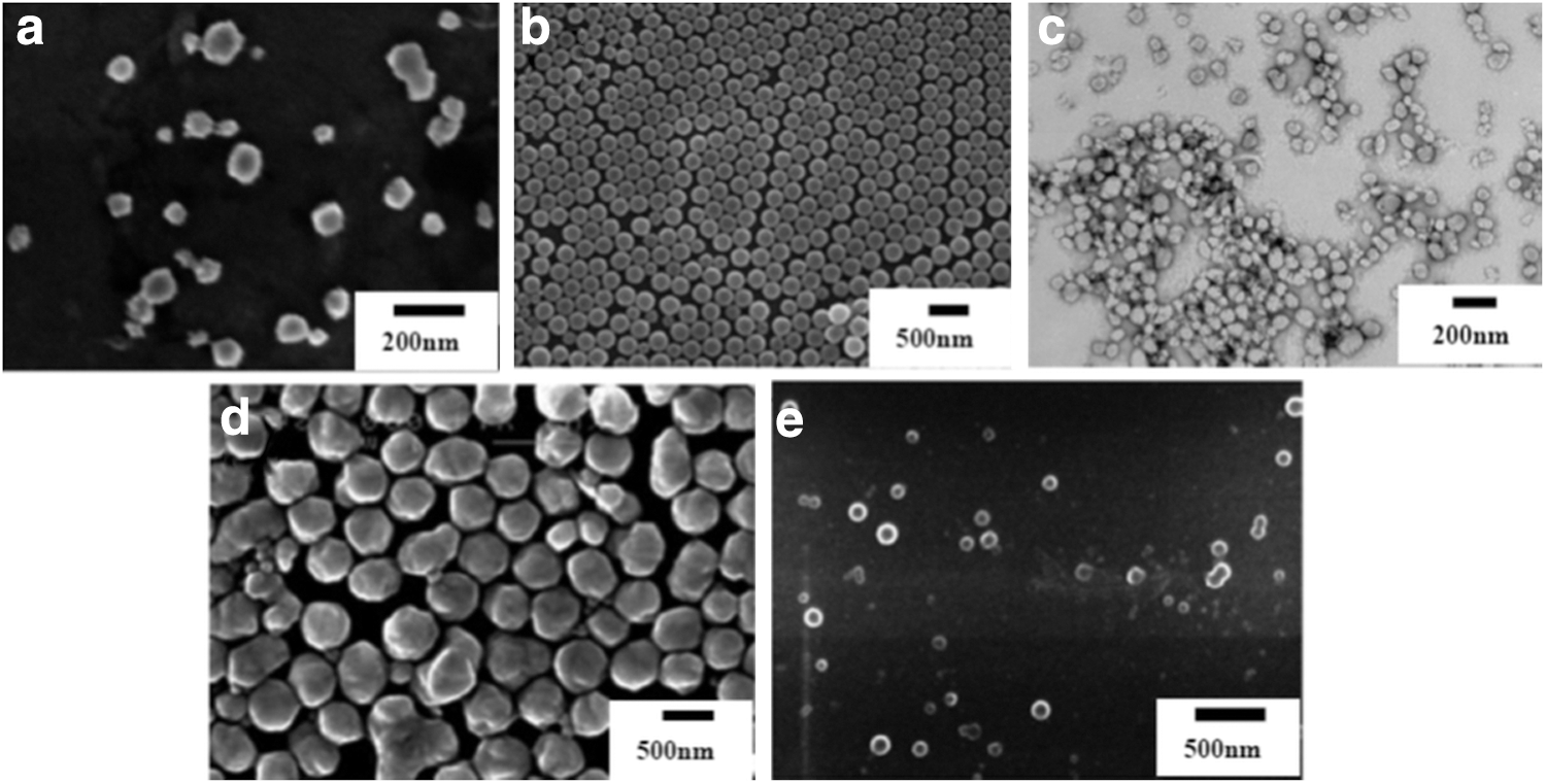
Scanning electron microscope images of cellulose nanospheres prepared by various methods. (
Methods of Preparing Cellulose Nanospheres
To classify the various kinds of spherical cellulose nanoparticles, we have opted to group experiments by method of particle formation, with subgrouping based on the chemistry of the cellulose or derivative. Most reports in this field focus on improvements in process yield/efficiency coupled with environmentally responsible considerations, such as reduction of energy consumption, reduction/elimination of organic solvents (particularly volatile ones), and the ability to control particle size and its distribution precisely and reproducibly.
Since most proposed applications for cellulose-based nanospheres are as controlled-release carriers of drugs, fragrances, or chemicals, such factors become dominant when considering direct production of cellulose nanospheres by adapting the acid hydrolysis methods used to make nanocrystals, as well as for emulsion-evaporation-, nanoprecipitation-, microfluidic-, and self-assembly-based processes involving cellulose derivatives. Table 1 summarizes specific examples with the general method, formation size, polymer, DS (if applicable), molecular weight (Mw) or viscosity data (when available), surfactant (if any), and concentration, particle size, polydispersity index (typically from DLS), and relevant bibliographic references. 19,21 –50
Methods for the Formation of Cellulose-Based Nanospheres Using Different Starting Materials and Surfactants
NS, no surfactant used.
Values obtained from particles within encapsulated molecules.
Centipoise (cps), is the unit of dynamic viscosity of viscous fluid. 1 cps=1 10−3 Pa·s. Dynamic viscosity is an indirect way to measure the molecular weight of polymer in solution.
NA, not applicable.
In-phase inversion methods—the concentration of the surfactant mixture is based on the whole system. In other methods the concentration of surfactant dissolved in single phase.
size measurements are obtained from solids (dry) by TEM and from dispersions by DLS.
Spherical Cellulose Nanocrystals (SCNs)
Approximately spherical, semi-crystalline, all-cellulose nanospheres prepared by acid hydrolysis with mixed-acid (hydrochloric and sulfuric acid) hydrolysis of native cellulose under sonication were first reported by Li et al. 17 After swelling cellulose fibers from a commercial pulp in dilute NaOH, Zhang et al. used mixed-acid hydrolysis with sonication to prepare spherical SCNs with a mean diameter of 85 nm. 18 However, reproducible and consistent research results in this field are sparse, perhaps due to the absence of application targets. Since spheres, with an aspect ratio equal to 1, offer the smallest specific surface area (SSA) (m2/g), and asymmetric particles such as needles or disks have increasingly large SSAs, surface loading per gram of particles correlates with the minimum particle dimension and the deviation of the aspect ratio from unity.
The influence of cellulose origin and variations in process conditions on SCN formation are discussed in the literature. 51 –53 Ibrahim et al. report plant species-dependent differences in nanosphere diameter, while Thakore, using only sulfuric acid without sonication, reports the formation of fibrous cellulose, which upon acetylation formed cellulose triacetate nanospheres (d=25–30 nm). 51,52 Xiong and coworkers prepared SCNs with a mean diameter of 35 nm (range 10–65 nm), which was obtained by converting cotton fabric to microcrystalline cellulose via additional acid hydrolysis with mixed sulfuric and nitric acids to form the spherical product. 53 Degree of polymerization (DP) of their product was 144, while that of the starting cotton was 1,200. Their data indicate that the nanospheres are significantly larger than the crystalline domains, suggesting that the spheres are polycrystalline. Still another group reported that neither mixed acid hydrolysis nor sonication is required to form spherical nanocellulose particles. 54 In summary, the use of this approach for preparing cellulose nanospheres has been limited and, to date, results are not in agreement with one another as to the optimal procedure. This method has not attracted as much interest as the porous nanospheres discussed in the next sections. This may be because large surface areas for solid particles benefit from asymmetry, since the greater the ratio of length to transverse dimension deviates from unity, the larger the SSA. The absence of an application for these spheres seems to have limited interest in their preparation and characterization.
Dispersion-Based Methods for Cellulose Nanoparticle Preparation
Emulsion-solvent evaporation
Surfactants, with their hydrophilic and hydrophobic domains, provide a facile route to phase separation of coated particles dispersed in a fluid and are a common method for the preparation of polymeric nanoparticles. Although simple in concept, the process allows efficient encapsulation of numerous lipophilic and hydrophobic compounds. 55,56 The conventional solvent-evaporation process involves the formation of an oil-in-water (O/W) emulsion of a water-immiscible organic solution of a hydrophobic cellulose derivative and an aqueous solution of surfactant at a level exceeding the critical micelle concentration. During the evaporation of organic solvent under reduced pressure, the polymer chains then organize as surfactant-encapsulated nanospheres dispersed in the continuous aqueous phase. The nanospheres are further purified by centrifugation to remove the extra polymer and surfactants. This technique has been applied to both cellulose ethers and esters. In general, this process results in cellulose-based nanospheres dispersed in water with diameters in the range of 50–300 nm, with spherical shape and relatively narrow size distribution (Table 1). 13,19,24,25,57
Desgouilles indicated that final nanospheres prepared from the solvent evaporation process are formed through a dynamic process involving the possible coalescence between emulsion droplets when the organic solvent diffuses from the inside of the particles. 13 Thus, the diameter of the final nanospheres is determined by the size of firstly formed emulsion droplets and the polymer chain conformation in the solvent. 13 Studies report that the diameter of nanospheres is independent of molecular weight and chemical structure of cellulose derivatives or the entrapped guest molecules. 13,19,24,57 The most important factors regulating droplet size and distribution are surfactant choice and input energy during emulsion formation.
Increases in sonication energy and duration led to decreases in the mean diameter of cellulose ester nanospheres, from ∼250 nm to ∼206 nm, while maintaining a narrow size distribution—a polydispersity index (PDI)=0.049. 19 Decreases in sonication energy favor broader particle size distribution. 28 Hydrophilic drugs benefit from the use of alternative methods such as oil-in-oil (O/O) emulsions, since hydrophilic molecules do not dissolve in either oil phase. 28 For example, ethyl cellulose (EC) and metformin HCl are both methanol-soluble. Nanoparticles were formed when their co-solution was mixed with paraffin light oil under vigorous stirring. After mixing the material with hexane, the oil is then removed by centrifugation with 90% encapsulation efficiency, meaning that 90% of the metformin mass is entrapped in the EC particles. 25
Another method for entrapping hydrophilic molecules is the multiple emulsification process. In the initial step, an aqueous solution of water-soluble molecules is emulsified in a lipophilic organic polymer phase, such as EC in methylene chloride. Next, this primary emulsion is dispersed in an aqueous solution of surfactants forming a water-oil-water (W/O/W) emulsion. Nanosphere formation occurs as the evaporation of organic solvent progresses and particles are purified by centrifugation. A high-pressure homogenization process was applied in both steps to ensure particle sizes in a nanometer range and nearly uniform size. EC nanoparticles were prepared containing a low molecular-weight hydrophilic drug with 67% encapsulation efficiency. 24
To reduce the use of chlorinated organic solvents, O/W emulsification can be replaced by the salting-out method of emulsion formation, in which a water-miscible organic phase, such as a polymer dissolved in acetone, is dispersed into a concentrated salt solution. Since miscibility of the miscible organic with water will increase with decreasing concentration of dissolved salt in the water, the initial emulsion is formed by mixing the water-miscible organic solution of polymer with a salt-saturated aqueous phase, such as CaCl2, and a suitable surfactant. The organic solvent was then extracted from the droplets in the emulsion by dilution of the primary emulsion with large amounts of water, leading to the formation of nanospheres. EC nanospheres loaded with sunscreen agent were prepared by this process. 28 However, the long dialysis time needed to remove the salt could also cause a loss of guest molecule.
In order to reduce the energy input, one may employ a phase-inversion method where, at constant temperature, water is added drop-wise into the organic phase with polymer while stirring. When the aqueous fraction is much larger than the organic phase, an O/W emulsion is formed due to the change of surfactant curvature at the interface. 29,30 Calderó and coworkers noted that besides the constraints on the rate of water addition, there is a limited region within the three-component phase diagram where phase inversion occurs. 31
Nanoprecipitation
Although the emulsion-solvent evaporation method is well established, it suffers from several drawbacks related to internalization in the human body. Large amounts of surfactant are required for the emulsion formation (Table 1). The use of organic, particularly chlorinated, solvents are discouraged by Environmental Protection Agency (EPA) regulations and may be harmful to the environment and human health. Energy requirements for extensive mixing are costly, as is purification by centrifugation. The latter may also cause damage to the integrity of the capsules and leakage of the guest molecules. Nanoprecipitation has become an increasingly attractive alternative to the emulsion process as a first stage in particle formation. The technique offers excellent reproducibility, simplicity, and low cost due to the reduced energy requirements due to room temperature mixing and low viscosity systems as well as the absence of a surfactant requirement. Nanoprecipitation is also described as solvent displacement, the Ouzo process, and solvent shifting. 58 –60
Nanoprecipitation is based on rapid nucleation of hydrophobic polymer chains into nanospheres by adding large amounts of a hydrophilic liquid, such as water, into a totally miscible organic phase, such as acetone or ethanol, containing polymer, possibly a cellulose ester. The mixing process can be controlled either by dialysis or drop-wise addition of water. 59 It has been suggested that nanoprecipitation occurs only in a dilute region of polymer in the ternary system. 61 Increases in polymer concentrations lead to a mixture of micro- and nanoparticles, which guarantees a broad distribution of diameters. 60 Nanospheres composed of cellulose ethers, esters, their mixture, and even unsubstituted hydrophilic cellulose have been obtained by nanoprecipitation with high reproducibility and nearly uniform size in the range of 100–250 nm (Table 1).
Since the mechanism of the nanoprecipitation process is incompletely understood and usually described in terms of nucleation and growth steps, the control of particle size utilizes empirical adjustment of process parameters. Current practice is based on the expectation that mixing parameters, volume fraction of organic solvent, and the chemical structure of the polymer are the key elements in controlling the final product. Thorough mixing is essential to forming homogeneously supersaturated batches—a prerequisite for both narrow size distributions and smaller particle sizes. 33 The volume fraction of the organic solvent determines the characteristics of the final system. However, correlations between particle size and solvent fraction differ between individual laboratories. This may arise from variation in polymer solutes, such as molecular mass or DS. A recent study by Kulterer reported that increasing the tetrahydrofuran fraction after mixing decreases the particle size of cellulose acetate (CA) (Mw∼30,000). 33 Finally, differences in derivitization of cellulose lead to changes in polymer solubility, which alters the degree of super-saturation for a given mass concentration. Using CAs with DS values between 1.65 and 3.0, Hornig and Heinze found that, in general, particle size increases with DS. 32 However, since the control of DP and DS at the same time involves two different hydrolysis reactions, it is difficult to quantify the function of chemical structure in size control during nanoprecipitation. They also discovered that cellulose triacetate could not be applied in the nanoprecipitation process, which may result from the possible crystallization of the triacetate. 32
Today, stable nanospheres, including modified or unmodified cellulose, attract substantial interest in biotechnology. Stable, hydrophobic nanospheres made from CA and nanocomposites composed of hydrophobic CA and other hydrophilic polysaccharides with mean diameters between 80 and 160 nm and a particle PDI smaller than 0.2 were prepared by nanoprecipitation. 62 Regenerated pure cellulose nanospheres were prepared from analogous processes using cellulose solutions, such as 50% w/w n-methylmorpholine-n-oxide, and NaOH/urea, while the supercritical antisolvent (SAS) process, based on the nanoprecipitation mechanism, was employed to prepare derivatized cellulose nanospheres. 38 –40,63
Wondraczek et al. compared conventional solvent evaporation with nanoprecipitation by characterizing the resulting nanospheres. 19 Both methods produced narrow reproducible dispersions of spherical nanoparticles. However, nanoprecipitation is preferred for its lower energy and surfactant requirements and greater flexibility with respect to polymer derivitization and process time. 19
Microfluidic methods based on hydrodynamic flow focusing
Microfluidics offers an alternative method for nanoprecipitation using hydrodynamic flow focusing in a microfluidic device to control mixing, where the polymer stream is focused into a thin stream to meet and mix rapidly with one or more intersecting nonsolvent and polymer miscible solute streams. 64 Such devices offer homogeneous and rapid, millisecond-scale, mixing in a tunable and continuous manner. 65 Conventional nanoprecipitation utilizes dialysis or drop-wise addition, both of which are slower processes with limited control over heterogeneous mixing conditions. In microfluidic devices, a mixing time below 1 ms can be achieved by increasing the width of the channel where streams of organic and aqueous phases are injected and mixed. The mixing time is tunable by regulating the ratio of flow rates for individual streams of two or more incoming fluids (flow ratio). 64 This technique, already used in preparing poly (lactic-co-glycolic acid) (PLGA) nanoparticles but rarely used in the preparation of cellulose-based nanoparticles, has been employed in producing chitosan and modified chitosan nanospheres in microfluidic systems. 41,42, 64
Using PLGA-polyethylene glycol (PEG) as modal biopolymer, Karnik et al. demonstrated by DLS that changes in the flow ratio altered particle diameters with an increased mixing rate correlating with decreased particle size (below100 nm) and size distribution (PDI approaching 0.1). 64 Similar trends were observed when microfluidics was applied to production of modified chitosan. Majedi et. al prepared chitosan nanospheres with a diameter of 110 nm and PDI below 0.2—among the lowest values reported for chitosans. 41 With increasing mixing time, diameter increased to 210 nm, approaching the value obtained from conventional nanoprecipitation methods. However, the PDI observed for the latter approaches 0.4. Modified hydrophobic chitosan nanospheres loaded with a hydrophobic anticancer drug through a microfluidic method have diameters in the range of 50–180 nm; the larger values correlate with increasing flow ratio. 42
Thanks to rapid, millisecond-scale mixing, the microfluidics approach with three or possibly more independent input flows can produce nanospheres with complex architectures and compositions as composite and polyelectrolyte. It is difficult to control the formation of complex multicomponent nanospheres through bulk synthesis. 66 For example, nanocomposites of PLGA and PLGA-PEG diblock polymer are nearly uniform 30-nm diameter, compared to a wide size distribution (50–300 nm) obtained in a conventional nanoprecipitation process. 63 Elsewhere, the cellulosic polyelectrolyte sodium carboxy methyl cellulose was reproducibly self-assembled into nanospheres and stabilized by crosslinking with diameters in the 50–300 nm range. 43
Polymer Self-Assembly
Cellulose esters and ethers are amphiphilic by nature, since the polymer chains contain both hydrophilic and hydrophobic segments. Therefore, in aqueous solution, these polymers self-associate to form metastable nanocoils with parameters sensitive to changes in temperature. As in nanoprecipitation, solubility of cellulose derivatives decreases due to increased interactions among hydrophobic portions of the polymer. Lu et al. first discovered such amphiphilic temperature-dependent behavior with a hydroxypropyl cellulose/water system. 67 In a narrow temperature range, 41–44 °C, slightly higher than the lower critical solution temperature (LCST) of 41°C, hydroxyl propyl cellulose (HPC) self-assembled into metastable nanospheres (150–200 nm), which were further stabilized by covalent cross-linking and formed a spherical nano/microgel (∼300 nm). 44 Further study of self-assembly of nanospheres focused on temperature-sensitive HPC indicated that the formation of attractive interactions involving hydroxyl groups of cellulose derivatives, other proton-donating or -accepting groups, and ionic groups such as salts or carboxylate groups all decreased the LCST of the polymer. 45,47,49,50 Thus, nanospheres were obtained at room temperature when water was replaced by salt solution (1.5 M) or with the addition of carboxylates-containing molecules, such as acrylic acid. However, removal of residual chemical reagents required lengthy dialyses and, in some instances, loss of hydrophilic drugs carried in the particle.
Chemical modification of HPC has been used to aid nanosphere production through specific interactions without further crosslinking. Nanoparticles, between 88–160 nm in diameter, of thiolated HPC were prepared, while longer thiol groups improved stabilization by crosslinking without any extra reagents. 49 Ionic derivatives of HPC, modified with cationic trimethylammonium groups and anionic styrenesulfonate groups, have been employed to form nanoparticles through polycation-polyanion interactions. The particles spontaneously self-assemble in water with average diameters of 150 nm. 21 Such charged polymer chains can carry biomolecules, such as proteins or DNA. Bagheri and Pourmirzaei employed a similar methodology using modified amphiphilic HPC to obtain nanospheres through atom transfer radical polymerization. 26 The increased water solubility of grafted HPC may increase the stability of nanospheres in vivo and further enhance its utility as a drug carrier. 25
Evaluation of Cellulose-Based Nanospheres
Since specific properties of cellulose-based nanospheres are application-dependent, an understanding of both chemical features, such as surface chemistry and cross-linking within nanospheres, as well as physical properties, such as particle size and its distribution, porosity, and particle morphology, is required (Table 2). 13,19,21 –26,32 –50,62 –64 Size distribution is particularly important, since narrower distributions favor control of secondary properties such as controllable release rates. Successful nanosphere applications meeting expected performance criteria require largely empirical correlations between measured particle properties and responses in specific applications. Here we shall review, briefly, methods for characterizing particle morphology, size distribution, encapsulation efficiency, and stability.
Advantages and Disadvantages of Cellulose-Based Nanospheres Prepared by Various Methods
Nanomedical research has shown that both size (typically below 200 nm) and narrow size distributions are key factors influencing the biodistribution of nanospheres. 68 Biodistribution refers to areas where particles of a specified size and chemistry may accumulate within bodily tissues. Particle composition and morphology may also influence the binding capacity of a particle for specific guest molecules. This specificity makes it difficult to establish general guidelines for design. Although size ranges are reported routinely for laboratory-scale nanoparticle preparations, different techniques, e.g., DLS of dispersions and TEM of dry samples under vacuum, will not agree, since they are made under different hydration conditions. Scanning electron microscopy (SEM), TEM, and atomic force microscopy (AFM) are powerful techniques, providing images of individual particles under vacuum and enabling measurements of size, shape, and morphology, but the particles are smaller than those seen by DLS. 68 Number-average radii, obtained from statistical analyses of well-resolved particle images, are significantly smaller than the weight-average hydrodynamic radii obtained by DLS from an assessment of diffusion coefficients for the same preparations in their original dispersions. The use of cryo-TEM methods might produce samples whose dimensions are closer to the DLS values, but this has yet to be reported. Electron microscopy of cellulosic nanospheres confirms spherical or near-spherical shapes, while elliptical particles, when observed, may be artifacts of drying stresses associated with evacuation of the sample chamber or aggregation of unstable nanoparticles. 63 Agglomerated particles often exhibit fused edges between particles, resulting in a broader size dispersion. Any investigator seeking this data will have to judge which techniques are most relevant to their specific application.
Particle size and distribution are measured rapidly by DLS, which provides an indirect measurement of hydrodynamic volume based on the distribution of diffusion coefficients. The quality of DLS measurements depends on the particle concentration, particle size distribution, and particle interactions. In acquiring DLS data, concentrations should be sufficiently dilute to minimize multiple scattering effects and particle interactions since they lead to an underestimation of size. 20 Reproducible DLS measurements, for identically prepared batches, demonstrate the batch-to-batch reproducibility for a given protocol. While cryo-TEM methods might shield the nanoparticle with a protective ice layer, current sample preparation for TEM studies of colloidal nanospheres expose the particle to high vacuum and an anticipated reduction in diameter from the DLS value due to loss of volatiles. However, DLS is experimentally simpler than TEM, does not require lengthy sample preparation, and measures particle dimensions in the liquid phase. But, both solvated and dry measurements are required to understand the character of the dispersed nanoparticles.
Entrapment efficiency (EE) and release kinetics are important parameters for evaluating nanospheres as carriers of drugs, fragrances, etc. After redissolving loaded nanospheres, the residual drug is extracted with water or ethanol. The ratio of the mass of the drug detected in the supernatant to the mass of polymer remaining defines the EE. 19 This value depends on both particle-preparation methods and chemical properties of the drug molecule. Release characteristics are evaluated under experimental conditions that are both sample-specific and defined by individual laboratory protocols. Release behavior is helpful to understand and evaluate the structure of the carrier matrix in which the drug molecules are entrapped. 24,35 For example, a rapid initial release indicates that the guest molecules are adsorbed on the surface of nanospheres while a progressive decrease in release at the final stage depends on diffusion from and dissolution in the polymer. Typically these behaviors are defined by fitting data to kinetic models.
Finally, stability is an important factor for shelf life, transportation, and delivery of particles to their ultimate target, all of which are critical to the economics of each system. A turbidity test is a simple quantitative method for monitoring the stability of nanoparticle suspensions. Aggregation of nanoparticles will lead to reduced numbers of particles and a concomitant decrease in scattered light intensity and turbidity. Either DLS or visible light transmittance at a wavelength where absorption is negligible may be used for this measurement.
Applications
Cellulose and many of its derivatives are biocompatible and biodegradable, as discussed above. Cellulose acetate, ethyl cellulose, and hydroxypropyl cellulose are considered to be nontoxic and are already employed in pharmaceutical applications. Those factors, and the fact that many cellulose and cellulose derivatives sell for below one dollar per pound in bulk, suggest that cellulose and cellulosic nanoparticles should compete effectively with polylactic acid and synthetic polymers.
Controlled-release characteristics of cellulosic microspheres have been studied extensively for drug loading and controlled drug release. 69 Recently, based on the development of technology and the size limitation for drug delivery, nanospheres made from cellulose derivatives by the methods mentioned before have been examined for drug loading of both hydrophobic and hydrophilic drug molecules.
EC nanospheres have been reported as an effective delivery system with excellent sustained release characteristics for both hydrophobic and hydrophilic drugs with high encapsulation efficiency (>80%). 25,27 Such high drug-to-polymer mass ratios are an important factor for release control. Ubrich did a comparative study for encapsulation of hydrophilic drugs by PLGA, polycaprolactone (PCL), and EC nanospheres. 24 The EC displayed the slowest drug release. X-ray and differential scanning calorimetry demonstrate that small organic drug molecules are dispersed molecularly across the polymeric matrix. Such in vitro tests do not guarantee in vivo success. Recently, in vivo testing in C57BL/6 mice of clarithromycin-loaded EC nanospheres vs. free clarithromycin showed enhanced activity of the nanoparticles against infection by Helicobacter pylori, a common human-specific pathogen. It was suggested that drug-loaded nanospheres prevented the bacterial infection by adhering to the cell wall, thus restricting bacteria adhesion while also providing sustained drug release. 36 Cellulose nanospheres with covalently bound fluorescent dye molecules were taken up by human fibroblast cells, albeit without any attachment to a selective receptor, and persisted for seven days. Rapid mixing typical of microfluidic processes is likely to exhibit slower, longer-lasting drug release than is observed with similar particles prepared by larger-scale batch mixing. 42
Multifunctional nanospheres offer advanced nanodelivery systems that combine properties such as simultaneous drug loading, targeting, and release in a single particle. Such nanocomposites were prepared from both solvent evaporation and nanoprecipitation methods using cellulose or cellulose derivatives. 62,70,71 Iron/EC nanospheres in a core/shell morphology were prepared by emulsion evaporation with internally loaded 5-fluorouracil, a widely used component of colon cancer drugs. Iron provides a magnetic field response for targeting delivery to tumor cells. The EC shell provided a polymeric matrix that enhanced drug capacity and controlled release. Loading efficiency was increased from 5% to 20% relative to bare iron surfaces. A slower drug-release profile was achieved from this nanocomposite. 70 Nanoprecipitated, functional nanocomposites of hydrophobic cellulose acetate with hydrophilic polysaccharide nanocomposite were stable in water for 28 days. 62
As a nanocarrier, cellulose-based nanospheres have provided improved resistance to photo-degradation in preparation of advanced functionality materials, enhancement of the absorption of target molecules to composite sensors, modification of chemical properties of entrapped sensor molecules, and generally improved the performance of nanocomposite-based systems relative to traditional materials. 4,28,30,34,39,57,62
Cellulosic nanospheres with entrapped pH-sensitive fluorescence sensors were prepared and used to detect dissolved ammonia at sub μg/L levels. The high surface-to-volume ratio and small diameters characteristic of nanospheres enhanced ammonia diffusion rates into the sensor membrane and reduced the path length, i.e., detection time, compared to traditional materials. The detection limit for dissolved ammonia at or below 1ug/L involved a 30-min response time. With faster response times, near real-time monitoring by a sensor assembly is feasible. 34 Finally, in a recent study, the use of nanosized cellulose esters using lauric acid substituents was shown to exhibit super-hydrophobicity mimicking that of lotus leaves. 7 The potential for these tiny, stable, sustainable packages is limited largely by our own willingness to explore their potential.
Conclusions
This review has shown that precise, reproducible control of nanosphere formation based on cellulosic compounds is readily possible. Such control is important because it provides the manufacturer facile, economic routes to control particle size, size distribution, surface chemistry, and porosity and has been widely demonstrated at bench scale.
The next step will be to explore the scalability of these and other, yet-to-be-developed methods to pilot plant and commercial levels. Precise control/optimization of mixing conditions throughout larger vessels, as well as of temperature and temperature gradients within those batch mixers, are two important areas of study. The feasibility of employing continuous flow microfluidic systems on a large scale is another area of major concern. Hopefully, this review introduces some of the “why bother” factors for incorporating cellulosics into the nanosphere arsenal. The ultimate proof will lie in the work of others to achieve commercial scale and to select and tune these methods for their particular applications.
Footnotes
Acknowledgments
The authors wish to acknowledge partial financial support by a SUNY-ESF Seed Grant. Xing Fei Zhao would also like to thank the SUNY-ESF Department of Chemistry for help during the course of preparing this review.
Author Disclosure Statement
No competing financial interests exist.