
Abstract
This study aimed to investigate the effect of an ethanolic extract of propolis (EEP) on the expression of genes associated with the adherence of Streptococcus mutans (S. mutans) to the tooth surface. A total of 10 S. mutans isolates, obtained from individuals with and without dental caries, were included in the study and identified using biochemical methods. The biofilm-forming ability was assessed using the microtiter plate method. Then, the antibacterial effect of the EEP was evaluated through the agar well diffusion and broth microdilution assay. Real-time polymerase chain reaction as employed to investigate the impact of sub-MIC (minimum inhibitory concentration) of EEP on the expression of gtfB, gtfC, gbpB, and ftf genes. In the standard microtiter plate method, 30% of the isolates produced strong biofilms (n = 3), 50% produced moderate biofilms (n = 5), and 20% produced weak biofilms (n = 2). The EEP demonstrated significant antimicrobial activity against S. mutans, with concentrations ranging from 30% to 100% (1.2–4 mg/mL) producing a notable increase in inhibition zone size compared with the control (p < 0.05), as measured by the well diffusion method. The MIC for all strong biofilm-producing isolates was found to be 1.2 mg/mL (30% EEP concentration). Gene expression analysis revealed a reduction in the expression of all target genes in the presence of EEP. Among the genes studied, gtfB exhibited the most substantial reduction with a 4.63-fold decrease (p < 0.001), while ftf showed the smallest decrease at 2-fold (p < 0.05). These findings suggest that the EEP has a notable antimicrobial effect, particularly against biofilm-producing strains. Moreover, the observed downregulation of key genes involved in biofilm formation and adhesion (gtfB, gtfC, ftf, and gbpB) highlights the potential of propolis as a natural agent for combating S. mutans infections and inhibiting biofilm formation.
Introduction
Among the numerous bacterial species that inhabit and endure in the oral cavity, S. mutans (streptococcus mutans) stands out as one of the few species consistently associated with the development of dental caries. 1 In low-pH conditions typical of plaque environments, S. mutans initiates an acid tolerance response, aiding its survival. This adaptation enables the bacterium to thrive within carious lesions by lowering plaque pH to levels inhibitory to other oral microbes. Furthermore, the bacterium’s capacity to produce extracellular polysaccharides that facilitate plaque biofilm formation enhances its pathogenicity. 2
The capacity of S. mutans to adhere to and colonize teeth is crucial for the onset and progression of dental caries. 3 Key among the surface-associated proteins of S. mutans that facilitate adherence and colonization are glucosyltransferases (gtfB, gtfC, and gtfD) and fructosyltransferase (ftf). These enzymes play a role in cleaving sucrose to produce extracellular glucan and fructan polymers. GtfB and gtfC specifically generate water-insoluble glucans, serving as adhesive molecules that anchor bacteria to the tooth pellicle. In addition, interactions between surface-associated glucan-binding proteins (gbp) and glucans contribute to oral bacterial aggregation, promoting the formation of plaque. Altogether, these enzymes play a crucial role in the pathogenicity of S. mutans. 4,5
As of now, the management of dental caries involves various preventive measures, such as the utilization of fluoride-containing toothpaste, topical application of fluorides, regular monitoring, adherence to a low-sucrose diet, application of dental sealants, and the use of fluoride-containing solutions for at-home rinsing. 6 Nevertheless, certain cases necessitate more robust control measures. Hence, it is comprehensible that researchers are presently intrigued by the promising prospects natural substances present as alternatives for controlling caries disease, particularly with respect to their antimicrobial efficacy and reduced associated risks. 7
Propolis, an innocuous resinous organic material, displays antimicrobial, anticancer, and anti-inflammatory attributes, thereby capturing the interest of professionals in the fields of dentistry and medicine. 8 The chemical elements present in propolis discovered in regions with mild weather conditions encompass galangin, chrysin, pinobanksin, and pinocembrin whereas its primary constituent is caffeic acid phenethyl ester. The utilization of propolis extract restrains the formation of plaque on tooth surfaces, thereby indirectly mitigating the occurrence of dental caries. The cariostatic effect of propolis is attributed to its fatty acids, which diminish the tolerance of microorganisms to low pH and decelerate acid production. The efficacy of ethanolic extract of propolis (EEP) against gram-positive bacteria was demonstrated to be superior to its efficacy against gram-negative bacteria in the planktonic state. Furthermore, EEP is recognized as a highly efficacious agent with antimicrobial and anti-inflammatory properties and is commonly employed commercially as a constituent of toothpaste, mouthwash, and lozenges. 8,9 However, the mechanisms of propolis on the biofilm of S. mutans remain unclear. Therefore, the objective of this study is to examine the impact of EEP on the expression of genes responsible for S. mutans adherence to tooth surfaces.
Material and Method
BACTERIAL ISOLATES
This cross-sectional study was conducted between February 2021 and June 2022 and focused on two distinct cohorts: individuals exhibiting susceptibility to dental caries (cases) and those demonstrating resistance to dental caries (controls). Participants were recruited from patients seeking healthcare services at clinics and medical facilities throughout the urban landscape of Isfahan. A total of six individuals with dental caries and four individuals without dental caries were included in the study. The age range for individuals with dental caries was 12–60 years, while those without dental caries fell within the age range of 26–32 years. Before study enrollment, individuals with dental caries underwent confirmation by a qualified dentist. Conversely, participants without dental caries exhibited a complete absence of decayed, missing, or filled teeth. To commence the study, all participants were required to provide their informed consent. In addition, the ethics review board of Islamic Azad University, Science and Research University, Tehran, Iran, approved the study. Sampling from decayed teeth was performed by a qualified dental practitioner. In the case of individuals without dental caries, dental plaque sampling was undertaken. The obtained samples were then introduced into tubes containing 2 mL of phosphate buffered saline (PBS) and transported to the laboratory. In order to recover S. mutans from the samples, a single loopful from each was streaked onto a Mitis Salivarius Medium (Merck, Germany) and subsequently incubated at a temperature of 37°C for a duration of 48 hours. Then, identification of the isolates was performed using standard microbiology and biochemical tests. Moreover, S. mutans ATCC 35668, sourced from the microbial bank of Isfahan University of Sciences, served as the standardized reference.
BIOFILM FORMATION ASSAY USING MICROTITER PLATE METHOD
A purified colony from each studied isolate, as well as S. mutans ATCC 35668, was added to a sterile tube containing 3 mL of Brain Heart Infusion (BHI) broth medium (Merck, Germany) enriched with 2% sucrose. The tubes were incubated at 37°C for a duration of 48 hours. Following incubation, 200 μL of the inoculum was added to three wells of a flat-bottomed 96-well plate and incubated again for 24 hours at 37°C. The samples were then washed three times with PBS (Merck, Germany) (pH = 7.3) to remove non-adherent bacteria from the plate wall. Subsequently, the samples were fixed by adding 250 μL of methanol (Merck, Germany) for 15 minutes. Following fixation, 200 μL of a 2% crystal violet solution (Sigma-Aldrich, USA) was added for 5 minutes to perform the staining procedure, followed by washing with PBS. After air-drying the plate, 200 μL of glacial acetic acid (33% v/v, Merck, Germany) was added to each well to resolubilize the formed biofilm. The optical density (OD) of each well was measured at a wavelength of 630 nm using an ELISA reader (Stat Fax—2100, USA). To enhance experimental accuracy, the experiment for each isolate was repeated three times, and the obtained average and standard deviation were considered as the final result. 10 Moreover, Escherichia coli K12 and Pseudomonas aeruginosa ATCC 27853 were utilized as negative and positive controls, respectively. The biofilm formation capacity of the isolates was assessed based on the following criteria: OD >0.240 indicating strong biofilm producers, OD <0.240 indicating biofilm producers, and OD <0.120 indicating non-biofilm producers. 11
EEP EXTRACTION
Fresh beehives were collected from the pol sefid region of Mazandaran Province, Savad Kooh County, during the summer season. The beehives were dissolved in 250 mL of 99% ethanol (Merck, Germany) (to effectively extract a broad range of bioactive compounds due to its ability to break down cell walls and dissolve both polar and non-polar substances) by employing a 150 rpm orbital shaking at room temperature for 48 hours. Subsequently, the resulting extract underwent filtration using Whatman No. 4 filter paper (Whatman, UK) and was then restored to its original volume by the addition of 80% ethanol (Merck, Germany) (to enhance the solubility of the extracted compounds, ensuring a manageable consistency for further analysis). The liquid filtrate was subjected to a vacuum evaporator (Rotavapor R-100, Buchi, Switzerland) for a period of 8 hours. Finally, a brownish-dark resinous propolis extract was obtained. This extract was subsequently stored in a cool and dry location.
Evaluating Antibacterial Activity of EEP
AGAR WELL DIFFUSION METHOD
To conduct an antimicrobial assay of EEP against S. mutans, 0.5 mL of a suspension of inoculums containing 1.5 × 10⁸ CFU/mL of the bacterium was applied using the streak method to BHI. Using a sterile punch apparatus (Harris Uni-core, USA), wells with a diameter of 6 mm were created on the culture medium. Subsequently, 0.3 mL of various dilutions (1.2–4 mg/mL) of the extract were introduced into each well. The plates were then placed in the refrigerator for a duration of 10 minutes to facilitate the diffusion of the extracts into the agar. After this, the plates were incubated at 37°C for 24 hours, after which the diameters of the halos surrounding each well were measured. Dimethyl sulfoxide [(DMSO), Merck, Germany] was employed as the negative control, and 2% chlorhexidine [(CHX) Iran Najou, Iran] served as the positive control. The methodology for utilizing these controls was analogous to the aforementioned procedure, substituting the extract with DMSO and CHX.
BROTH MICRODILUTION METHOD
The determination of the MIC of EEP was conducted using the broth microdilution method in 96-well plates. The MIC value was determined for isolates that were strong biofilm producers. Initially, 100 μL of BHI broth medium was dispensed into each well, followed by the addition of 100 μL of various concentrations (ranging from 0.4 to 4 mg/mL) of EEP to each well. Subsequently, 10 μL of a bacterial culture containing 1 × 10⁵ CFU/mL was introduced into each well. As a positive control, CHX 0.2% was employed. Following a 48-hour incubation at 37°C, bacterial growth inhibition was assessed by measuring OD at 620 nm using an ELISA reader (Stat Fax—2100, USA). The comparison of OD between control wells and treatment provided the growth inhibition rate.
EFFECT OF SUB-MIC CONCENTRATION OF EEP ADHERENCE GENE EXPRESSION
To assess the efficacy of EEP on genes responsible for the adherence of S. mutans to the tooth surface, planktonic cells of S. mutans, which were strong biofilm producers based on biofilm assay results, were cultured in trypticase soy broth. The culture was supplemented with a sub-MIC concentration of EEP and then incubated at 37°C for a duration of 24 hours. Following this incubation period, the cell suspension underwent RNA extraction using SinaPure ONE (Cinacolon, Iran), adhering to the manufacturer’s protocol. Subsequent to this step, the complementary DNA (cDNA) synthesized utilizing the SinaClon First Strand cDNA synthesis kit (Cinacolon, Iran). 10,12 gtfB, gtfC, gbpB, and ftf genes were amplified using real-time polymerase chain reaction (PCR) in a Qiagen Real-Time PCR System (Qiagen, Germany). In this study, the 16S rRNA gene was utilized as the internal control, serving as a reference with stable expression to ensure the reliability of the quantitative analysis.
For the amplification process, 0.5 μL of both the forward and reverse primers (Table 1) at a concentration of 200 nM, 12.5 μL of Ampliqon SYBR® Green Master Mix (Ampliqon, Denmark), 1 μL of the cDNA sample, and RNases-free water to achieve a final volume of 25 μL. The program was executed under the following conditions: an initial denaturation step at 95°C for a duration of 10 minutes, followed by 40 cycles of 10 seconds at 95°C, and a subsequent extension stage at 60°C for 1 minute. The findings were examined through the application of the comparative quantification cycle (Cq) technique (2− ΔΔ Cq). 15
Primers Used in the Study
STATISTICAL ANALYSIS
The data were analyzed using SPSS version 20 software, and statistical analysis was performed using the Duncan test.
Results
IDENTIFICATION OF S. MUTANS ISOLATES
The streptococci identification tests revealed that the studied strains exhibited chain-shaped cocci with long chains and a gram-positive violet color. They were characterized by negative catalase activity and resistance to optochin and bacitracin, while also displaying alpha hemolysis. The colonies formed after 24 hours of incubation on Mitis Salivarius agar appeared small and smooth, ranging in color from dark to pale blue. On the 5% sucrose agar, the colonies were dry, sticky, and shiny. The streptococci showed enhanced growth in anaerobic conditions with 5% CO2, resulting in larger colony formation compared with aerobic conditions.
BACTERIAL CULTURE AND COLONY COUNTING
The study utilized a serial dilution method and Mitis Salivarius bacitracin agar to determine the bacterial population of S. mutans. Colonies related to S. mutans were counted (Table 2), and the average population in the caries group was found to be 62.49 × 10⁴ CFU/mL. This population was significantly higher compared with the control group (p < 0.05) (Fig. 1).
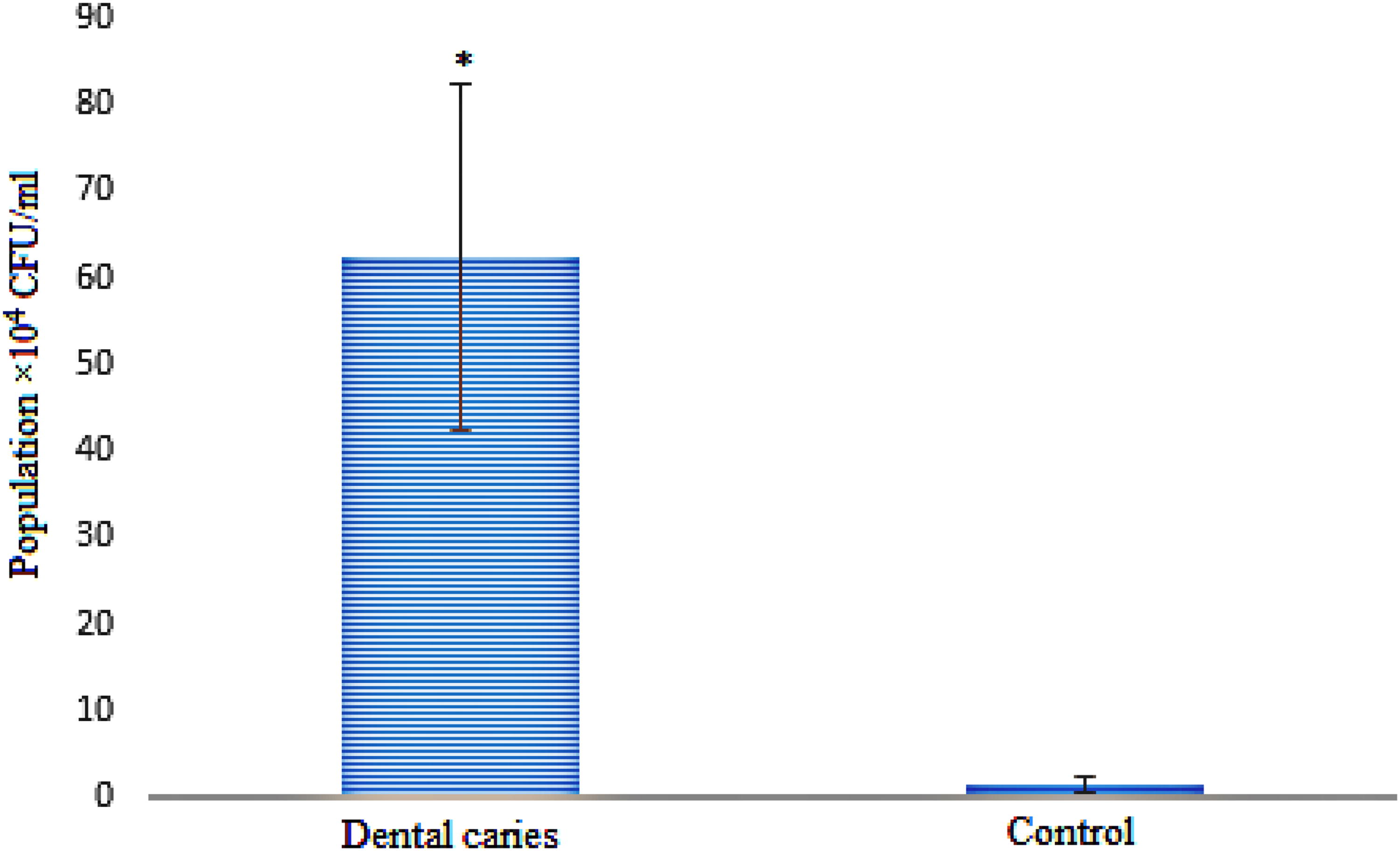
Comparison of the average population of S. mutans in individuals with and without caries. The bar representing the caries group shows a significantly higher S. mutans population compared with the control group, indicated by a single asterisk (*), reflecting statistical significance (p < 0.05). S. mutans, Streptococcus mutans.
The Results of Bacterial Culture and Colony Counting
DETERMINING THE ANTIBACTERIAL EFFECT OF THE PROPOLIS EXTRACT BY WELL DIFFUSION METHOD
In the investigation of the antimicrobial effect of EEP using the good diffusion method, it was observed that concentrations ranging from 30 to 100% (1.2–4 mg/mL) of the extract created a significant difference in the inhibition zone compared with the control (p < 0.05) (Table 3). Furthermore, an increase in the concentration of EEP resulted in a significant increase in the diameter of the inhibition zone (p < 0.05) (Fig. 2).
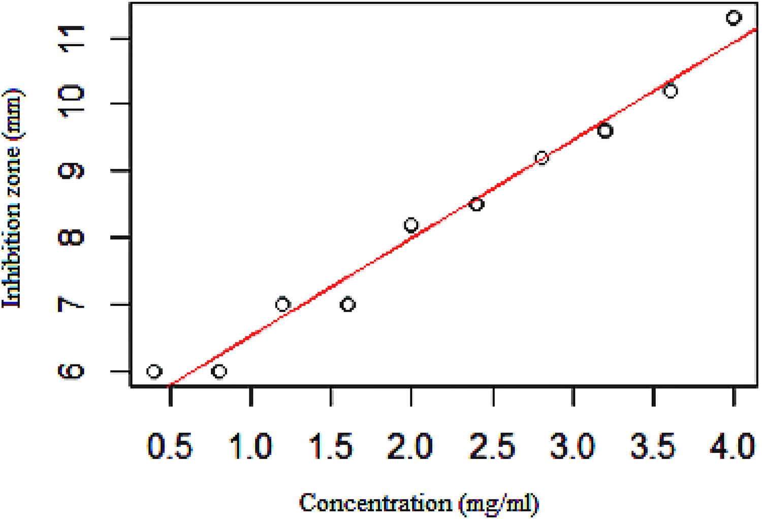
The relationship between the mean diameter of the inhibition zone of S. mutans strains with different concentrations of EEP. This figure demonstrates the linear relationship between the concentration of EEP and the corresponding mean diameter of the inhibition zone (mm) for S. mutans strains. As EEP concentration increases from 0.5 mg/mL to 4.0 mg/mL, the inhibition zone diameter shows a proportional increase, indicating enhanced antimicrobial activity. The red line represents the best-fit linear regression, underscoring the positive correlation between EEP concentration and the inhibition effect on S. mutans. S. mutans, Streptococcus mutans; EEP, ethanolic extract of propolis.
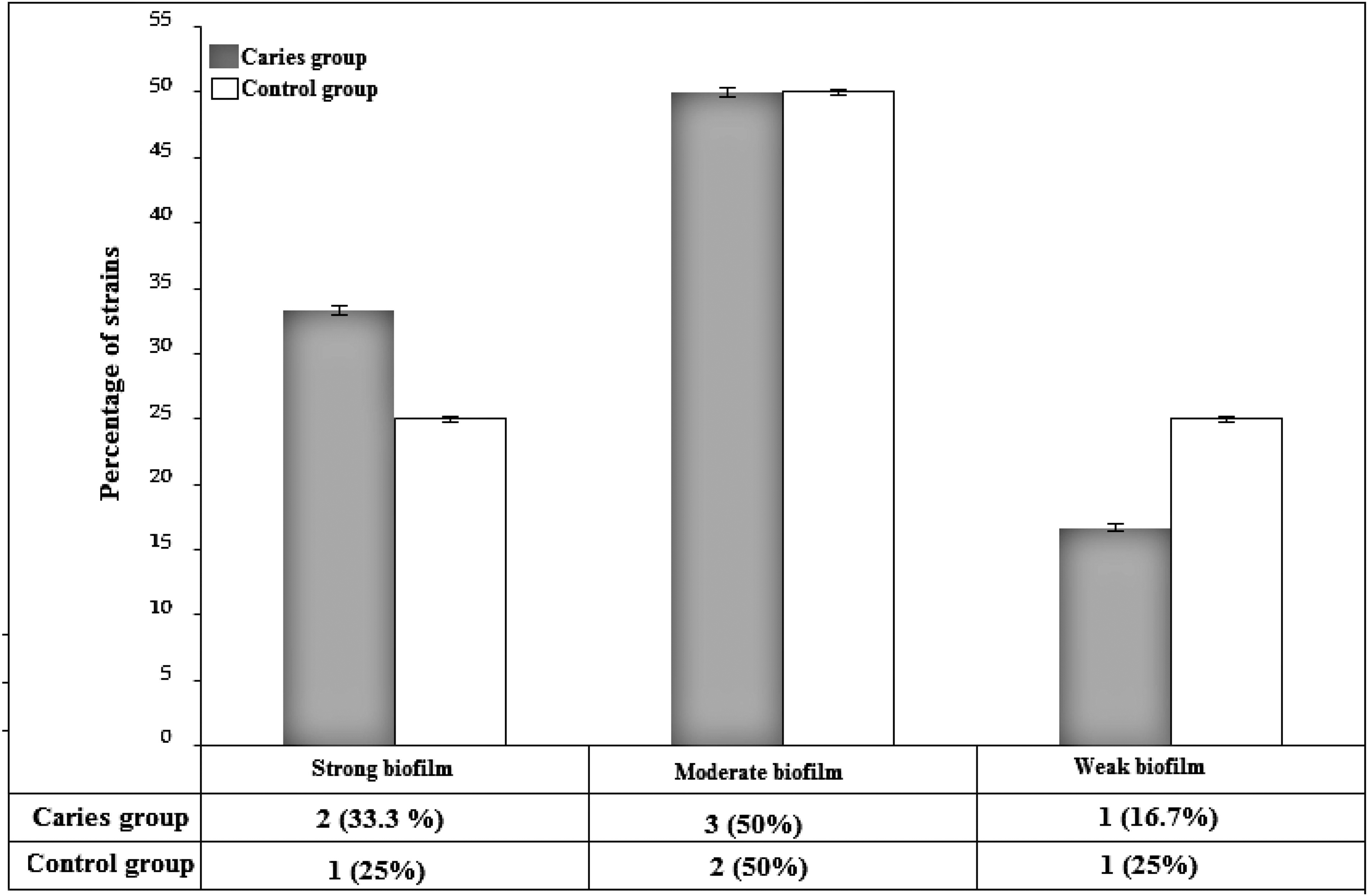
Comparative assessment of biofilm production in strains isolated from individuals with dental caries and a control group. This figure highlights the distribution of biofilm production capacity, categorized as strong, moderate, and weak, among bacterial strains isolated from individuals with dental caries (gray bars) and controls (white bars). The caries group exhibits a higher percentage of strains producing strong biofilms compared with the control group, while moderate biofilm production is similarly distributed in both groups.
Antimicrobial Effect of EEP by Well Diffusion Method
EEP, ethanolic extract of propolis; DMSO, Dimethyl sulfoxide; CHX, chlorhexidine.
BIOFILM FORMATION
In the standard microtiter plate method, 30% of the isolates produced strong biofilms (n = 3), 50% produced moderate biofilms (n = 5), and 20% produced weak biofilms (n = 2) (Fig. 3). Statistical analysis revealed no significant relationship between the strength of biofilm formation and the occurrence of dental caries (p = 0.93).
DETERMINATION OF THE MIC OF EEP
The sensitivity of three isolates of S. mutans to EEP was assessed using the broth microdilution method. All investigated strains were found to be strong biofilm producers. The MIC for all isolates was determined to be 1.2 mg/mL of the propolis extract, which corresponded to a concentration of 30%.
EXPRESSION OF BIOFILM RELATED GENES
The expression levels of the gtfB, gtfC, ftf, and gbpB genes were assessed both with and without exposure to EEP. In the presence of EEP, all studied genes exhibited reduced expression compared with the control group. Specifically, gtfB showed the highest reduction with a 4.63-fold decrease (p < 0.001), while ftf exhibited the lowest reduction at 2-fold (p < 0.05) (Fig. 4).
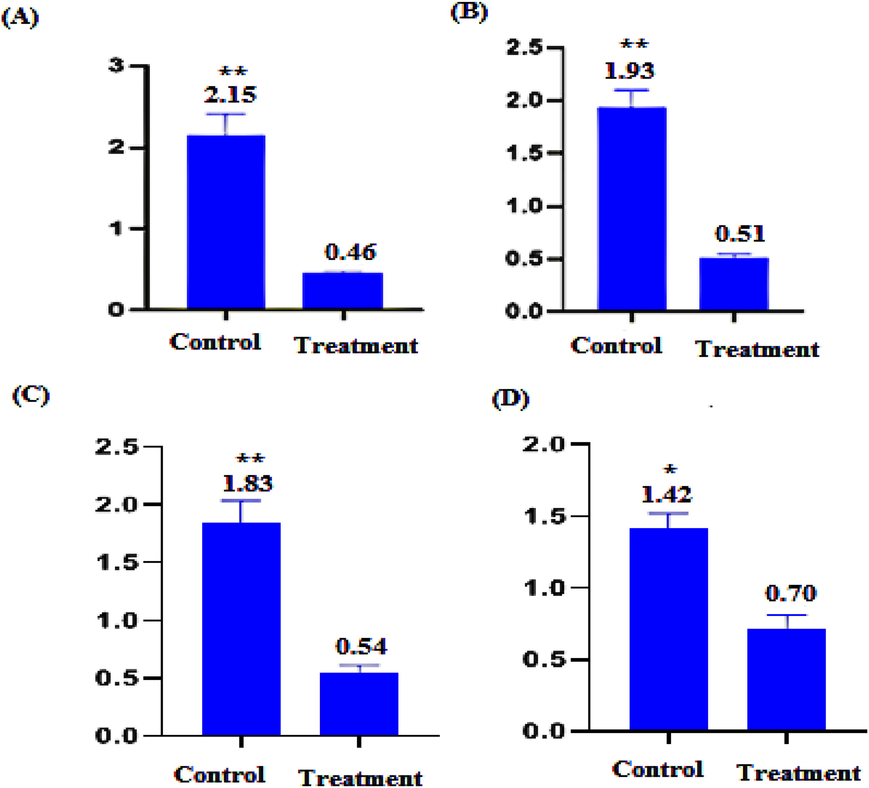
Relative expression of biofilm-associated genes in S. mutans following EEP treatment. Graphs show the fold change in gene expression for
Discussion
In this study, we selected the EEP based on existing literature that demonstrates its superior antimicrobial efficacy compared with water extracts. Previous research has established that EEP exhibits significantly stronger antimicrobial activity against gram-positive bacteria, such as S. mutans, than against gram-negative bacteria. 8,16 The potent antimicrobial properties of propolis against various oral bacteria, particularly S. mutans, underscore its relevance in oral health. Given that biofilm formation is a critical factor in the virulence of S. mutans, our study aimed to evaluate the expression levels of key genes associated with biofilm formation, specifically gtfB, gtfC, ftf, and gbpB. We compared the isolates treated with EEP to control isolates to assess any effects on gene expression. The results of our real-time PCR analysis indicated a significant downregulation of gene expression in the EEP-treated samples, with decreases of 2-fold for ftf, 3.34-fold for gbpB, 4.63-fold for gtfB, and 3.74-fold for gtfC. These findings suggest that EEP may effectively inhibit biofilm formation in S. mutans by targeting and reducing the expression of key genes involved in its pathogenicity.
Similarly, Gabe et al., 14 demonstrated that ethyl gallate, a polyphenol-rich extract similar to propolis, altered the expression of three genes-gtfC, gtfB, and gbpB, and exhibited the potential to inhibit S. mutans biofilm formation. The gtf genes play a crucial role in the production of glucan polymers and the attachment of bacteria to surfaces. The upregulation of these genes in the biofilm state compared with the planktonic form further supports their significance. In a study by Veloz et al., 17 a propolis extract abundant in polyphenols was found to inhibit the expression of glycosyltransferase genes (gtfB, gtfC, and gtfD). The expression of these genes decreased by 40%, 60%, and 40%, respectively. The gtf genes play a crucial role in the production of glucan polymers and the attachment of bacteria to surfaces. The increased expression of these genes in the biofilm state, compared with the planktonic form, confirms their significance in this process. In a study conducted by Yoshida et al., it was demonstrated that the expression of the gtfB gene increases 4-fold in the biofilm state compared with the planktonic state. 18 The findings of the present study align with those of Hyungho et al. 19 who investigated the impact of apigenin extracted from propolis on the expression of gtf genes in S. mutans. They reported that the presence of apigenin led to approximately a 50% decrease in the expression of both the gtfB and gtfC genes. The findings from these studies align with the results of the present study, indicating the inhibitory effect of EEP on the expression of genes involved in biofilm synthesis in S. mutans strains.
Notably, studies have shown that rats infected with S. mutans mutants lacking gtfBCD or ftf genes exhibited significantly reduced dental caries, indicating the importance of these enzymes in the development of tooth decay. 5
In the current study, we examined the ability of S. mutans strains isolated from individuals with and without caries to form biofilms. Our findings revealed that the frequency of strong biofilm-forming isolates was higher among individuals with caries compared with those without caries. Specifically, 66.7% of the strains that formed strong biofilms were obtained from individuals with caries, while 33.3% of the strong biofilm-forming strains were isolated from individuals without caries. The MIC for strains that produce strong biofilms was consistent, with a value of 1.2 mg/mL of EEP at a concentration of 30%. This value is higher than what has been reported in some previous studies. 20,21 The difference in results could potentially be attributed to the source of propolis used. According to the findings of previous studies, 21 the plant source, the season of production, and the solvent used in propolis preparation can significantly impact its properties and determine its level of antibacterial effectiveness.
The observed MIC value of 1.2 mg/mL was consistent across all samples, indicating that this concentration effectively inhibited bacterial growth. However, the expression levels of biofilm-related genes demonstrated variability, ranging from 4.63- to 2-fold changes. This discrepancy suggests that, while the MIC was sufficient to inhibit planktonic bacterial growth, the biofilm regulatory mechanisms may respond differently to the antimicrobial treatment. The differential expression could be attributed to various factors, such as strain-specific responses to the antimicrobial agent, differences in metabolic states, or variations in biofilm-forming potential among samples. Furthermore, the complexity of biofilm formation—involving multiple regulatory pathways—might contribute to heterogeneous gene expression in response to the same antimicrobial concentration. This variability underscores the complexity of targeting biofilm-related pathways, which may not uniformly correlate with MIC values alone, as biofilm regulation involves distinct stress response mechanisms beyond those affecting planktonic bacterial survival.
Among the various findings of the present study, it is worth mentioning the significant prevalence of S. mutans in individuals with caries compared with those without caries. Numerous studies conducted across different age groups and populations, examining samples from various areas of the mouth, saliva, and dental plaque, consistently indicate a positive correlation between the severity of tooth decay and levels of S.mutans. 22 –24 In addition, other studies have reported that the presence of decayed teeth significantly increases the abundance of S. mutans. 25,26 The results obtained from this investigation provide further support for the infectious theory of dental caries and establish a clear association between S.mutans and caries. 22
Previous publications have extensively discussed the medications used for treating and preventing oral cavity diseases, along with their potential side effects and the causes of tooth decay. 27 Furthermore, there is a growing body of in vitro and in vivo studies that have explored the potential use of propolis in managing dental caries. It is worth noting that propolis has already been integrated into commercially available oral care products, underscoring its importance in this field. 15,17,28,29 The antimicrobial properties of propolis have also been well-documented and supported by scientific evidence. 30,31 In our study, we focused on investigating the antibacterial effects of EEP against S. mutans biofilm formation. Dental plaque, a type of oral bacterial biofilm, is a significant factor in the development of dental caries. Biofilms contribute to bacterial drug resistance, making it essential to find ways to inhibit or destroy them for effective dental caries prevention and treatment. 32 Our findings demonstrate that EEP effectively suppresses the genes involved in S. mutans biofilm formation. This disruption of the biofilm structure may lead to increased permeability of the bacterial membrane, resulting in the release of Lactic Dehydrogenase (LDH) and calcium ions while inhibiting bacterial proliferation. 32 These results highlight the potential of EEP as a promising agent for targeting and controlling S. mutans biofilms, which can contribute to the prevention and treatment of dental caries.
Conclusion
The finding of the present study suggests that the alcoholic extract of propolis has a notable antimicrobial effect, particularly against biofilm-producing S. mutans isolates. Moreover, the observed downregulation of key genes involved in biofilm formation and adhesion (gtfB, gtfC, ftf, and gbpB) highlights the potential of propolis as a natural agent for combating bacterial infections and inhibiting biofilm formation.
Footnotes
Acknowledgments
All the authors acknowledge the staff of the Department of Microbiology, Science and Research Branch, Islamic Azad University, Tehran, Iran.
Authors’ Contributions
S.T.: writing—original draft, methodology, formal analysis, software, data curation, and investigation, A.S., and S.S.: software, writing—original draft, review, and editing. S.S. and A.T.: supervision, conceptualization, resources, project administration, validation, funding acquisition, writing—review, and editing.
Ethical Statement
Each author confirms that this research is supported by the Science and Research Branch, Islamic Azad University, Tehran, Iran, an institution primarily involved in education and research. All procedures were approved by the institution’s Ethics Committee and conducted in accordance with ethical standards.
Consent for Publication
All authors have read and agreed to the published version of the article
Data Availability Statement
The data that support the findings of this study are available from the corresponding author upon reasonable request.
Author Disclosure Statement
No competing financial interests exist.
Funding Statement
No financial support was received for this study.