
Abstract
Nanotechnology involves the study of matter at the nanoscale level. Significant developments in nanotechnology have drawn a lot of interest in a range of biomedical applications. Synthesis of Syzygium cumini copper nanoparticles (SC-CuNPs) using S. cumini seed extract was primarily confirmed by the color change where light brown turns into dark brown, with sharp absorption maxima at 360 nm. Fourier Transform Infrared Spectroscopy revealed the various functional groups corresponding to the secondary metabolites that are responsible for the stabilization of SC-CuNPs, while X-ray diffraction analysis indicated a crystalline structure with an average particle size of 80 nm. SC-CuNPs were spherical, as revealed by the Scanning Electron Microscope analysis. At 125 μg/mL concentration, the SC-CuNPs exhibited significant antioxidant activity, as evidenced by 53% of hydrogen peroxide scavenging and 55% of free radical scavenging activity as measured by the phosphomolybdenum method. The antibacterial activity of SC-CuNPs revealed maximum zone of inhibition against Escherichia coli (28 ± 0.97 mm) and Staphylococcus aureus (32 ± 0.89 mm). Furthermore, SC-CuNPs demonstrated 43% anti-inflammatory activity at a 500 μg/mL concentration. The current work concludes by emphasizing the non-toxic and cost-effective properties of SC-CuNPs. Additionally, it presents an alluring and promising avenue for future applications in the biomedical field.

Introduction
Nanotechnology has the potential to revolutionize a number of scientific sectors. Because of their distinct size and shape, special qualities, including their high absorption capacity and numerous chemical compounds, especially medicinal particles, 1 nanomaterials have a variety of uses and have been the focus of extensive material science and biological research. 2 Nanotechnology is anticipated to unveil new avenues for combating and preventing diseases through atomic-scale tailoring of materials. Nanobiotechnology equips engineers with the capability to develop nanoscale material properties for biological applications. 3 The widespread and indiscriminate use of broad-spectrum antibiotics has resulted in microbial resistance, posing challenges in the treatment of infectious diseases. However, advances in nanotechnology have opened up new domains for the synthesis and applications of nanoparticles against multidrug-resistant pathogens. 4
The exceptional chemical and physical characteristics of nano-sized semiconductors have made them an approach in recent years. However, this method does have several drawbacks, such as being costly, time-consuming, and labor-intensive. Therefore, there is an urgent need for an alternative approach, such as nanoparticle (NP) synthesis, that is safe, environmentally friendly, and economical to overcome these limitations. 5 In recent years, there has been an increase in the use of plant-mediated nanoparticle synthesis due to its simplicity, speed, and environmental friendliness. These methods help to avoid the harsh chemicals that are costly, as well as the stringent conditions necessary for reduction and stabilization. 6 Green synthesis approaches utilize bacteria, fungi, algae, and plant parts such as fruits, stems, roots, leaves, and seed extracts. 7
Biosynthetic processes, particularly those using plants, are gaining attention as a simplified and scalable way for creating impurity-free metal oxide particles. The process of producing different metallic nanoparticles (MNPs) using bioactive chemicals is referred to as “green nanomaterial synthesis.” Compared with earlier physicochemical methods, these biosynthetic processes provide nanoparticles with more precise sizes and shapes. 8 Secondary metabolites present in biological systems are crucial for NP production. In the environmentally friendly synthesis of stable MNPs, the individual biomolecules present in this plant extract act as reducing, capping, and stabilizing agents when utilizing their corresponding metal ion precursor solution. MNPs have recently gained attention in cancer research due to several characteristics, such as their small, defined volume and increased reactive surfaces, which make them suitable for conventional small molecules and pharmaceutical agents. Previous studies have shown that using plants offers numerous advantages than other biosystems, due to their ease of availability and production of more stable NPs. In addition to this, plant-mediated synthesis was a sustainable and environmentally beneficial method. 9,10
Over the past 10 years, nanotechnology has enabled the development of modern methods for producing copper at the nanoscale. In contrast to other noble metals, such as silver, gold, and platinum, which are relatively easy to work with and resistant to oxidation but can be expensive. MNPs include copper nanoparticles (CuNPs), silver nanoparticles, and gold nanoparticles, 11,12 which were used for antibacterial, antifungal, and antiviral applications.
CuNPs are gaining popularity due their accessibility and economic viability. Their effective light-to-heat conversion when exposed to near-infrared laser radiation, makes them frequently used in cancer imaging. 13 Additional applications include improving sensors, photonic devices, electrochemical devices, and heat transfer liquids. 14 The unique properties of CuNPs have led to their widespread use across various sectors, including construction, food, textiles, agriculture, cosmetics, 15 drug delivery. 16 Previous reports on the synthesis of CuNPs using plant extracts include Momordica cymbalaria, 17 Rosmarinus officinalis, 18 Curcuma longa, 19 Tridax procumbens, 20 Hyptis suaveolens (L.), 21 Ceiba pentendra, 22 Parthenium hysterophorus, 23 and Solanum xanthocarpum. 24
Syzygium cumini, a fruit-bearing plant in the Myrtaceae family, is commonly known as Jamun in Asia (Fig. 1). It has long been utilized as a therapeutic herb. Various parts of the plant, including fruit, seeds, bark, and leaves, have been employed to treat a range of diseases. The fruit juice from S. cumini has been taken orally to treat diabetes, diarrhea, and stomach issues. A high concentration of tannins imparts an astringent flavor to S. cumini fruit, while anthocyanins contribute to its purple hue. 25 Moreover, delphinidin, 3,5-diglucosides of malvidin and petunidin, has been identified in the peels of S. cumini. 26 Bioactive components such as phenolic acids, gallic acid, ellagic acid, carotenoids, flavonoids, myricetin, and their derivatives have been detected in the fruit pulp. 27 The flavor of the purple fruits is contributed by around 30 different compounds, including dihydrocarvyl acetate, geranyl butyrate, and terpinyl isovalerate. 28 Additionally, a qualitative analysis of S. cumini seeds revealed the presence of β-sitosterol, gallic acid, corilagin, ellagic acid, quercetin, and jambosine. 29

Syzygium cumini tree
Previous studies of S. cumini seed extract copper oxide nanoparticles exhibited spherical shape with size ranging from 42 to 90 nm, demonstrating antibacterial properties against both Gram-positive Staphylococcus (2 mm) and Gram-negative Klebsiella (6 mm), as well as antioxidant assay, as demonstrated by the 2,2-diphenyl-1-picrylhydrazyl (DPPH) method. 30 In another study, manganese oxide nanoparticles (MgONPs) synthesized from the methanolic extract of S. cumini seed extract showed antioxidant and antimicrobial activity. 31 This is the first report on hydrogen peroxide scavenging and antibacterial activities against Escherichia coli, Salmonella typhimurium, Proteus vulgaris, and Staphylococcus aureus as well as anti-inflammatory activity using S. cumini seed extract CuNPs (Table 1).
The current study concentrated on the phytomediated production of inexpensive and environmentally acceptable bioactive CuNPs utilizing a S. cumini seed extract. The antibacterial, antioxidant, and anti-inflammatory properties of this green biosynthesized nanoparticle were evaluated and assessed in vitro. This will provide preliminary information regarding the probable application of green synthesized nanoparticles in nanomedicine as well as their in vivo performance.
Materials and Methods
PLANT MATERIAL COLLECTION
The S. cumini fruit was collected from the Botanical Garden at Yogi Vemana University in Kadapa, Andhra Pradesh, India. S. cumini fruits were washed first with tap water and then with distilled water to remove any surface debris. The fruit core was collected, and the processed seed samples were shade-dried for 8–10 days, then finely powdered and kept in airtight glass jars at 4°C for further studies.
PREPARATION OF S. CUMINI SEED EXTRACT
In total, 10 g of dried seed powder and 150 mL of deionized water were mixed and heated at 80°C in a beaker for 30 minutes. The solution was filtered using a Whatman No.1 filter paper and then stored in the refrigerator at 4°C for future examination. During the synthesis of CuNPs, seed extract acts as a reducing agent.
BIOSYNTHESIS OF CUNPs FROM S. CUMINI SEED EXTRACT
In total, 25 mL of S. cumini seed aqueous extract was mixed with 4 g of 0.1 M copper sulfate pentahydrate in 100 mL of distilled water and stirred at 70°C employing a magnetic stirrer for 4 hours. The reduction of copper ions from Cu (II) to Cu metal caused the blue solution of copper sulfate pentahydrate to turn brown, indicating the synthesis of CuNPs. To obtain the pellet, the solution was then centrifuged for 10 minutes at 9 grpm. The resulting CuNPs were stored at 4°C for further analysis after being dried overnight at 70°C in a hot air oven. 32
CHARACTERIZATION OF SC-CUNPS
Physicochemical characterization.
To determine the physicochemical characterization of S. cumini CuNPs (SC-CuNPs), UV-spectroscopy, X-ray diffraction (XRD), Fourier Transform Infrared Spectroscopy (FTIR), and Scanning Electron Microscope (SEM)-Energy Dispersive X-ray (EDAX) analysis were conducted. The formation of synthesized SC-CuNPs was confirmed by recording spectra from 200 to 800 nm by using a UV-visible spectrophotometer (SHIMADZU; Model UV-1800 240v, Japan), equipped with a 1.0-cm quartz cell, and all experiments were done at room temperature. A small quantity of SC-CuNPs solution was prepared to coat the glass slide and measured using an X-ray diffractometer model (Flex-600, Rigaku Mini) with 40 kV, 30 mA with Cu k radiation at 2θ angle. 22 FTIR in the range of 4000–400 cm−1 wavenumber was employed to detect the functional groups responsible for reducing the copper in the SC-CuNPs. Scanning Electron Microscopy was utilized to determine the external morphology of the SC-CuNPs (Model: JSM-IT 500; JEOL, Japan), and EDAX was used to analyze the elemental composition. Furthermore, to determine the crystal structure of the prepared SC-CuNPs, XRD analysis was conducted.
Biological characterization.
To evaluate the biological characterization of SC-CuNPs, antioxidant, antibacterial, and anti-inflammatory activities were assessed.
Antioxidant activity of SC-CuNPs and S. cumini seed extract.
In vitro antioxidant activity of SC-CuNPs was evaluated by using phosphomolybdenum assay and hydrogen peroxide scavenging assay.
Phosphomolybdenum assay of SC-CuNPs and S. cumini seed extract.
Phosphomolybdenum assay was conducted to determine the total antioxidant activity of SC-CuNPs. For this assay, 1 mL of molybdate reagent (0.6 M sulfuric acid, 28 mL of sodium phosphate, and 4 mL of ammonium molybdate), 3 mL of distilled water were prepared in each test tube, followed by various concentrations of seed extract, SC-CuNPs, and ascorbic acid (50, 100, 150, 200, and 250 µg/mL) were added separately to each test tube. The test tubes were incubated at 95°C for 90 minutes in a water bath. After incubation, test tubes were cooled down to room temperature for about 20–30 minutes. Ascorbic acid was used as a positive control. Reaction mixture’s absorbance was recorded at 695 nm. The means of three replicates of the samples were computed.
Hydrogen peroxide scavenging assay of SC-CuNPs and S. cumini seed extract.
Different concentrations of (50, 100, 150, 200 and 250 µg/mL) seed extract, SC-CuNPs and ascorbic acid (positive control) were mixed with 0.6 mL of 40 mM H2O2 solution and 0.4 mL phosphate buffer (prepared in 50 mM phosphate buffer, pH 7.4) and incubated for 10 minutes at room temperature. At 230 nm, the absorbance was measured. The percentage of hydrogen peroxide scavenging activity of seed extract, CuNPs, and the positive control was recorded (23).
A0 = absorbance of control; A1 = absorbance of test.
Antibacterial activity of SC-CuNPs and S. cumini seed extract.
Applying the agar well diffusion method, the antibacterial efficacy of SC-CuNPs was calculated against Gram-positive (S. aureus) and Gram-negative (E. coli, P. vulgaris, and S. typhimurium) bacteria. Sterilized Nutrient agar media plates were prepared, and the bacterial strains were spread on petri plates. Various concentrations of seed extract and SC-CuNPs (25, 50, 75, and 100 µg/mL) were loaded along with ampicillin (positive control) and incubated at 37°C overnight. The zone of inhibition was measured around the well to calculate the antibacterial activity. 33
Anti-inflammatory activity of SC-CuNPs and S. cumini seed extract.
Different concentrations of seed extract, SC-CuNPs and diclofenac sodium (positive control) (100, 200, 300, 400, and 500 µg/mL), were mixed with fresh hen egg albumin (0.2 mL) and phosphate-buffered saline (pH 6.4) 2.8 mL. After 15 minutes of Biological Oxygen Demand (BOD) incubator incubation at 37°C, the mixtures were heated at 70°C for 5 minutes. After cooling the mixtures, the absorbance was measured at 660 nm.
A0 = absorbance of control; A1 = absorbance of test.
STATISTICAL ANALYSIS
Each test was repeated three times. The mean and standard deviation (SD) of each test were evaluated. The findings of the three experiments were presented as mean ± standard error of the mean. A two-way ANOVA and Dunnett’s test were employed to evaluate the statistical significance of each concentration with a positive control. ** indicates the significant difference of positive control with test sample at p ≤ 0.01 and * indicates the significant difference of positive control with test sample at p ≤ 0.05.
Results
BIOSYNTHESIS AND UV-VIS SPECTRAL ANALYSIS OF SC-CUNPS
S. cumini seed extract and aqueous copper sulfate solution were mixed in a 1:4 (v/v) ratio, followed by continuous stirring for 30 minutes. This resulted in a color change from light brown to dark brown, indicating the CuNPs formation. UV-spectral analysis detected the formation of SC-CuNPs at an absorption maximum of 360 nm, corresponding to the characteristic band pattern of CuNPs (Fig. 2).

UV-visible spectrum of SC-CuNPs synthesized using S. cumini seed extract. The inset figure illustrates the color change from light brown to dark brown. SC-CuNPs, Syzygium cumini copper nanoparticles.
XRD ANALYSIS OF SC-CUNPS
Figure 3 illustrates the XRD pattern of SC-CuNPs synthesized using S. cumini seed extracts. In order to confirm the synthesized SC-CuNPs crystalline size, structure, and purity, XRD investigation was performed. Distinct peaks at 30°, 35°, 43°, 63°, and 74.4° were visible in the SC-CuNPs XRD pattern in the 2θ range. These peaks matched the (002), (111), (202), (020), and (202) planes. These peaks define the face-centered cubic lattice structure of SC-CuNPs. The synthesized SC-CuNPs had average crystalline diameters of 80 nm.
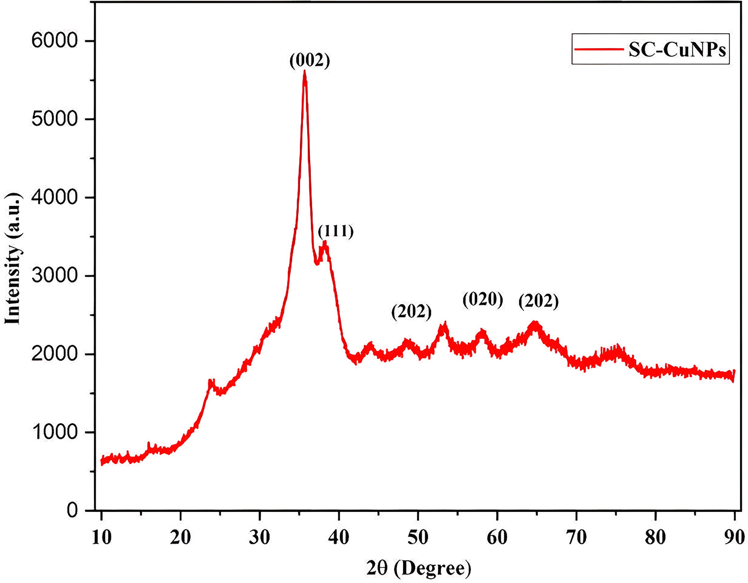
XRD spectrum of SC-CuNPs synthesized using seed extract. XRD, X-ray diffraction.
FTIR OF SC-CUNPS AND S. CUMINI SEED EXTRACT
FTIR measures the vibrational frequencies of a molecule’s bonds to identify associated functional groups (Fig. 4; Table 2). FTIR spectra of SC-CuNPs showed characteristic peaks at 1094 cm−1 corresponding to C-O stretching, indicating the presence of alcoholic and ether compounds; 1049 cm−1 corresponding to C-N groups, indicating presence of nitriles, amines and amides; 698 cm−1 often signifies the presence of a single, out-of-plane C-H bending vibration in a monosubstituted benzene ring; 537 cm−1 stretching indicates the presence of out-of-plane C = O bending vibration, suggesting the presence of an amide VI; 480 cm−1 stretching corresponds to Si-O-M (M = Mg, Al, or Fe) indicating the presence of metals.
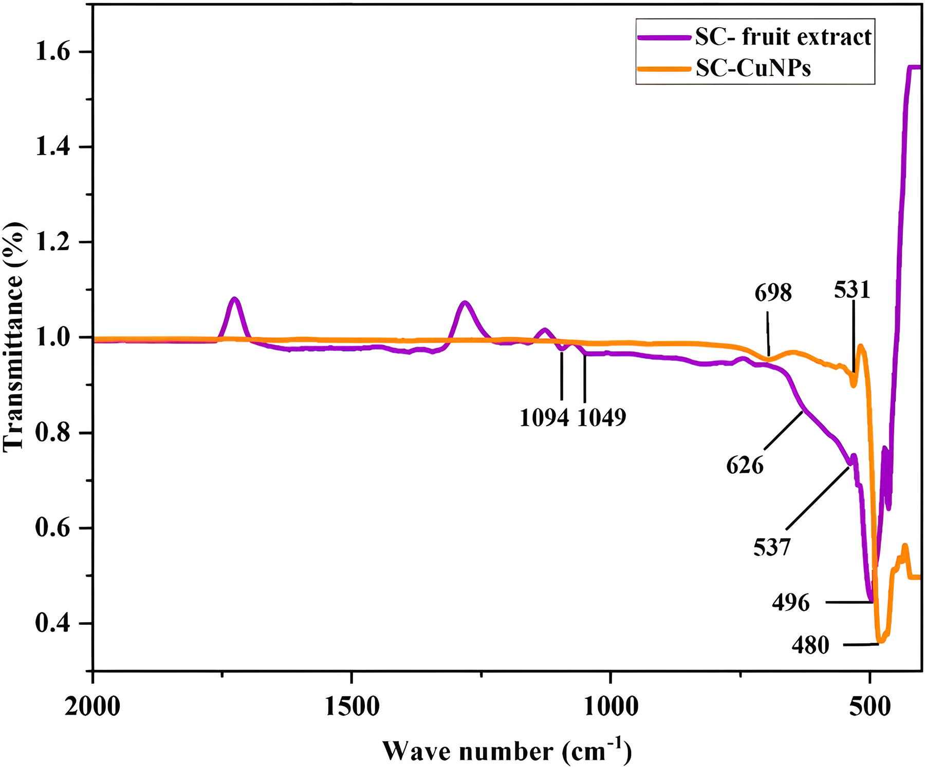
FTIR spectra of SC-CuNPs and S.cumini seed extract. FTIR, Fourier Transform Infrared Spectroscopy.
Metal and Metal Oxide Nanoparticles Synthesized Using Different Plant Parts of Syzygium Cumini and Their Applications
Possible Functional Groups Involved in Reduction and Stabilization of SC-CuNPs Identified Through FTIR Analysis
FTIR, Fourier Transform Infrared Spectroscopy; SC-CuNPs, Syzygium cumini copper nanoparticles.
SCANNING ELECTRON MICROSCOPY AND ENERGY DISPERSIVE X-RAY ANALYSIS OF SC-CUNPS
SEM was used to examine the morphological properties of the synthesized SC-CuNPs. The surface morphology of SC-CuNPs is illustrated in a SEM micrograph, showing a spherical shape and some degree of aggregation (Fig. 5A). The SEM image revealed the size of CuNPs in the range of 20–80 nm with an average particle diameter of ∼ 75 nm. Particle aggregation was noted as an interconnected spherical structure. 28 SC-CuNPs were further analyzed using EDAX. The EDAX spectra of the Cu metal primary elemental peak at 3 keV is presented in Figure 5B along with the composition percentages. Additional faint peaks were detected for the biomolecules that capped the SC-CuNPs. The inset table in Figure 5B lists the proportions of Cu and other elements.
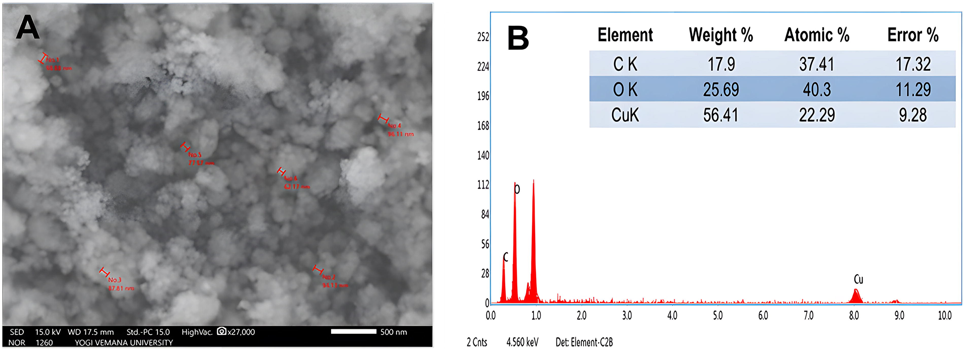
SEM analysis of SC-CuNPs
BIOLOGICAL CHARACTERIZATION OF SC-CUNPS
Antioxidant activity of SC-CuNPs and S. cumini seed extract.
Antioxidant potential of SC-CuNPs was determined using Total Antioxidant Assay (TAA) and H2O2 scavenging assays. SC-CuNPs demonstrated enhanced antioxidant compared with S. cumini seed extract. An increase in the concentration of SC-CuNPs resulted in a higher percentage of free radical scavenging activity. At a concentration of 125 µg/mL, SC-CuNPs exhibited 55% TAA activity (Fig. 6A) while H2O2 scavenging activity was approximately 53%. SC-CuNPs exhibited significant antioxidant activity at p ≤ 0.01 and p ≤ 0.05 (Fig. 6B).
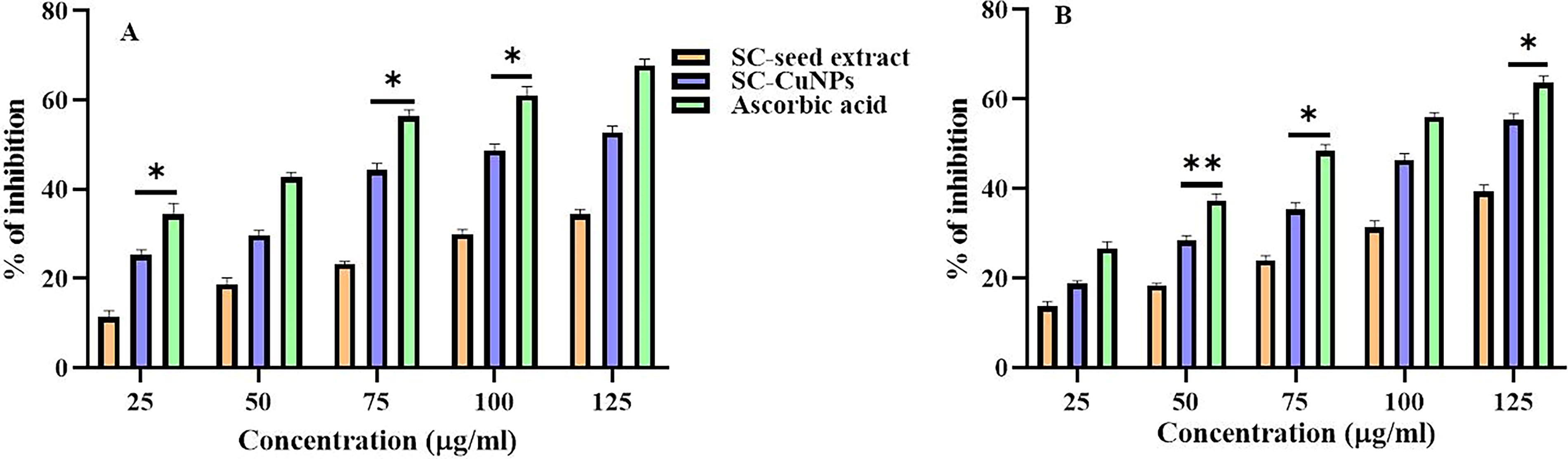
Antioxidant activity of S. cumini seed extract and synthesized SC-CuNPs along with positive control (ascorbic acid). Hydrogen peroxide assay
Antibacterial activity of SC-CuNPs and S. cumini seed extract.
To assess the effectiveness of SC-CuNPs against particular bacterial strains, including both Gram-positive and Gram-negative strains were used. The significant Zone Of Inhibition (ZOI) was detected in S. aureus (32 ± 0.89 mm), E. coli (28 ± 0.97 mm), P. vulgaris (27 ± 0.79 mm), and S. typhimurium (21 ± 0.81 mm) as indicated in Figure 7. Both Gram-positive and Gram-negative bacteria were significantly inhibited by SC-CuNPs, demonstrating their strong antibacterial activity. At a dosage of 100 μg/mL, SC-CuNPs showed the best bactericidal effectiveness against every pathogenic bacterial strain tested, surpassing the positive control. Furthermore, there was a strong dose-dependent correlation between the growth of harmful bacteria and the inhibitory activity of synthesized SC-CuNPs. SC-CuNPs exhibited significant antibacterial activity at p ≤ 0.01 and p ≤ 0.05.

Antibacterial activity of S. cumini seed extract and synthesized SC-CuNPs along with positive control (ampicillin). E. coli
Anti-inflammatory activity of SC-CuNPs and S. cumini seed extract.
In the present study, heat induced denaturation of egg albumin, which expresses antigens linked to type III hypersensitivity. One theory for the cause of inflammation is the denaturation of proteins. Denaturation of proteins in vivo may be the cause of auto-antigen formation in inflammatory illnesses such as rheumatoid arthritis. SC-CuNPs exhibited the highest percentage of anti-inflammatory activity when compared with seed extract. SC-CuNPs exhibited significant anti-inflammatory activity at p ≤ 0.01 and p ≤ 0.05. At a concentration of 500 µg/mL, SC-CuNPs exhibited the highest 43% anti-inflammatory activity. With increasing concentration of SC-CuNPs, there was an increase in anti-inflammatory activity (Fig. 8).
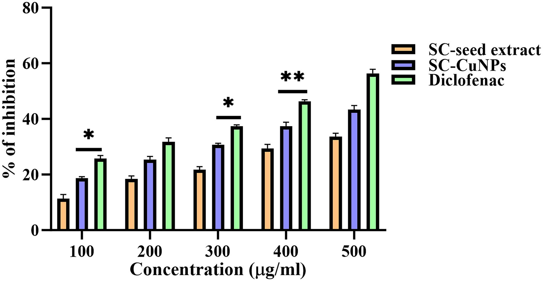
Anti-inflammatory activity of S. cumini seed extract and synthesized SC-CuNPs along with positive control (diclofenac). **Indicates the significant difference of positive control with test sample at p ≤ 0.001 and *indicates the significant difference of positive control with test sample at p ≤ 0.05.
Discussion
The investigation of plant-mediated nanomaterials and medicinal plants for potential antibacterial properties has intensified in recent years. 34 Numerous bioactive compounds with proven therapeutic benefits are derived from these natural sources. 35 S. cumini, a fruit-bearing plant in the Myrtaceae family, is commonly known as Jamun in Asia (Fig. 1). It has long been utilized as a therapeutic herb. Various parts of the plant, including fruit, seeds, bark, and leaves, have been employed to treat a range of diseases. S. cumini seeds revealed the presence of β-sitosterol, gallic acid, corilagin, ellagic acid, quercetin, and jambosine. 29 Among various synthesis methods available, the present work focuses on green synthesis of S. cumini seed extract for the production of bioactive CuNPs due to their eco-friendly nature and a simple, reproducible, and cost-effective method. 36
Green synthesis of CuNPs can be visually seen by the color change from light brown to dark brown when the S. cumini seed extract and aqueous copper sulfate solution were mixed at a 1:4 (v/v) ratio and continuous stirring for 30 minutes. The interaction between conduction electrons of metal NPs and incident photons was responsible for the color change. 32 UV-spectral analysis of SC-CuNPs reveals absorption maxima at 360 nm is due to surface plasmon resonance (Fig. 2). 30 S. cumini seed extract contains glucoside, isoquercetin, ellagic acid, anthocyanins, myrecetin, jambosine, jambolin, and kaempferol, which stabilize the SC-AgNPs and also reduce the Cu+ to Cu0. XRD analysis reveals that the SC-CuNPs were crystalline with diameters of 49 nm. The crystals exhibited a preferred orientation toward this specific plane, 37 as indicated by this significant discovery (Fig. 3). FTIR analysis reveals that S. cumini seed extract secondary metabolites functional groups (Fig. 4; Table 2) are responsible for capping and stabilizing of CuNPs. 38 According to Narayana et al., 39 the carbonyl groups of amino acid residues and peptides exhibit a significant affinity for copper ions. Proteins can bind to NPs via free amine or cysteine groups, thereby stabilizing the CuNPs. The proteins that cover the surface of the SC-CuNPs serve as capping agents. These peaks confirm the formation of Cu nanostructures in the present study. The moderate band at 1609 cm−1 corresponds to C = C stretching (alkene), while that at 1398 cm−1 denotes O-H stretching (alcohol), band at 2831 cm−1, represent C-H stretching (alkane). These groups, particularly C-H, C = C, O-H, and C-O, suggest the presence of reducing agents facilitating CuNPs synthesis. It was also revealed that functional groups of C-H, C = C, and O-H play a role as a reducing agent and capping agent. 40 Surface morphology of SC-CuNPs revealed by SEM, showing a spherical shape and some degree of aggregation (Fig. 5A). 30 Particle aggregation was noted as an interconnected spherical structure. 2 The EDAX spectra of the Cu metal primary elemental peak at 3 keV are presented in Figure 5B along with the composition percentages.
The phytochemicals found in S. cumini seeds, such as anthocyanins, flavonoids (quercetin, catechin, and kaempferol), phenolic acids (gallic acid, ellagic acid), and tannins are responsible for various pharmacological properties, including antioxidant, antibacterial and anti-inflammatory activities. 29 Nanoparticles interact with these phytochemicals in different ways, thereby increasing the stability, bioavailability, and therapeutic efficiency. 36 The morphology of nanoparticles significantly influences their biological activity and functioning. SC-CuNPs demonstrated enhanced antioxidant with an increase in the concentration of SC-CuNPs. Secondary metabolites contribute to the elevated antioxidant potential of SC-CuNPs, along with their antibacterial and anti-inflammatory potential. 41 SC-CuNPs display strong antioxidant activity and due remarkable ability to eliminate free radicals (Fig. 6). This provides compelling evidence for their potential applications, particularly in biological research (Table 1).
Both Gram-positive and Gram-negative bacteria were significantly inhibited by SC-CuNPs, demonstrating their strong antibacterial activity due to elevated antioxidant activity (Fig. 7). CuNPs exert their antibacterial action through a multifaceted mechanism, primarily by disrupting bacterial cell structure and function. They damage the cell membrane, leading to leakage of intracellular components, and also induce the generation of reactive oxygen species, which further damage cellular components. Additionally, CuNPs can interact with sulfur-containing biomolecules, disrupting their structure and causing protein denaturation. 42 The most potent bactericidal responses are known to be significantly influenced by nanoparticle size, with smaller CuNPs displaying the highest levels of antibacterial activity. SC-CuNPs inhibit bacterial growth by penetrating the bacterial cell walls. The shape and size influence the SC-CuNPs affecting the antibacterial activity. Both the pH inside the cells and the particles’ free surface energy vary with size and shape. 43 Selenium nanoparticles synthesized by using S. cumini leaf extract exhibited significant antibacterial, antifungal, and anticancer properties. 44
The immune system’s natural reaction to infections, damaged cells, irritants, and dangerous stimuli is inflammation. 45 Chronic inflammation, however, can occasionally result from an excessive immune response. Certain MNPs and secondary metabolites from plants have been shown by researchers to have anti-inflammatory properties. 46 CuNPs are utilized to preserve flavonoids such as hydroxycinnamic acid and isovitexin derivatives like chlorogenic acid, ferulic acid, and carboxylic acid. These chemicals have demonstrated potential for decreasing inflammation through several mechanisms. 40 In the present study, heat was used to induce denaturation of egg albumin, which expresses antigens linked to type III hypersensitivity. One theory for the cause of inflammation is the denaturation of proteins. Denaturation of proteins in vivo may be the cause of auto-antigen formation in inflammatory illnesses such as rheumatoid arthritis. With increasing concentration of SC-CuNPs, there was an increase in anti-inflammatory activity (Fig. 8). The potential mechanism for SC-CuNPs anti-inflammatory activity could be attributed to their stabilizing effect on the lysosomal membrane, which inhibits the release of inflammatory mediators that cause inflammation. Therefore, new avenues for locating and treating inflammatory and damaged body parts have been made possible by the application of nanotechnology in medicine. 47 Treatment efficacy can be increased by employing nanocarriers such as SC-CuNPs to improve drug penetration at the active site of microbial infections.
Conclusion
Aqueous seed extract of S. cumini was used to synthesize SC-CuNPs through a simple, economical, and eco-friendly method. During synthesis, the solution’s color changed from light brown to dark brown, indicating the formation of SC-CuNPs, which was validated by UV-vis spectroscopic analysis. The absorption peak at 360 nm observed in UV-visible spectroscopy confirmed the formation of stable SC-CuNPs. The green synthesis of the SC-CuNPs was further confirmed by FTIR spectroscopic studies. The spherical shape and crystalline structure of the generated nanoparticles were verified by SEM and XRD analyses, with particle sizes of the SC-CuNPs ranging from 20 to 80 nm. SC-CuNPs displayed significant biomedical applications, including antioxidant, antibacterial, and anti-inflammatory properties. CuNPs were synthesized via a plant-based method with an extract from the fruit of S. cumini. Therefore, the methods employed in this investigation provide several noteworthy advantages, such as economic feasibility, multifunctionality, and biocompatibility. Further molecular investigation will elucidate the detailed mechanisms underlying antibacterial and anti-inflammatory activities.
Footnotes
Authors’ Contributions
Conceptualization: R.K. Methodology: U.K.C. and M.C. Formal analysis: R.K., H.K.C., and R.N. Writing original draft: U.K.C., M.C., and R.N. Writing review and editing: R.K. and H.K.C.
Author Disclosure Statement
The authors certify that there is no conflict of interest with any financial organization regarding the material discussed in the article.
Funding Information
No external funding is received for this work.
Supplementary Material
Supplementary Figure S1
References
Supplementary Material
Please find the following supplemental material available below.
For Open Access articles published under a Creative Commons License, all supplemental material carries the same license as the article it is associated with.
For non-Open Access articles published, all supplemental material carries a non-exclusive license, and permission requests for re-use of supplemental material or any part of supplemental material shall be sent directly to the copyright owner as specified in the copyright notice associated with the article.