
Abstract
Abstract
Background:
The pulmonary route is very promising for drug delivery by inhalation. In this regard, nanoparticulate drug delivery systems are discussed, and one very promising nano carrier example is gold nanoparticles (Au NP). Directly after their deposition, inhaled Au NP come into contact with pulmonary surfactant protein D (SP-D). SP-D can agglomerate Au NP in vitro, and this may influence the clearance as well as the systemic translocation in vivo. The aim of the present study was to investigate the clearance and translocation of Au NP at a very early time point after inhalation, as well as the influence of SP-D.
Methods:
Aerosolized 20-nm radioactively labeled Au NP were inhaled by healthy adult female mice. One group of mice received dissolved 10 μg of SP-D by intratracheal instillation prior to the Au NP inhalation. After a 2-hr Au NP inhalation period, the mice were killed immediately, and the clearance and translocation to the blood stream were investigated.
Results:
The highest amount of Au NP was associated with the lung tissue. In the bronchoalveolar lavage fluid (BALF), more Au NP remained free compared with the amount associated with the BALF cells. The amount of Au NP cleared by the mucociliary escalator was low, probably because of this very early time point. Instillation of SP-D prior to Au NP inhalation had no statistically significant effect on the biodistribution of the Au NP.
Conclusion:
Our data show that inhaled Au NP are retained in the mouse lungs and are translocated after a short time, and that SP-D has only a minor effect on Au NP translocation and clearance at a very early time point.
Introduction
The main function of the phospholipid fraction in conjunction with the hydrophobic SP-B (∼8 kDa) and SP-C (∼4 kDa) is to decrease surface tension during breathing and thereby prevent alveolar collapse. SP-A (∼28–36 kDa) and SP-D (∼43 kDa) are large hydrophilic proteins and belong to the family of collectins (collagen-containing lectins). They participate primarily in immune responses. Importantly, it is known that SP-D can bind to various inhaled particulates, including allergen particles.(12,13) This conjugate of Au NP and SP-D may modulate the interaction with lung cells. In particular, SP-D is able to modulate the uptake in macrophages and thereby may enhance clearance.(13) Importantly, SP-D can bind to NP,(11) and especially Au NP, leading to agglomeration of the NP in vitro.(14) Besides SP-D, SP-A also is able to interact with particulate material and especially with NP, which influences the uptake by immune cells.(15)
The aim of the present study was to perform a proof-of-concept study for the clearance as well as translocation of inhaled Au NP across lung barriers to blood as a model for a particulate drug delivery system. For this purpose, we used a very early time point to study Au NP biodistribution, immediately after a 2-hr inhalation period. The second aim was to determine the effect of SP-D on the clearance pathways in general, including translocation of inhaled Au NP to blood. Clearance and translocation were investigated after a 2-hr inhalation of 20-nm spherical radioactively labeled 195Au NP by healthy adult female mice. Prior to the inhalation, one group of mice received 10 μg of SP-D by intratracheal instillation to increase the amount of natural SP-D in the lungs. Directly after inhalation, the lung retention, as well as clearance and systemic translocation, of the Au NP was analyzed and compared with that of control mice that did not receive additional SP-D, but only the vehicle [phosphate-buffered saline (PBS)]. For this analysis, we used our quantitative biodistribution assay.(16)
Materials and Methods
Animals
Female C57BL/6 mice (10–12 weeks old, body weight ∼21.5 g) were obtained from Charles River (Sulzfeld, Germany) and housed in the HMGU animal facility in isolated ventilated cages (VentiRack, Biozone, Margate, UK). They received a standard pellet diet and tap water ad libitum. The study was conducted under the guidelines of the Helsinki Convention for the Use and Care of Laboratory Animals and was approved by the Government of the District of Upper Bavaria (approval no. 55.2-1-54-2531-26-10) and the Animal Care and Use Committee of the Helmholtz Center Munich. All chemicals were purchased from Sigma-Aldrich (Deisenhofen, Germany), unless otherwise specified.
Aerosol production and characterization
Ultrafine 195Au aerosol was produced with a spark ignition generator as previously described.(17) For radiolabeling, the gold electrodes (3.1-mm diameter, 4-mm length) of the spark ignition generator were proton irradiated at one end in the Scanditronix MC40 cyclotron at the Joint Research Center (Ispra, Italy), using the highest proton energy available.
195Au has several gamma energies below 100 keV and a long half-time of 186 days, allowing gamma spectroscopy for measurement of organ activities.
The 195Au activity concentration of the aerosol was 1.2 kBq L−1 at the reference date. Aerosol mass concentration was calculated to be 1.6 mg m−3 from the radioactivity measurement of an integral filter sample of 0.3 L/min aerosol flow during the entire exposure and the sampled aerosol volume. Size distribution parameters were the following: particle number concentration, 2±0.5×10⁷/cm3; count median diameter (CMD), 22 nm; geometric standard deviation, 1.6; the first being continuously monitored with a condensation particle counter (CPC 3022A; TSI, Aachen, Germany) and the second by a differential particle mobility analyzer (Classifier 3071+CPC 3010; TSI). As the aerosol was heat-treated at 600°C online in a tube furnace immediately after generation and prior to exposure, the agglomerated gold particles were melted to spherical-shaped particles of 22-nm CMD. Morphologically, Au NP were analyzed by TEM collecting Au NP of the freshly generated aerosol after heat treatment with an electrostatic TEM sampler (University of Applied Sciences, Windisch, Switzerland).
Instillation of SP-D
Directly prior to the 195Au NP inhalation, mice were anesthetized using medetomidine 0.5 mg/kg, midazolam 5 mg/kg, and fentanyl 0.05 mg/kg. Afterwards, the anesthetized mouse was fixed with its incisors to a rubber band on a board at an angle of 60° to the lab bench. A flexible cannula (BD Vasculon Plus; 26 G, 0.75 in) was placed under visual control into the upper third of the trachea. Proper placement in the trachea, and not misplacement into the esophagus, was controlled with a pneumotachograph connected to the cannula. Directly before the start of the Au NP aerosol inhalation, the suspension (50 μl of PBS or 50 μl of PBS containing dissolved 10 μg of SP-D; recombinant human SP-D, R&D Systems, Minneapolis, MN) was gently instilled into the trachea of the mouse during the inspiration phase, followed by 400 μl of air using a 1-ml insulin syringe. The instilled air prevented liquid losses in the syringe and supported deeper penetration of the instillate into the lungs. Intratracheal instillation was aligned with the inspiration of the mouse.
195Au nanoparticle inhalation
Immediately after SP-D instillation, the intubated mouse was placed in a plethysmograph and connected with the endotracheal cannula to the aerosol line outside of the plethysmograph, as described earlier.(17,18) Ventilation was computer-controlled by a negative pressure of 1.5 kPa applied to the plethysmograph chamber during 0.25 sec of inspiration followed by 0.25 sec of expiration at ambient air pressure, resulting in a breathing frequency of 120 min−1. This ventilation pattern usually caused an inspiration of >50% of total lung capacity of the mice; therefore, animals were slightly hyperventilated and did not breathe spontaneously, but followed the computer-controlled breathing pattern. Four mice were ventilated simultaneously, each placed in an individual plethysmograph chamber. For radiation protection, the entire aerosol system was maintained at −30 Pa low pressure and additionally installed in a glove box, which was highly ventilated through an absolute particle filter providing an additional low pressure in the glove box of about −20 Pa.
Sample preparation
Directly after inhalation, mice were kept anesthetized (5% isoflurane inhalation) and euthanized by exsanguination via the abdominal aorta. Approximately 70% of the total blood volume was withdrawn. For 195Au radioanalysis, the following samples were taken by careful necropsy after euthanasia of the animals: blood sample, lungs, esophagus, larynx, stomach, gastrointestinal tract (GIT) (stated as fast clearance via the mucociliary escalator), total skin including fur, bronchoalveolar lavage (BAL), and the remainder (all remaining organs, tissue, muscles, skeleton).
During dissection, no organs were cut in order to avoid any cross contamination.
Afterwards, the organs and tissues were directly frozen and radioanalyzed as quickly as possible without any further preparation step.(17,19)
By this approach, we quantitatively determined the entire NP dose in the animal by analyzing each organ and tissue in a 100% balance of the biodistribution of the applied NP. As there were no excreta at this early time point immediately after inhalation, no excreta samples were collected.
Bronchoalveolar lavage
A BAL was performed by applying 5×1 ml of PBS without Ca2+ or Mg2+ under gentle massage of the thorax. The recovered BAL fluid (BALF) (∼90% of instilled saline) was centrifuged at 500 g for 20 min to separate the lavaged cells from the supernatant.
195Au radioactivityanalysis
The 195Au radioactivity of all samples was measured by gamma spectroscopy without any further physicochemical preparation in either a lead-shielded 10-mL or a lead-shielded 1-L well-type NaI (Tl) scintillation detector. The count rates were corrected for physical decay and background radiation. In addition, count rates were calibrated to a 195Au reference source in order to correlate 195Au radioactivities to the numbers and masses of the Au NP. Samples yielding net counts (i.e., background-corrected counts) less than three standard deviations of the totally measured counts in the photopeak region of interest of the 195Au gamma spectrum were defined as below the detection limit. For a complete balance of the administered 195Au radioactivity within each mouse, 195Au radioactivities of all samples were summed up for each mouse (except the skin and fur sample); this was considered to be the initial lung dose (ILD) and was used as a denominator for the calculation of the 195Au percentage of each sample. Additionally, all data (except those of mucociliary clearance) were corrected for the fast cleared Au NP determined in the esophagus and GIT. Using this approach, we attempted to normalize the translocated amount of Au NP to the initial peripheral lung dose.
Calculations and statistical analysis
Four animals per group were used. Calculated values are given as a percentage of the relevant integral 195Au radioactivity (calculated for a reference date) of all samples in each animal with the standard error of the mean (SEM). All radioactivities were correlated with the corresponding mass of gold NP in each animal. Data of animals with and without SP-D were compared using an unpaired t test, and differences were considered to be significant at p<0.05.
Results
The aerosol and inhalation data are shown in Table 1. A representative image of the inhaled Au NP is shown in Figure 1.
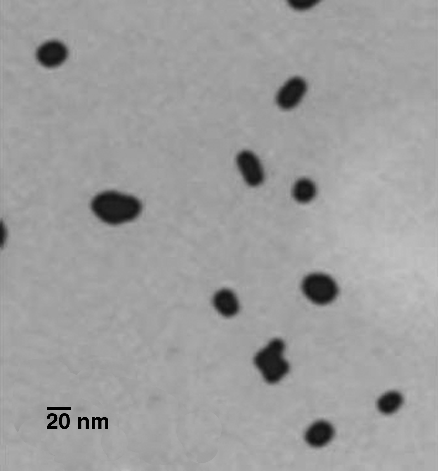
Transmission electron micrograph (TEM) image of freshly generated Au NP collected with an electrostatic TEM sampler (University of Applied Sciences). Au NP had been heat-treated at 600°C in the airborne state to melt and form spherically shaped NP.
Note: the surface area of a 22-nm sphericalAu NP is 1.5×10³ nm2.
Directly after the 2-hr inhalation period, 3.6±1.2% of the deposited Au NP within the control group were cleared via the mucociliary escalator (Fig. 2). Instillation of SP-D directly before the inhalation did not significantly change this amount, because 3.3±0.3% of the Au NP were measured with prior SP-D instillation. Likewise, the uptake of the Au NP within the BAL cells, which consisted mainly of macrophages, was not changed due to SP-D instillation (Fig. 3). Within lavage cells for the control group and the SP-D group, 12.4±1.1% and 12.6±0.9%, respectively, of the Au NP could be detected. As expected, the amount of Au NP that was found free in the BALF was much higher compared with the amount of Au NP within the cells of the BALF (Fig. 3). In this regard, 29.9±1.8% of the deposited Au NP were found free in the BALF supernatant within the control group. Previous instillation of SP-D led to 26.3±1.1% free Au NP in the BALF supernatant. High amounts of Au NP associated with the lung tissue (not lavageable) were detected (Fig. 4). In the control group, 57.5±1.9% of the deposited Au NP were lung tissue associated compared with 60.8±1.4% of the Au NP in the SP-D group (no significant difference). Interestingly, a trend toward a different translocation to the blood was detected within the two groups: 1.2±0.5% of the deposited Au NP within the control group versus 0.4±0.1% within the SP-D group translocated to the blood (Fig. 5; yet no significant difference).
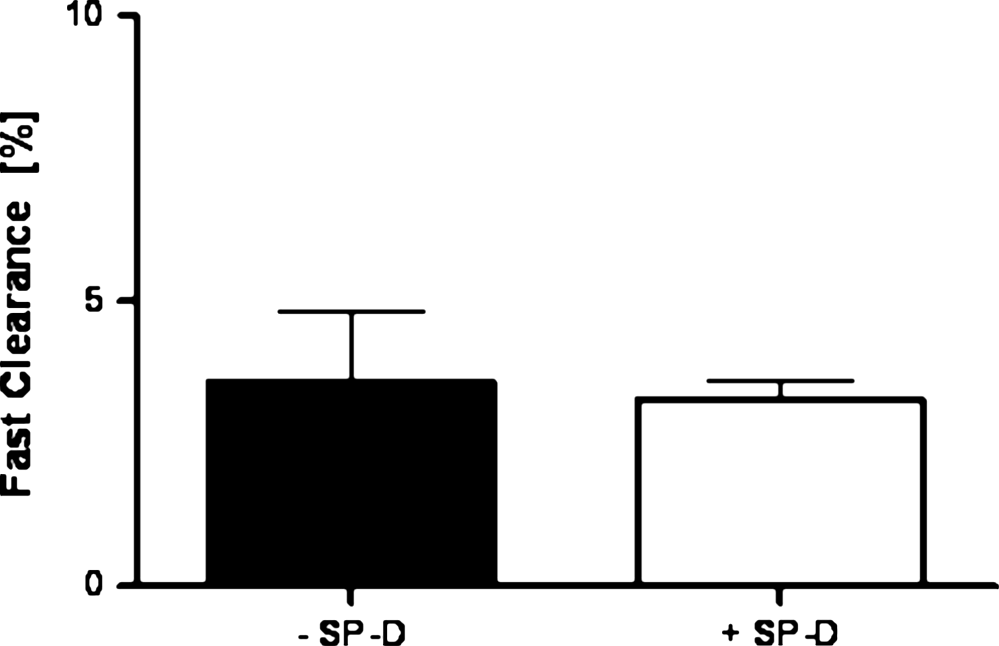
Fast-cleared percentage of Au NP relative to ILD in SP-D–exposed and control mice (means±SEM; n=4). Au NP, at 0 hr after a 2-hr inhalation, were transported via the mucociliary escalator to the larynx and swallowed into the GIT.
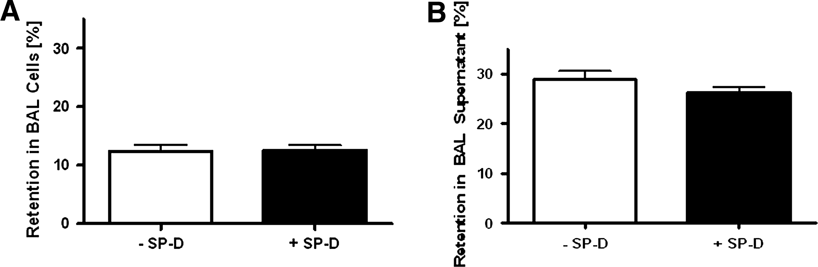
Retention of Au NP within cells of BAL and in supernatant. Retention of Au NP within BAL cells
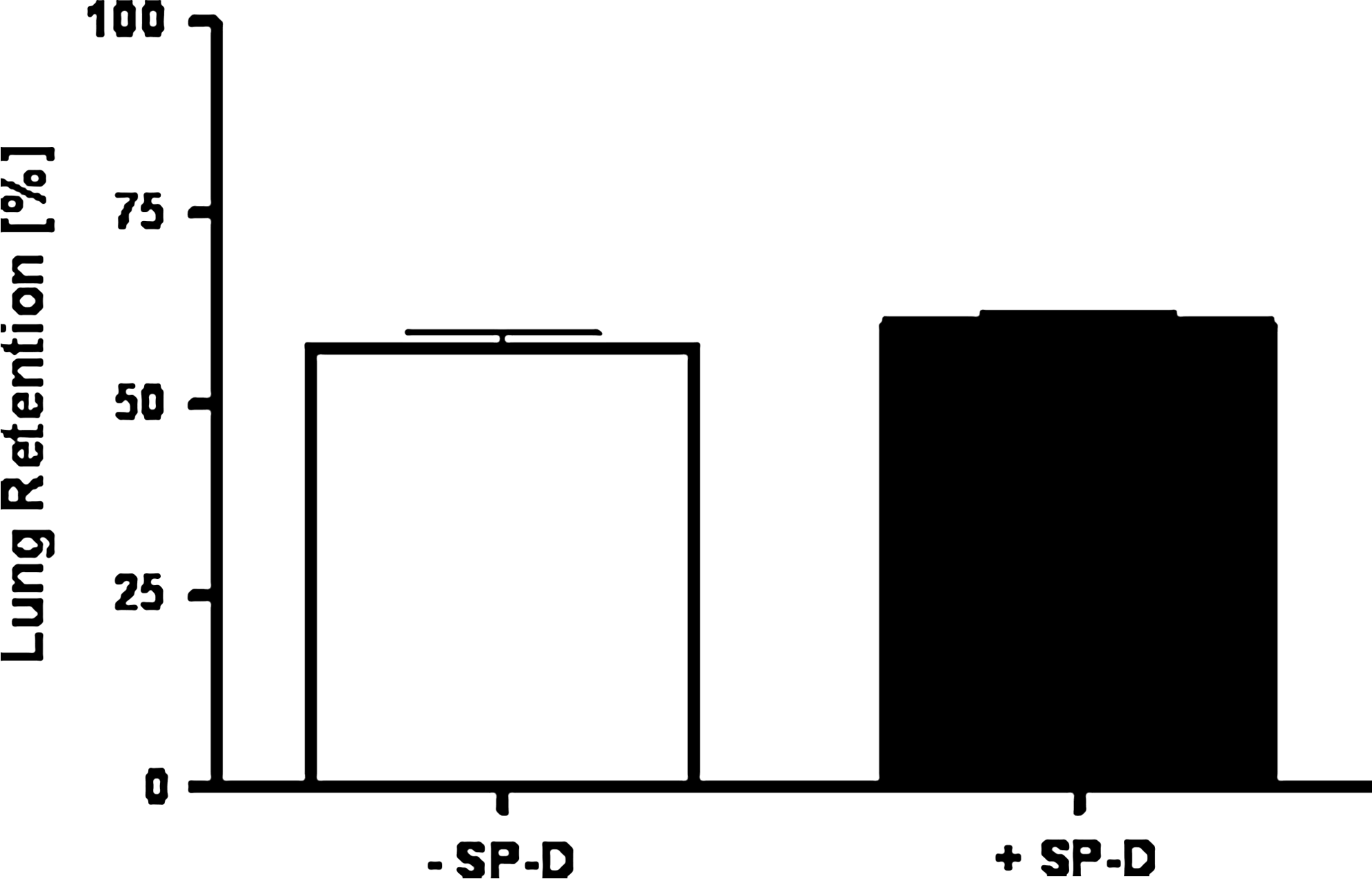
Retention of Au NP within the lung tissue. Retention of Au NP within the lungs at 0 hr after a 2-hr inhalation after performing BAL is shown. Data are corrected for fast clearance.
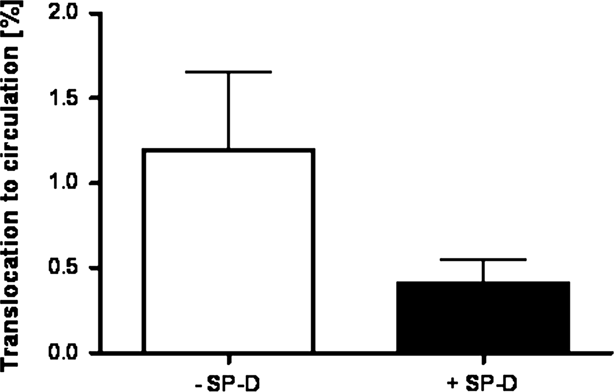
Translocation to circulation of Au NP. Au NP with and without SP-D, at 0 hr after a 2-hr inhalation, were transported to the circulation. Data are corrected for fast clearance.
Discussion
In the present study, we performed a proof-of-principle study to investigate the clearance and translocation of inhaled Au NP as a model of a nanoparticulate drug delivery system at a very early time point (directly after the 2-hr inhalation period). Furthermore, the effect of SP-D on clearance as well as translocation through the air–blood barrier was investigated.
Fast clearance via the mucociliary escalator
Immediately after the end of the 2-hr Au NP inhalation period, the fast mucociliary clearance was low and related to only 3.6±1.2% of the deposited Au NP of the control group. However, this is not astonishing, because the animals were intubated during Au NP inhalation, allowing only minor particle transport into the stomach; only NP that deposited in the lung at the very beginning of the inhalation period were able to get cleared and swallowed into the stomach. Hence, we would expect more mucociliary clearance of the Au NP at longer duration after NP inhalation. This is consistent with a former study using inhaled iridium NP.(20)
Au NP within the BALF
Using centrifugation, we separated the cells of the BALF from the lavage fluid. Hence, we could distinguish the Au NP taken up by macrophages from the Au NP remaining free in the BALF.
Directly after inhalation, more “free” Au NP (29.9±1.8% of lung retained) were found within the BAL supernatant compared with the fraction associated with BAL cells (12.4±1.1% of ILD). As these are the results directly after the 2-hr inhalation period, we believe that this is mainly due to the short time interval between NP deposition and the collection of the BALF. Hence, we hypothesize that at later time points more NP will be taken up by BAL cells compared with free Au NP in the BALF. Similar results were also found with iridium NP,(21,22) where directly after the inhalation more free NP were found in BALF compared with the amount of NP in cells. Importantly, a rapid increase of NP in the BAL cells within the first day was observed previously.
Au NP associated with the lung tissue
A high fraction of the lung-retained Au NP was associated with the lung tissue (57.5±1.9%) directly after the inhalation period. In this regard, it is important to note that we did not distinguish between NP taken up by lung cells (i.e., epithelium) and NP bound to the lung tissue. However, as it is known that NP can be easily taken up by lung cells in vitro and in vivo,(23–25) we assume that both uptake and binding occur. Due to the investigated time frame (directly after inhalation), we assume that most of the particles may be attached to the cell surface of the lung tissue.
Au NP translocation via the air–blood barrier
We found a rather high amount of Au NP translocated to the blood and accumulated in secondary organs and tissues directly after inhalation. In this regard, 1.2±0.5% were detected beyond the air–blood barrier. Importantly, we can exclude that uptake via the GIT was responsible for this translocation. On the one hand, there was very limited time for the NP to reach the GIT, and just a limited amount of approximately 3.6% NP was found within these organs. On the other hand, we know from recent gavage and instillation studies that absorption of NP via the GIT is very small.(19,26,27) Hence, we conclude that the uptake into blood described in the present article was almost exclusively across the air–blood barrier in the lung. A previous publication of our laboratory from Lipka and co-workers described a translocation rate of 2.6% 1 hr after intratracheal instillation of 5-nm Au NP.(27) The twice-higher translocation is expected for the smaller 5-nm Au NP compared with the 22-nm Au NP used in this study. However, to compare these results (directly after inhalation and intratracheal instillation) much better, more time points—especially 1 hr after inhalation—need to be performed in future studies.
The influence of SP-D
No significant effect of SP-D on Au NP clearance and translocation was detected—besides the trend of decreased translocation combined with a slightly higher increased cellular binding. This may be due to several reasons.
One possibility is that the instilled SP-D just increased the physiologic pool, which does not have any additional influence compared with the already residing SP-D. Hence, one could speculate that the effect of SP-D on NP in the lungs is already “saturated” in healthy animals. Even if we cannot completely exclude this, we do not believe that this is the case, because it is known that the effect of SP-D is strongly dose-dependent.(12,14) However, this has to be investigated further, which could be done, for example, by using knockout mice.
A second explanation could be that the early time point of measurement in this proof-of-concept study was too short to detect SP-D modulating effects. We do not expect that the in vivo effect of additional instilled SP-D is very prominent. Hence, later time points would be much more suitable to detect these slight SP-D–modulated influences. This will be considered in a further study.
A third very reasonable explanation is directly linked to the mechanism by which SP-D modulates the clearance of particulates. It is known that SP-D induces agglomeration of various particulates like allergen particles,(13) E. coli,(28) A. fumigatus conidia,(29) S. pneumoniae, S. aureus,(30) and even Au NP.(14) By this agglomeration, more particles are taken up by macrophages. Importantly, in our present study, SP-D was simply not able to agglomerate the deposited Au NP after inhalation. To become agglomerated, a single Au NP has to come into close contact with another single NP. This can be excluded in our study. By calculation of the total projected area of the deposited Au NP (Table 1) and due to a rather homogeneous Au NP deposition on the entire lung epithelium (due to diffusion), the Au NP coverage of the lung surface is so sparse (<10–12) with huge free spaces in between that no NP can coagulate—even if SP-D binds to the surface of the NP. Hence, this estimation, as well as our results, is consistent with the assumption that the Au NP modulation by SP-D is only dependent on agglomeration and not on receptor-specific modulation. However, as it is known that SP-D binding to other particulates (e.g., P. aeruginosa) does not induce agglomeration, but enhances phagocytosis by alveolar macrophages,(31) it has to be proven if this effect is really dependent on the specific type of particulate material or not.
Although there was a trend of decreased translocation from the lungs of SP-D–treated mice to blood, the results are not significant; this interesting tendency needs to be investigated in more detail, for example, by investigating later time points. A more detailed investigation could evaluate whether SP-D mediates binding to resident lung cells like epithelial cells in vivo.
Footnotes
Acknowledgments
The authors appreciate the technical assistance of Sebastian Kaidel and Nadine Senger. This work was partially supported by the German Research Foundation (SPP-1313) and the EU FP7 projects NeuroNano (NMP4-SL-2008-214547) and ENPRA (NMP4-SL-2009-228789).
Author Disclosure Statement
No conflicts of interest exist.