




Abstract
Abstract
Background:
The lung is the primary entry site and target for Mycobacterium tuberculosis; more than 80% of the cases reported worldwide are of pulmonary tuberculosis. Hence, direct delivery of anti-tubercular drugs to the lung would be beneficial in reducing both, the dose required, as well as the duration of therapy for pulmonary tuberculosis. In the present study, microsphere-based dry powder inhalation systems of the anti-tubercular drugs, rifampicin and rifabutin, were developed and evaluated, with a view to achieve localized and targeted delivery of these drugs to the lung.
Methods:
The drug-loaded chitosan microparticles were prepared by an ionic gelation method, followed by spray-drying to obtain respirable particles. The microparticles were evaluated for particle size and drug release. The drug-loaded microparticles were then adsorbed onto an inhalable lactose carrier and characterized for in vitro lung deposition on an Andersen Cascade Impactor (ACI) followed by in vitro uptake study in U937 human macrophage cell lines. In vivo toxicity of the developed formulations was evaluated using Sprague Dawley rats.
Results:
Both rifampicin and rifabutin-loaded microparticles had MMAD close to 5 μm and FPF values of 21.46% and 29.97%, respectively. In vitro release study in simulated lung fluid pH 7.4 showed sustained release for 12 hours for rifampicin microparticles and up to 96 hours for rifabutin microparticles, the release being dependent on both swelling of the polymer and solubility of the drugs in the dissolution medium. In vitro uptake studies in U937 human macrophage cell line suggested that microparticles were internalized within the macrophages. In vivo acute toxicity study of the microparticles in Sprague Dawley rats revealed no significant evidence for local adverse effects.
Conclusion:
Thus, spray-dried microparticles of the anti-tubercular drugs, rifampicin and rifabutin, could prove to be an improved, targeted, and efficient system for treatment of tuberculosis.
Introduction
T
Oral therapy using the currently employed antitubercular drugs (ATDs) is effective, but associated with several major drawbacks. More than 80% of TB cases are of pulmonary TB, which require high drug doses to be administered, because only a small fraction of the total oral dose reaches the lungs that are cleared in a few hours, making it necessary to administer multiple ATDs on a regular basis for long periods (6 to 9 months), a regimen which the majority of TB patients find difficult to adhere to.(1,3,4)
Despite the availability of highly effective drugs for TB, cure rates have not improved.(5–7) This is largely due to problems relating to drugs, formulations, and duration of therapy. Although the first-line drugs for TB have been highly effective in curtailing and curing the disease, the daily dose of these therapeutic agents range from 300–600 mg for rifampicin, rifabutin, and isoniazid, and to 900–1500 mg for ethambutol and pyrazinamide.(3) The World Health Organization (WHO) suggests treatment of tuberculosis with a standard multi-drug therapy called DOTS (Directly Observed Therapy, Short course) under which a patient is required to take two or more (200–300 doses) of the anti-TB drugs for a period of 6–12 months under observation.(1)
Such high doses formulated as solid oral formulations such as tablets and capsules, along with the long duration of treatment, make it inconvenient for the patients to complete the course of therapy. Moreover, relatively few ATDs have been discovered in the past 3 decades because of complex, lengthy, and expensive process of drug development.(8) Further to this, the emergence of resistant strains (multidrug resistant strains) to existing drugs is a growing concern.(6) Thus, considering the present scenario, due to difficulties in obtaining new drugs for TB, it is important to look into the existing drugs and develop novel and targeted delivery systems of these drugs, which will overcome the drawbacks of current conventional oral formulations.(9–12)
Direct delivery to the lungs of antibiotics, anti-infective agents, and other drugs has been used for treatment of several diseases of the lungs such as asthma, pneumonia, and cystic fibrosis. There is potential for administration of ATDs in a similar way to overcome the drawbacks of oral delivery.(13) Since lung is the primary entry site and target for Mycobacterium tuberculosis, direct delivery to the lung can be expected to reduce the dose required and also significantly reduce the duration of therapy. This can also overcome the toxicity issue by reducing the amount of drug required by delivering it directly to the site of action.
Mycobacterium tuberculosis is an intracellular pathogen and is difficult to treat because infections are localized within phagocytic cells (alveolar macrophages) where they remain in a dormant stage. Most antibiotics, although highly active in vitro, do not actively pass through cellular membranes, making it difficult to achieve the relatively high concentrations of the drugs within the infected cells. The main challenge for intracellular chemotherapy is to design and develop a carrier system for antibiotics and antifungals that could be efficiently endocytosed by phagocytic cells and, once inside the cells, should deliver large concentrations for prolonged periods, so that the number of doses and associated drug toxicity can be reduced. It has long been established that macromolecular drugs and particulate or vesicular drug delivery systems introduced into the deep lung are likely to be taken up by alveolar macrophages (AM).
The particulate carrier systems (micro-particles, liposomes, and nanoparticles) have been found to be effective in delivering ATDs at the site of infection.(14,15) Among the first line of drugs used, rifampicin and rifabutin would be the drug of choice as they act against the intracellular, nonreplicating dormant organisms, thereby reducing duration of therapy and preventing a relapse of the disease.(1)
The objective of the present study was to develop microsphere-based dry powder for inhalation (DPI) system of ATDs, using methods free from organic solvents and toxic cross-linking agents, with a view to achieve localized and targeted delivery of these drugs to the alveolar macrophages within the lungs. Chitosan is a high molecular weight polysaccharide linked by β-1, 4 glycoside. It is composed of N-acetyl-glucosamine and glucosamine. It is considered to be the most widely distributed biopolymer, having huge resources.(16) It is a cationic polyelectrolyte which is nontoxic, biocompatible, biodegradable, and has been shown to be enzymatically degraded by the body, including in organs like the lungs.(16,17) The nontoxicity of chitosan has been previously established by many authors in lung epithelial cell lines as well as in vivo.(18,19)
The primary amine groups in chitosan renders it a positive charge and contributes to mucoadhesive properties that make chitosan very useful in drug delivery applications.(20) Chitosan has attracted a great deal of attention in pharmaceutical drug delivery applications, including colon-targeted drug delivery, mucosal delivery, cancer therapy, vaccine delivery, gene delivery, and nasal and pulmonary delivery.(21,22) Today chitosan-based inhalation delivery systems have become the focus of researchers for developing treatment strategies for asthma, COPD, and even diabetes.(13,23) Thus, there is potential for administration of ATDs in a similar way to overcome the drawbacks of oral delivery.
Materials and Methods
Rifampicin (RIF) and rifabutin (RFB) were obtained as gift samples from Lupin Ltd, India. Chitosan (CHT) having degree of deacetylation value of 90% was obtained as gift sample from Esvee Agro and Feeds, Pune, India. Tripolyphosphate (TPP) was procured from Sigma Aldrich, India. Lactohale® 100 and Lactohale® 300 were obtained as gift samples from DFE Pharma, Germany. Hard gelatin capsules were obtained as gift samples from Associated Capsules Ltd, India. All other chemicals and reagents used were of analytical grade.
Preparation of drug-loaded chitosan-TPP microparticles
The chitosan microparticles were prepared by ionic gelation technique described by Ko et al.(24) and further isolated by spray drying. Briefly, accurately weighed quantities of the drugs were dissolved in 1% (w/v) chitosan solution prepared in 0.1 N acetic acid. Different concentrations (0.5%–2% w/v) of TPP solution were added dropwise to the chitosan-drug solution under high-speed homogenization (Silverson homogenizer, Sri Ram Industries, India) at 2000 rpm for 15 min. The suspension of chitosan–TPP–drug microparticles was then spray dried (LSD-48 spray dryer, JISL, India) to obtain TPP cross-linked drug loaded microparticles. The parameters for spray drying are given in Table 1. Batches with different ratios of chitosan and TPP were prepared and characterized in order to evaluate its effect on properties of microparticles.
RH, Relative humidity; All values are expressed as mean ± SD
Preparation of microparticle-based dry powder inhalation (DPI) system
The microparticles obtained after spray drying were adsorbed onto an inhalable lactose carrier. A blend of a coarser grade lactose (Lactohale 100) and fine grade lactose (Lactohale 300) was prepared in the ratio of 70:30 and used as the carrier system for the microparticles. Briefly, 400 mg of drug-loaded microparticles were mixed well with 2000 mg of pre-formed Lactohale blend. The admixture was then passed through ASTM (American Society for Testing and Materials) 60 sieve (250 micron) and mixed well again. Samples were taken from three different locations from the bulk blend and analyzed for drug content in order to ascertain content uniformity. The remaining blend was filled into size 3 hard gelatin capsules (average fill weight: 25 mg/capsule).
Characterization of drug-loaded chitosan-TPP microparticles
Moisture content
The moisture content of the microparticles was determined by the Karl-Fisher titration method. Accurately weighed quantities of microspheres (100 mg) were titrated on a Mettler DL40GP Memotitrator using Karl Fisher reagent. The percent moisture content was determined using the titer reading by the following formula:
Particle size and zeta potential
Particle size and zeta potential were determined using a Malvern ZetaSizer (Nano-ZS, U.S.A). For determination of particle size, the microparticles were suspended in methanol containing 0.1% Tween 80 to prevent aggregation, and the mean particle size and polydispersity index were determined. For determination of zeta potential, the microparticles were suspended in de-ionized water and placed in disposable polystyrene electrophoretic cells. The total zeta runs were 100 and the count rate was 250 particles/sec. The experiments were performed in triplicate.
X ray diffraction (XRD)
XRD patterns of the free drug, excipients, and drug-loaded microparticles were recorded to assess their solid state characteristics. The samples were prepared and analyzed using a Phillips X' Pert MPD diffractometer (TIFR, Mumbai) with a copper target, operated at a voltage of 40 kV and 20 mA current, at a scanning speed of 2° per minute. The experiment was performed in triplicate.
Differential scanning calorimetry (DSC)
The DSC thermograms of RIF, RFB, and their microparticles were recorded on a Diamond DSC (Perkin Elmer) thermal analyzer. The drug sample was heated in an aluminum crimped pan, sealed with a platinum lid at a heating rate of 20°C/min in the 50°–250°C temperature range under a nitrogen atmosphere purged at a flow of 50 mL/min. The experiment was performed in triplicate.
Surface morphology
Surface topography of the free drug, drug-loaded microparticles and the DPI system was visualized using a scanning electron microscope (Zeiss EVO LS10, Germany). The samples were prepared by plasma deposition method using a gold coating unit to make the surface of the microparticles conductive to the scanning electron beam. About 40°–100 A° thick coat of gold was applied on the microparticle surfaces. The gold coating was done under argon atmosphere. The gold-coated microparticles were scanned using secondary electron beam of 10 KV intensity.
Drug content/loading efficiency
Drug content was determined by UV spectrophotometry. The drug from 10 mg of accurately weighed microparticles was extracted in 5 mL of methanol by sonication in a bath sonicator (Ultrasonic Systems, India) for 15 min, followed by centrifugation (Remi, India) at 1000 g for 10 min. The sediment was given two washings with 5 mL of filtered distilled water and the above steps of sonication and centrifugation were repeated. The supernatant and washings were combined and the absorbance was recorded on a UV-Vis spectrophotometer (Jasco 530V, Japan) at 239 nm for RIF and 277 nm for RFB, using appropriate blanks as the reference. The experiment was performed in triplicate.
Swelling index
The swelling properties of the chitosan microparticles was evaluated by the difference in weight of the microparticles before and after incubation in simulated lung fluid (SLF), pH 7.4 for a period of 24 h, at 37 ± 0.5°C in an incubator (Tempo Instruments and Equipments, India). The composition of simulated lung fluid, pH 7.4 is given in Table 2.(25) The experiment was performed in triplicate.
In vitro drug release
The in vitro drug release from the microparticles was evaluated by a static method in simulated lung fluid (SLF), pH 7.4 at 37 ± 0.5° C and compared to that of free drug under the same conditions. An accurately weighed quantity (10 mg) of drug-loaded microparticles was taken in a dialysis bag (Himedia; MWCO: 12000–14000 Daltons) containing 5 mL of SLF, pH 7.4 and tied at both ends. The dialysis bag was suspended in a 100 mL stoppered test tube containing 50 mL of SLF, pH 7.4, and the assembly was placed in an incubator (Tempo Instruments and Equipments, India) maintained at 37 ± 0.5°C.
At specific intervals of time, the whole dissolution medium was replaced with 50 mL of fresh pre-warmed medium (37 ± 0.5°C) and the dialysis bag was inverted each time to prevent the microparticles from adhering to the sides of the bag or aggregating. The aliquots were evaluated for drug content on a UV-Vis spectrophotometer (Jasco 530V, Japan) using SLF, pH 7.4, as the blank.
In order to further understand drug release mechanism, the drug release data was plotted into the Korsmeyer-Peppas equation as log cumulative percentage of drug released versus log time, and the value of diffusion exponent (n) was calculated using the slope of the straight line. The experiment was performed in triplicate.
In vitro lung deposition
In vitro lung deposition of drug-loaded microparticles, plain drug, and microparticles with lactose blends was evaluated on an Andersen Cascade Impactor(ACI) (Copley Scientific, U.K) at a flow rate of 28.3 L/min using a Lupihaler® device (Lupin, Ltd.). Fine particle dose (FPD), fine particle fraction (FPF), mass median aerodynamic diameter (MMAD), and geometric standard deviation (GSD) were calculated as per USP using CITDAS software application (Copley Scientific, U.K). The experiment was performed in triplicate.
Stability study
The stability study of selected batch (RIF2) of RIF-loaded microparticles, formulated as the dry powder inhaler blend in hard gelatin capsule (size 3) and sealed in tri-laminate pouches, was carried out at room temperature and at 40°C ± 2°C/75% RH ± 5% RH for a period of 3 months. At intervals of 1 month, the samples were evaluated to detect any changes with respect to visual appearance, drug content, moisture content, and in vitro lung deposition. The analysis was performed in triplicate
In vitro macrophage uptake study
In vitro uptake by macrophages was assessed on U937 human macrophage cell lines by the method mentioned by Evora et al.(26) The RIF2 and RFB3 batches, which showed highest in vitro lung deposition, were selected for the study. Different concentrations of the drug-loaded microparticles were added to a cell culture suspension of U937 human macrophage cells equivalent to 2 × 10⁶ cells per well and incubated in a 5% CO2 environment at 37°C for a period of 90 min in a CO2 incubator (Thermo Scientific, India). The macrophages were isolated by centrifugation (Remi, India) at 1000 g for 15 min and the supernatant was analyzed spectrophotometrically for drug content. The experiment was performed in triplicate.
In vivo acute toxicity study
Acute inhalation toxicity evaluation of drug-loaded microparticles of RIF and RFB was carried out by intratracheal instillation in female Sprague Dawley rats. The protocol followed for the study was approved by an independent animal ethics committee of Bombay College of Pharmacy (30/04/2010/CPCSEA/BCP/2010/04). The animals were divided into seven groups of three animals each as shown in Table 3.
PBS, phosphate buffered saline, pH 7.4; RFB, rifabutin; RIF, rifampicin; *Equivalent to 200 μg of RIF; #Equivalent to 200 μg of RFB.
The animals were anesthetized with an intra-peritoneal injection of 0.2 mL of ketamine hydrochloride. The animal was harnessed onto the wooden board with the help of stretch bands. A ventral incision of 0.5 cm was made over the tracheal region superior to the supraclavicular notch. The trachea was exposed through the longitudinal incision along the ventral aspect of the neck. 200 μL of suspension of microparticles (containing equivalent amount of 1 mg/mL of drug) or drug solutions (1 mg/mL) in phosphate buffered saline (PBS), pH 7.4, were administered intratracheally with a tuberculin syringe having 27-gauge needles.
Similarly, 200 μL of suspension of blank chitosan microparticles (0.5 % w/v) in PBS, pH 7.4, was administered to the respective control groups. The skin was then sutured with a sterile braided surgical silk thread and an antiseptic ointment (Betadine) was applied over the stitches. The animals were kept upright against a wooden board such that they remained upright till they regained consciousness.
The animals were then administered 200 μL of amoxicillin subcutaneously to prevent infection. The dosed animals were observed daily for any abnormal reactions or mortality for a period of 14 days. At the end of this period, the animals were humanely sacrificed by ketamine injection and their lung tissues were excised and subjected to histopathological examination to assess any bronchiolar epithelial hyperplasia, wall thickening, edema or accumulation of eosinophils, neutrophils, or mononuclear inflammatory cells.
Results and Discussion
Direct delivery of antibiotics, other anti-infective agents, and therapeutic agents to the lungs has been advocated for treatment of several locally occurring diseases of the lungs such as asthma, pneumonia, and cystic fibrosis. Thus there is a potential for administration of ATDs in a similar way to overcome the drawbacks of oral delivery.(15) Since the lung is the primary entry site and target for Mycobacterium tuberculosis, direct delivery to the lung would not only reduce the dose required, but also significantly reduce the duration of therapy.
Mycobacterium tuberculosis is known to reside in alveolar macrophages, thus delivery to the lung in the form of particulate or vesicular delivery systems such as microspheres, nanoparticles, and liposomes can also help in targeting the bacilli that remain dormant within these macrophages. Many researchers have reported delivery systems for targeting alveolar macrophages via the pulmonary route with promising results.(27) Inhalable microparticles containing ATDs fabricated as dry powder for inhalation (DPI) have great potential in overcoming the drawbacks of current therapy.(13)
Preparation of drug-loaded chitosan-TPP microparticles
Chitosan microparticles are prepared by several methods involving use of cross-linking agents such as formaldehyde, glutaraldehyde, sodium hydroxide, and ethylene glycol, di-glycidil ether. However, these chemical cross-linking agents are capable of imparting undesirable effects to the formulation, the active ingredient, and also to the mucosal membranes.(28,29) To overcome these disadvantages of chemical cross-linking, ionic cross-linking has been widely proposed.(30–32) Tripolyposphate is nontoxic in nature and thus a good alternative to the harsh cross-linking agents like formaldehyde.
Chitosan is insoluble in aqueous solutions; however it is soluble in aqueous acids. In acidic solutions, the −NH2 group of chitosan is protonated
This type of interaction between chitosan and TPP is reported to affect the characteristics of the formed microparticles like surface morphology, particle size, and release profile. Moreover the particle size and the release pattern of the microparticles can be easily controlled by varying the concentration of either chitosan or TPP solution.(34,35) The microparticles have to be isolated from the suspension by using suitable methods. Filtration and freeze drying techniques have been unsuccessful in isolating the formed microparticles owing to their gel like characteristics, and hence other methods like spray drying need to be explored.(35)
Spray drying is a one-stage continuous process widely used in pharmaceutical industries to produce powders for inhalation delivery. The microparticles obtained after spray drying are fine, light, and can be aerosolized easily, enabling efficient delivery to the lungs. The particle size obtained by spray drying is near to 1 μm and thus can reach the alveolar regions of the lungs.(10,36,37) In the present study, microparticles prepared using different ratios of chitosan and TPP were spray dried to assess the effect of concentration of TPP on particle size, process yield, and drug entrapment (Table 4).
CHT, chitosan; P.I., polydispersity index; TPP, tripolyphosphate.
All values are expressed as mean ± SD; n = 3.
The yield of the microparticles obtained after spray drying was found to be between 9%–30% for RIF-loaded microparticles (Table 4) and decreased with an increase in the amount of TPP. However, no significant difference was observed in the yield obtained for RFB-loaded microparticles for different concentrations of TPP used. The differences in the yield for different batches of RIF and RFB loaded microparticles were due to the different amounts of TPP used for cross-linking as seen from ratios in the Table 4. Higher concentration of TPP resulted in formation of a viscous dispersion prior to spray drying, which in turn resulted in low yield as compared to batches with less viscous dispersions. For RIF loaded microparticles, higher amount of TPP was used as compared to RFB loaded microparticles, resulting in viscous dispersion prior to spray drying and hence differences in the yield.
Characterization of microparticles
Moisture content
Moisture content is a critical parameter for microparticles to be administered by inhalation as it affects the deposition of particles in lungs. High residual moisture content can lead to aggregation of particles causing an increase in particle size, thus resulting in lesser fraction of inhaled drug reaching the alveolar region of lung.(38,39) Selected batches of microparticles (RIF2 and RFB3) were characterized for moisture content. RIF and RFB microparticles had moisture content of 1.43% and 1.58%, respectively, which were found to be relatively lower than previously reported for spray-dried formulations.(38,40,41)
Particle size and zeta potential
The particle sizes of the spray-dried microparticles with different concentrations of TPP are as shown in Table 4. The particle size increased with an increase in the concentration of TPP, for both RIF and RFB loaded microparticles. Many authors have reported increase in particle size of microparticles with an increase in the amount of cross-linking agent added.(24,31,35) This may be due to increase in the viscosity of the solution to be spray dried, resulting in a larger droplet size during spray drying and also due to increasing amount of cross-linking agent getting incorporated in the same volume of droplets which are produced during atomization before drying.(35)
The particle size showed a unimodal distribution that is also evident from low polydispersity indices. The particle sizes of all the batches were below 5 μm, which is critical for inhalation delivery. Particles below 5 μm are regarded as respirable fraction and defined as the fraction of the inhaled particles which can reach the alveolar regions of the lungs.
All spray-dried batches of microparticles showed positive zeta potential values, ranging from 18.1–29.4 mV (Table 4). No particular trend was observed in the zeta potential of spray-dried microparticles with respect to concentration of TPP. The results were as expected, since chitosan is a cationic polymer owing to the protonated
X-ray diffraction (XRD)
The X-ray diffractograms of free drugs and drug loaded microparticles are shown in Figure 1a. No sharp peaks, prominent for RIF, were observed in XRD spectra of RIF-loaded microparticles (RIF1), indicating that the drug was completely embedded within the polymer matrix and not adhering to the surface of the particles. RFB was obtained as an amorphous powder, thus the results for free RFB and RFB-loaded microparticles (RFB2) are consistent with the nature of RFB.

X-ray diffractograms of
Two diffused peaks were observed at 2θ values of 10° and 20° in both RIF and RFB-loaded microparticles. This pattern was similar to the XRD pattern of chitosan.(16) Thus, chitosan was found to retain its partial crystalline nature even after the spray drying process. The sharp peaks observed in the XRD pattern of TPP (Fig. 1b) were not seen in the XRD patterns of RIF as well as RFB-loaded microparticles, indicating absence of any residual TPP.
Differential scanning calorimetry (DSC)
The thermogram of RIF showed an endotherm having an onset at 186.49°C and reaching a peak value at 197.75°C, corresponding to its melting point, immediately followed by an exotherm corresponding to the recrystallization of the melt. The thermogram of RFB showed an endotherm having an onset at 137.63°C and reaching a peak at 143.06°C. The peaks corresponding to the endotherms of RIF and RFB were not observed in the thermograms of the drug-loaded microparticles, indicating that the drugs were embedded within the amorphous polymeric matrix (Fig. 2).

DSC thermograms of RIF, RFB, and their drug loaded microparticles.
Drug content/loading efficiency
The drug loading of microparticles with different concentrations of TPP was between 45%–60% for RIF loaded microparticles and 70%–89% for RFB loaded microparticles (Table 4). The increase in the concentration of TPP was found to affect the drug loading of the microparticles. The amount of drug loaded onto the microparticles decreased with an increase in the concentration of TPP added, which may be attributed to the increase in the extent of cross-linking, resulting in less accommodation of drug within the polymer matrix.(35) As discussed earlier, the interaction of chitosan with TPP results in the formation of a polymeric matrix. At lower concentrations of TPP, the chitosan network is loose and has high hydrodynamic free volume to accommodate more of the solvent molecules, whereas at higher concentrations of TPP, the chitosan network is dense, thus having low free volume for accommodation of solvent molecules. RIF has been reported to interact with the binding sites on chitosan. Thus the decrease in drug loading with an increase in amount of TPP may also be due to availability of lesser binding sites on chitosan polymer network, due to its interaction with TPP ions.(24,31,35)
The aliquots sampled from three different locations of the bulk blend demonstrated acceptable blend content uniformity with an RSD of less than 5%, indicating formation of a homogenous blend of microparticles and the carrier system prior to filling in hard gelatin capsules.
Surface morphology
The surface morphologies of the free drugs and selected batches of drug loaded microparticles (RIF2 and RFB3) were visualized using an EVO LS10 (Ziess, Germany) scanning electron microscope. The surface morphology of the spray-dried microparticles is important as it gives valuable information about the particulate nature of spray dried products. The properties of materials may change after the spray drying, leading to formation of agglomerates, skin-forming particles, crystalline structures, hollow or porous particles.(36)
Also, flow properties, release profile, and swelling behavior can depend on the nature of matrix or surface of the polymeric microparticles that can be ascertained using SEM.(40,42) The surface morphology of particles can also affect their deposition into the lungs. Particles that are spherical and have smooth surfaces have good aerodynamic properties and thus deposit deep into the lungs. However, irregularly shaped particles with rough surfaces deposit in the upper regions of the respiratory tract by impaction due to their poor aerodynamic properties.(43)
The SEM images of the free drugs are shown in Figure 3. RIF appears as distinct rod shaped, non-uniform crystals of size ranging from 10–100 μm, whereas RFB appears as fine agglomerated particles with un-uniform particle sizes. Such a morphology and particle size is not suitable for the lung delivery. This further justifies the need to incorporate these drugs into a more aerodynamically suitable system such as the microparticles for delivery to the lungs.
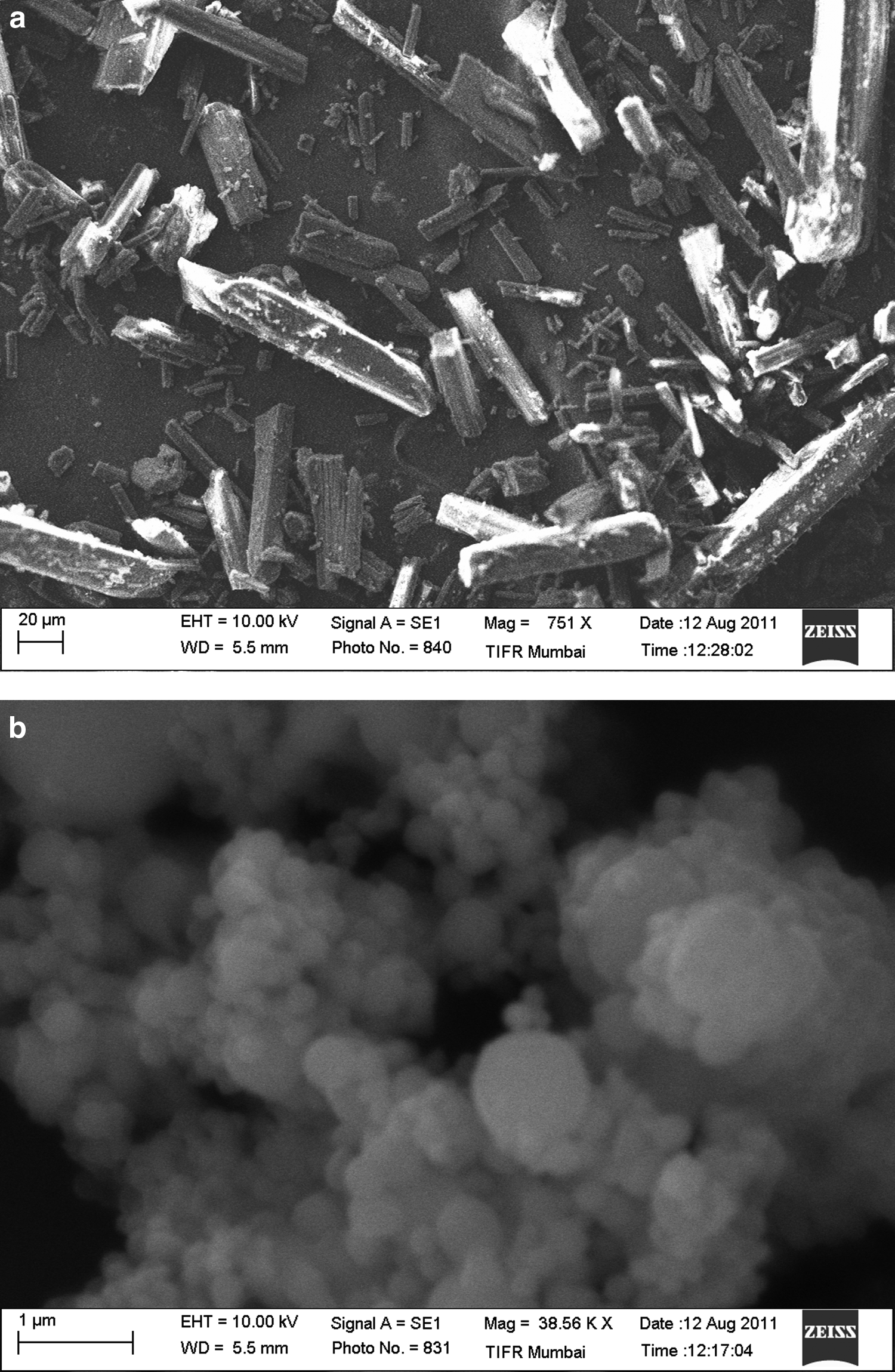
SEM images of
The SEM images for RIF and RFB loaded microparticles are shown in Figure 4. The sizes of the microparticles of both RIF and RFB were observed to be 1–2 μm, which was within the required size range of 1–5 μm, and hence suitable for lung delivery. The particle size was also in conformation with the particle size obtained from the zeta sizer.
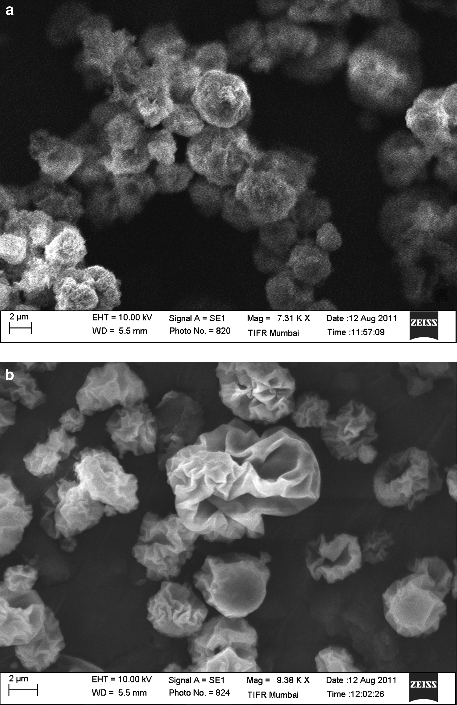
SEM images of
The concentration of TPP was seen to have an effect on the surface morphology of spray-dried chitosan microparticles. Lower concentrations of TPP resulted in formation of irregularly shaped microparticles with smooth surfaces, whereas higher concentrations of TPP resulted in formation of distinctly spherical particles having rough surfaces.(24) This can be seen from the SEM images of the drug-loaded microparticles of both the drugs. RIF-loaded microparticles have a rough surface and look more spherical than RFB-loaded microparticles, which have a wrinkled or corrugated surface. This is due to the difference in the amount of TPP used in their preparation (Table 4). The batch RIF2 of RIF loaded microparticles had more than twice the amount of TPP than batch RFB3 of RFB loaded microparticles, which may have resulted in formation of denser structure as observed from the images (Fig. 4).
The SEM images of DPI blends of drug loaded microparticles are as shown in Figure 5. The drug-loaded microparticles are seen adhering to the larger particles of the carrier system indicating formation of a homogenous blend for DPI.
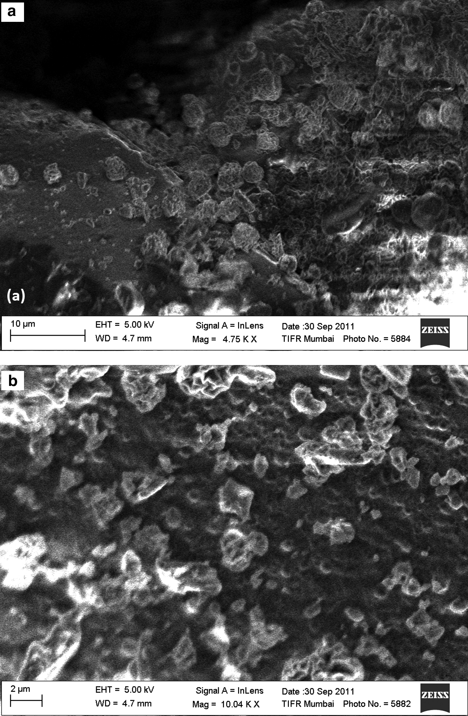
SEM images of DPI blend of
Swelling studies
Cross-linked chitosan has a matrix-like structure and thus has the ability to absorb water and swell. The extent of swelling is directly proportional to cross-linking density, which in turn depends on the amount of cross-linking agent used.(31) At higher concentrations of cross-linking agent, the polymer network is densely packed and thus can accommodate less volume of solvent molecules, whereas at lower concentrations of cross-linking agent, the polymer network is loosely held, resulting in voids and is able to accommodate higher volume of solvent molecules.(35)
All the batches of microparticles underwent swelling in aqueous medium as seen in Table 4. The swelling index decreased with an increase in concentration of TPP for both RIF as well as RFB-loaded microparticles. This difference in swelling indices between RIF and RFB-loaded microspheres is also evident from the differences in the surface morphology of the two systems, as seen in the SEM images (Fig. 4). The RIF loaded microparticles appear dense, spherical, and highly cross-linked, owing to higher concentration of TPP and thus would accommodate lesser volume of medium. RFB-loaded microparticles show a less rough surface, which is corrugated, and loosely cross-linked due to lesser amounts of TPP used and thus accommodates greater amount of the media in the study.
In vitro drug release
The release profiles of different batches of RIF-loaded microparticles are shown in Figure 6a. The RIF-loaded microparticles released 90% of the drug at the end of 12 hours. The release profile of RIF-loaded microparticles was found to be sustained as compared to the release of free RIF and no burst release of the drug was observed. The release of drug from the microparticles was found to increase with an increase in the amount of TPP.
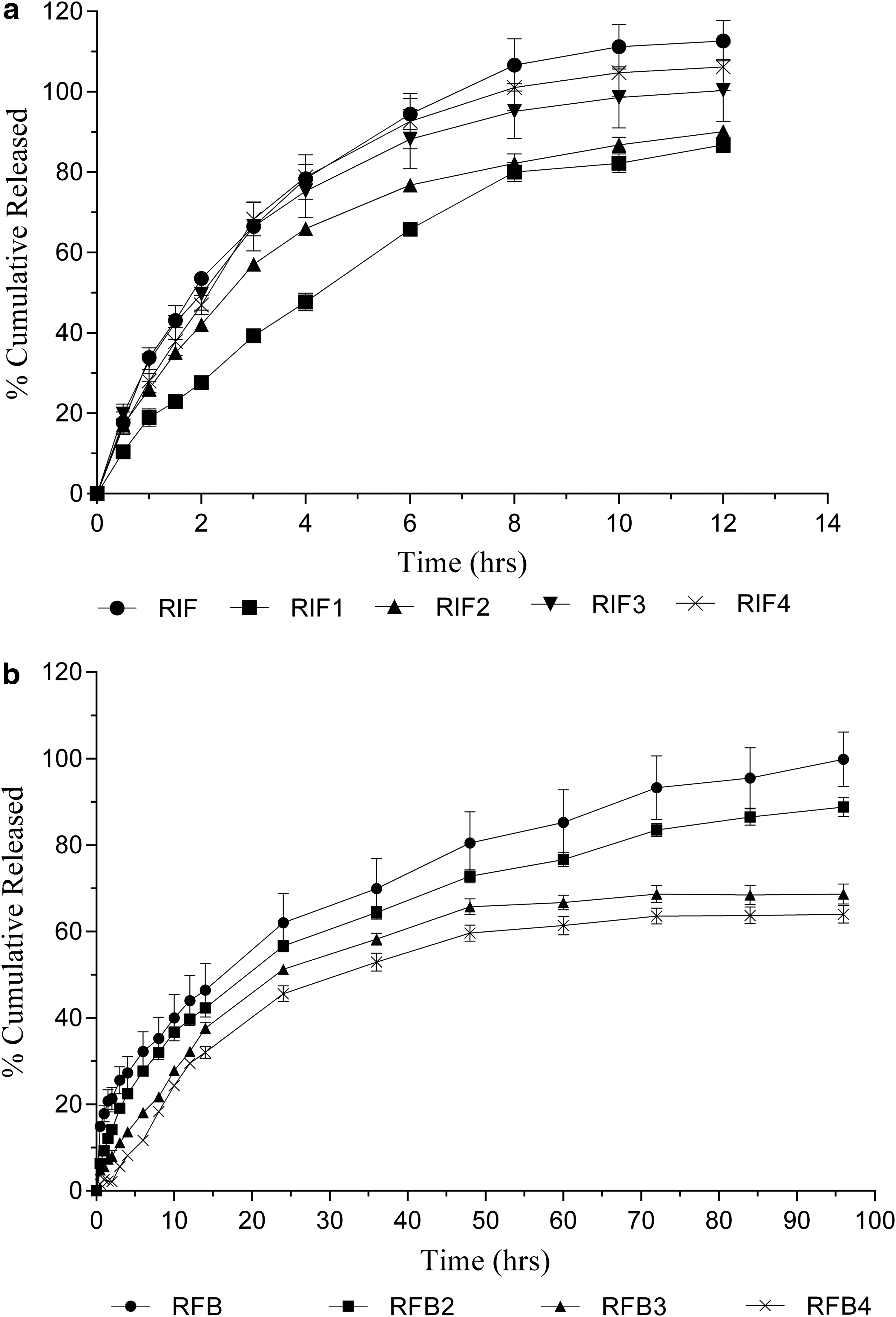
Release profile of
This was in contradiction to the findings reported by previous authors,(35) who found that release of drug from the chitosan-TPP microparticles decreased as the amount of cross-linking agent is increased as a result of formation of dense polymeric network, thus slowing drug release. However, for polymers such as chitosan that exhibit varied swelling behavior with varying amounts of cross-linking agent, the release pattern is different.
The swelling of microparticles affects the release profile by changing the path length followed by the drug, embedded in the microparticles, to diffuse into the surrounding medium. Greater extent of swelling results in longer path length, whereas lesser extent of swelling keeps the path length short. At higher concentrations of cross-linking agent, the polymer network is densely packed and hence enables swelling to a lesser extent, whereas at lower concentrations of cross-linking agent, the polymer network is loosely held resulting in swelling to a greater extent.(31,44) Hence, the RIF microparticles cross-linked with less amount of TPP (RIF1) showed slower release than those cross-linked with higher amounts of TPP (RIF4).
In order to further understand drug release mechanism, the drug release data was plotted into the Korsmeyer-Peppas equation as log cumulative percentage of drug released versus log time, and the value of diffusion exponent (n) was calculated using the slope of the straight line. If the exponent n is less than 0.45, then the drug release mechanism is by Fickian diffusion, and if n is 0.45 < n <0.89, then it is by non-Fickian or anomalous diffusion. An exponent value of 0.89 is indicative of Case-II Transport or typical zero-order release, while value higher than 0.89 is indicative of Super case-II Transport.(44,45) The values of n obtained from the slope of the equations, for different batches of Rifampicin loaded microparticles are shown in Table 5. All batches of rifampicin-loaded microparticles had n values less than 0.45 indicating Fickian diffusion.
The release profiles of different batches of RFB loaded microparticles are shown in Figure 6b. The RFB-loaded microparticles released 90% of the drug at the end of 96 hours. The release profile of RFB-loaded microparticles was more sustained as compared to the release of free RFB and no burst release of the drug was observed. The release of RFB-loaded microparticles decreased, with an increase in the amount of TPP added. This was unlike RIF-loaded microparticles, for which the drug release increased with an increase in the amount of TPP.
Although both RIF and RFB-loaded microparticles exhibited swelling in the media, the difference in the release profiles of the microparticles may be due to the differences in the drug solubility. RIF is more readily soluble in aqueous media than RFB, which is only slightly soluble. Thus the rate limiting step for release of RIF from the microparticles is diffusion through the chitosan matrix. In the case of RFB-loaded microparticles, the solubility of RFB being very low, the rate limiting step for release of drug from the microparticles is dissolution into the surrounding medium. Thus, release of RFB from the microparticles is a function of the drug present within the microparticles, hence slower release of RFB from microparticle as compared to RIF microparticles. This can also be seen also be seen from the values of diffusion exponent, obtained after plotting Korsmeyer-Peppas equation were greater than 0.45 but less than 0.89, indicating non-Fickian diffusion (Table 5).
In vitro lung deposition
Deposition on ACI
Particle size distribution is one of the important parameters for in vitro testing of DPI systems and is usually obtained with the help of cascade impactors. The ACI is comprised of different plates or stages corresponding to the different regions of the respiratory system, from the oral cavity to alveoli. The particulate matter is drawn at a pre-determined air-flow rate through the apparatus, resulting in deposition of the particles on different plates of ACI as a function of their particle size and aerodynamics. The ACI is similar to a twin stage impinger (TSI) in function. However in addition to particle size, it gives a detailed lung deposition data with additional parameters like MMAD, GSD, and FPF.
MMAD can be used as the predictor of the particle distribution in the lung. GSD is a measure of the variability of the particle diameters within the system. A DPI system with a GSD less than 1.2 is described as monodisperse. The FPF is generally regarded as the fraction of the total inhaled dose that reaches the stages corresponding to the cut-off diameter of 5 μm.
The batches of microparticles (RIF2, RIF3, RFB2, and RFB3), which showed promising results (data not shown) on TSI, were selected for a detailed study on an 8-stage Andersen cascade impactor. It was observed that free RIF and RFB, along with the carrier blend, did not show any appreciable results on the ACI as seen from the low FPF values (Fig. 7).
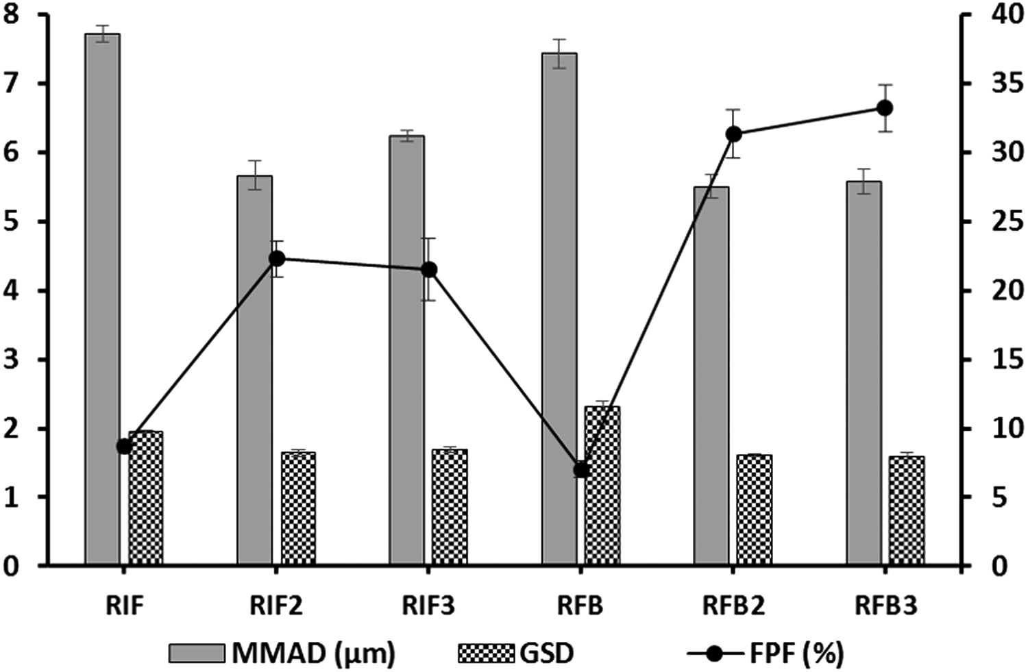
Parameters of lung deposition study of free drugs and their microparticles on ACI with respect to MMAD, GSD, and FPF. All values are expressed as mean ± SD; n = 3, FPF, fine particle fraction; GSD, geometric standard deviation; MMAD, mass median aerodynamic diameter.
A major portion of the emitted dose was found deposited on the pre-separator stage (oral cavity). This is due to the large particle size of the drug crystals, rod shaped morphology of RIF and agglomerated nature of RFB as observed in SEM images (Fig. 3). Among the two batches of RIF-loaded microparticles, no significant differences were observed with respect to MMAD, GSD, and FPF. However, the values of MMAD and GSD near to 5 μm and 1.2, respectively, are indicative of monodisperse DPI system suitable for deep lung delivery.
The results obtained for RFB-loaded microparticles were similar to those obtained for RIF-loaded microparticles (Fig. 7). However, as compared with RIF-loaded microparticles, RFB-loaded microparticles showed better deposition in the later stages of the ACI, indicating a greater FPF. This can again be attributed to the surface characteristics of the microparticles as seen in Figure 4. RFB microparticles appear smooth and have a corrugated surface as compared to RIF microparticles, which exhibited a rough surface. These differences in the surface morphology of the microparticles result in a change in their aerodynamic pattern, thus affecting in vitro deposition.(36.40)
Stability study
Spray-dried particles are prone to moisture uptake and aggregation during storage, which may further result in an increase in the particle size distribution, and thereby alter the lung deposition profile of the DPI.(41) Thus it is important to evaluate stability of spray-dried particles with moisture content and particle size and in vitro lung deposition.
The selected batch of RIF-loaded microparticles (RIF2), packed in trilaminate aluminum pouches, was subjected to storage at room temperature (ambient temperature) and at 40°C ± 2°C/75% RH ± 5% RH in order to evaluate stability. Different parameters were evaluated, such as color, appearance, particle size, moisture content, drug content, release profile, and in vitro lung deposition as shown in Table 6 and Table 7.
FPF, fine particle fraction; GSD, geometric standard deviation; M, month; MMAD, mass median aerodynamic diameter; RH, relative humidity; RT, room temperature.
All values are expressed as mean ± SD; n = 3.
FPF, fine particle fraction; GSD, geometric standard deviation; M, month; MMAD, mass median aerodynamic diameter; RT, room temperature; RH, relative humidity.
All values are expressed as mean ± SD; n = 3.
The microparticles were found to be stable at room temperature (ambient temperature), however at 40°C ± 2°C/75% RH ± 5% RH, an increase in moisture content and physical appearance was observed, indicating requirement of more robust packaging systems. The effect of moisture content on in vitro lung deposition of the microparticles is evident from the ACI deposition data. The RIF microparticles stored at 40°C ± 2°C/75% RH ± 5% RH for all time periods showed a decrease in the FPF as compared to RIF microparticles stored at room temperature. The increase in the particle size is confirmed by particle sizes and MMAD obtained on Malvern ZetaSizer and ACI, respectively (Table 7).
In vivo toxicity
Administration of free RIF and RFB to the rats by intratracheal instillation produced severe peribronchiolar infiltration of the inflammatory cells accompanied with hyperplasia of BALT (bronchus associated lymphoid tissue) and interalveolar septal thickening, evident of severe toxicity. The inflammatory cells were also present in bronchiolar and alveolar lumen (Fig. 8c and 8d).
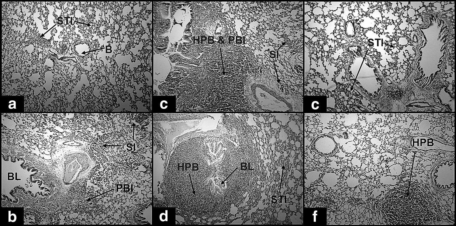
Photomicrographs of lungs of rats showing histopathological changes after administration of
In comparison to free drugs, the RIF and RFB-loaded microparticles produced only mild changes in the lung pathology, indicating no significant toxicity of the prepared microparticles of RIF and RFB to the lungs (Fig. 8e and 8f). The blank chitosan microparticles were found to be nontoxic to the lungs as previously reported.(19) The blank microparticles produced only mild septal thickening and infiltration, thus indicating no toxicity of chitosan microparticles produced by ionic interaction process (Fig. 8a and 8b).
In vitro macrophage uptake study
The causative agent for tuberculosis, Mycobacterium tuberculosis, is known to reside in the alveolar macrophages and remain dormant for years. Alveolar macrophages are reported to internalize microparticles when administered to the lungs. Particles within the size range of 1–3 μm are efficiently internalized, whereas particles less than 1 μm and larger than 10 μm escape internalization by alveolar macrophages.(46) Thus, size is an important parameter for microparticles meant for targeting Mycobacterium tuberculosis within alveolar macrophages. Many authors have reported internalization of microparticles by alveolar macrophages(47–49) and their visualization by fluorescence microscopy or staining techniques.(50)
In the present study, an indirect method was used, wherein the drug content in the supernatant was analyzed by UV spectrophotometry and the amount of drug entrapped within the macrophages was estimated (Table 8). The number of microparticles corresponding to the estimated concentration was determined based on the true density (0.453 gm/mL) and diameter of the microparticles (Table 4).
All values are expressed as mean ± SD; n = 3.
The percent internalization of the drug loaded microparticles is shown in Figure 9. It was observed that all concentrations of RIF and RFB-loaded microparticles were internalized to some extent at the end of 90 minutes. The internalization was found to increase with an increase in the number of particles for both RIF and RFB-loaded microparticles. However, RFB-loaded microparticles were internalized to a greater extent than RIF-loaded microparticles at all concentrations tried. This can be attributed to the differences in particle size between RIF and RFB-loaded microparticles. RFB microparticles being smaller in size than RFB microparticles (Table 4), would incorporate a larger number of particles at the same given concentration and volume than RIF-loaded microparticles.
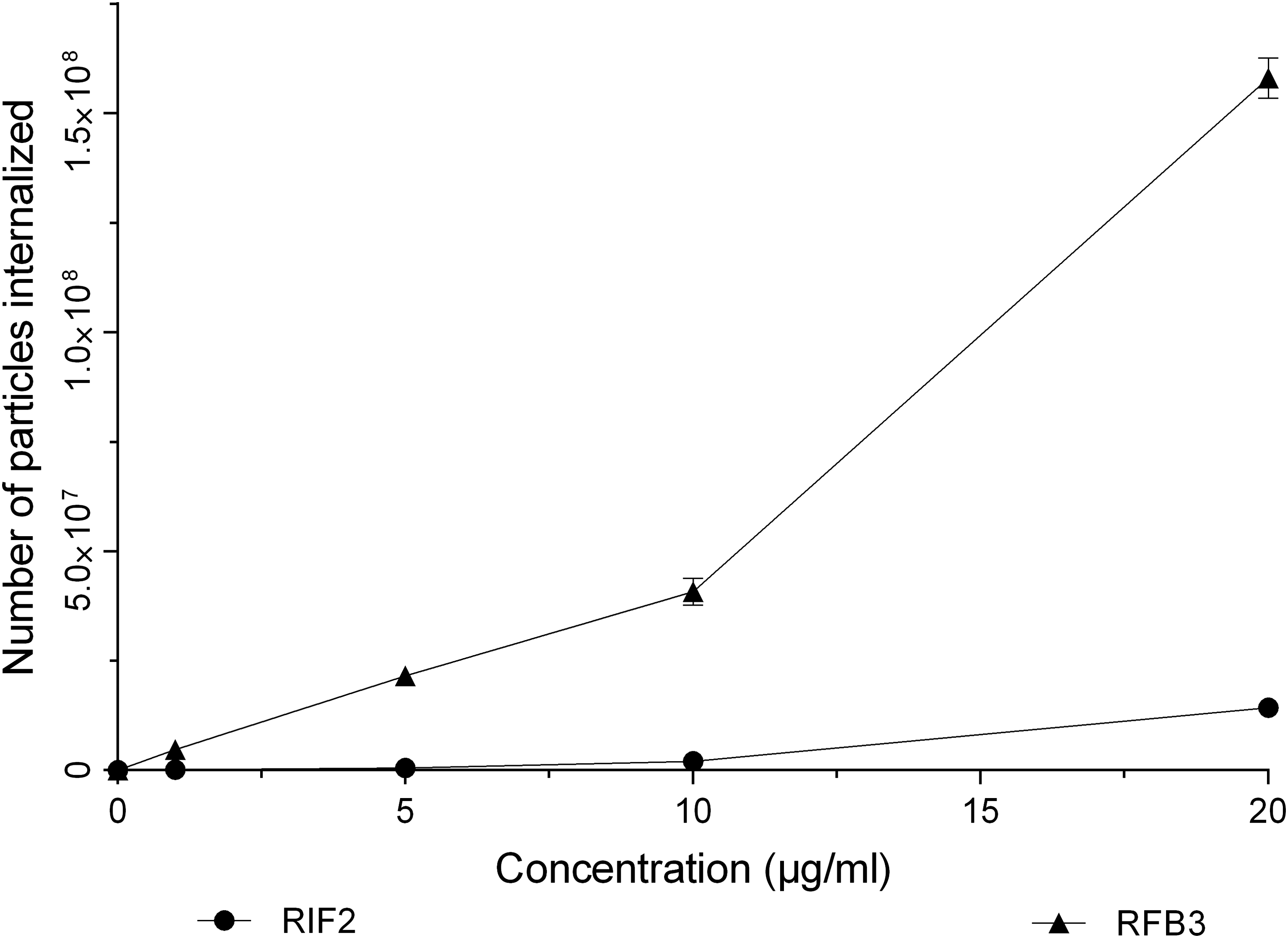
In vitro macrophage uptake of RIF and RFB microparticles. All values are expressed as mean ± SD; n = 3.
Within the tested levels, the internalization of microparticles was maximal at 20 μg/mL for both RIF and RFB-loaded microparticles, and no saturation in uptake was observed. Similar observations have been reported elsewhere in the literature for RIF.(27) The internalization of drug-loaded microparticles was found to be a volume-dependent process based on the observation that the internalization of the particles increased with the number of available particles.
Conclusion
The spray-drying method was efficient in producing microparticles suitable for lung delivery with good aerodynamic characteristics that were evident by their deposition on the later stages of the ACI. The chitosan-based microparticles containing anti-tubercular drugs are apparently nontoxic to the lung tissues within the scope of studies described; however, repeated-dose inhalation toxicology studies of these formulations will be needed to better assess their long-term safety. The microparticles are also taken up by alveolar macrophages, thus enabling targeting of the Mycobacterium tuberculosis residing within the macrophages.
Microparticles of the anti-tubercular drugs, RIF and RFB, prepared by a spray-drying technique, have a good potential for direct delivery to the lungs, when formulated as dry powder for inhalation (DPI) and can possibly provide the advantages of lower doses and shorter treatment times compared to the conventional oral delivery approach. However, further detailed studies are required to evaluate the uptake in alveolar macrophages and efficacy of these microparticles on Mycobacterium tuberculosis.
Footnotes
Acknowledgments
The authors thank Lupin Ltd, India for providing gift samples of rifampicin and rifabutin, and Mumbai University for providing financial assistance (Project No.314 & Ref. no.: APD/237/172 of 2011).
Author Disclosure Statement
The authors report no conflicts of interest. The authors alone are responsible for the content and writing of the publication.