
Abstract
Abstract
Background:
Current treatment regimens for inhalation injury are mainly supportive and rely on self-regeneration processes for recovery. Cell therapy with mesenchymal stromal cells (MSCs) is increasingly being investigated for the treatment of inhalation injury. Human amniotic MSCs (hAMSCs) were used in this study due to their potential use in inflammatory and fibrotic conditions of the lung. This study aimed at demonstrating that hAMSCs can be atomized with high viability, for the purpose of achieving a more uniform distribution of cells throughout the lung. Another aim of this study was to set ground for future application to healthy and diseased lungs by demonstrating that hAMSCs were able to survive after being sprayed onto substrates with different stiffness.
Methods:
Two methods of atomization were evaluated, and the LMA MAD780 device was selected for atomizing hAMSCs for optimized delivery. To mimic the stiffness of healthy and diseased lungs, gelatin gel (10% w/v) and tissue culture plastic were used as preliminary models. Poly-
Results:
The feasibility of atomizing hAMSCs was demonstrated with high cell viability (81 ± 3.1% and 79 ± 11.6% for cells sprayed onto plastic and gelatin, respectively, compared with 85 ± 4.8% for control/nonsprayed cells) that was unaffected by the different stiffness of substrates. The presence of the collagen I coating on which the sprayed cells were cultured yielded higher cell proliferation compared with both PLL and no coating. The morphology of sprayed cells was minimally compromised in the presence of the collagen I coating.
Conclusions:
This study demonstrated that hAMSCs are able to survive after being sprayed onto substrates with different stiffness, especially in the presence of collagen I. Further studies may advance the effectiveness of cell therapy for lung regeneration.
Introduction
C
A systematic review on smoke inhalation-associated acute lung injury reported that more than 70% of the patients with inhalation injury in a regional burn center were diagnosed with respiratory failure and 20% were diagnosed with acute respiratory distress syndrome.(8) Current treatment for inhalation injury is mainly supportive, with oxygen and fluid therapies,(1) medications to address the coexisting conditions such as bronchospasm and infections,(8,9) and heparin and N-acetylcysteine to prevent small airway obstructions.(9,10) These treatment regimens ultimately rely on self-regeneration processes for tissue recovery from the injury. However, inflammation caused by inhalation injury may become uncontrolled over time and cause ongoing lung injury,(11) leading to edema and fibrosis.(10,12,13) The endogenous MSCs, which are dispatched in the response to injury, support the repair of the damaged tissue. This highlights that cell therapy using MSCs holds promising potential for such injuries, as MSCs may reduce inflammation and facilitate the recovery process through the downregulation of pro-inflammatory factors and the anti-inflammatory cytokines (e.g., IL-4 and IL-10) released by MSCs.(14)
Furthermore, the growth factors secreted by the MSCs help maintain the functions of the endothelium and epithelium barriers after injury.(1,12,14) However, the ability of MSCs to reach all parts of the lung is limited, particularly when the extracellular matrix and vasculature are damaged, as has been observed for this type of injury. Hence, an adequate supply of MSCs at the site of injury is required. Furthermore, to exert effects at the level of the airway epithelium, where most of the immediate injury takes place in smoke inhalation, intratracheal administration of the MSCs directly delivered to the injury site may be beneficial. Therefore, there is a need to optimize the technology to deliver cells directly to airways.
Although the optimal source of MSCs for use in lung regeneration is increasingly being discussed, the most commonly investigated adult stem cells are those from bone marrow, adipose, and placental tissue.(15–17) MSCs from human amniotic membrane (hAMSCs) are progressively of interest, as they are easily accessible, available in large quantities, and have minimal ethical concerns as they can be obtained from term placentas that are usually discarded after delivery.(18–21) In a comparison study of different sources of cells utilized in a murine model of sustained lung injury induced by bleomycin, hAMSCs had demonstrated superior anti-inflammatory and immunomodulatory properties compared with bone marrow MSCs and amniotic epithelial cells.(22) The preclinical data of hAMSCs have provided a basis for the recent development of clinical investigations for the potential treatment of various lung inflammatory and fibrotic conditions, as well as for the liver dysfunction associated with cystic fibrosis.(19,22–25)
In comparison to the increasing number of studies that investigate the use of MSCs for lung injury, there are limited numbers of studies on the techniques to deliver MSCs to the lung. The intratracheal route for MSCs administration can be as effective as intravenous administration in some lung injury models,(15,26,27) and, thus, intratracheal cell administration is increasingly being studied.(28–33) These studies have demonstrated the efficiency of direct delivery of cells for potential treatment of various lung diseases. However, there is room for improvement in the intratracheal MSCs delivery technique, as most studies use a basic injection method for instillation of cells through the trachea for intratracheal administration of cells. Injection of cells through an intubation tube(32) or a catheter(33) or oropharyngeal aspiration(34,35) was used; however, no studies adopted a spraying technique to deliver cells intratracheally. Recent literature highlights the potential benefits of delivering cells directly to the lung by atomization, as it may promote and enhance the healing processes of various lung diseases.(36,37) Since the greatest challenge associated with the atomization of cells is to maintain their survival, our study focused on this aspect while evaluating for the first time other associated factors such as substrate stiffness and the use of co-factors to enhance cell proliferation.
Several spraying systems have been developed for topical application of MSCs, including the one recently patented to deliver human adipose tissue MSCs to wounds and burns.(38,39) In parallel, atomizing devices, including the PennCentury™ Microsprayer (model IA-1B), had been previously used to atomize fibroblasts for potential delivery to the lung.(36,40) The use of fibroblasts in these studies is not optimal for the treatment of lung injury and regeneration, as those cells are already differentiated and thus may not promote tissue regeneration. Some studies previously used an electrospraying device to atomize cells, including that of Braghirolli et al., which atomized MSCs from human deciduous teeth pulp.(41) However, this technique was designed for the application in repopulating scaffolds used for tissue engineering and is not applicable for delivering cells to lungs due to its complex design and the high voltage required to spray the cells.
Therefore, a new potential atomizing device, LMA® MADgic® MAD780 (LMA device), is proposed in this study and is compared with a PennCentury device. It should be highlighted that the LMA device we utilized to atomize the cells in our study is currently used as a medical device for airway intubation, and there are no previous reports that demonstrate the ability of the LMA device to atomize cells. This is a repurposing of the device that is clinically accepted for patients who require airway intubations, which highlights the translational value of this research. Furthermore, this adds significance for future application in patients with smoke inhalation injury, as airway intubations are one of the few current treatment options of inhalation injury in some patients at intensive care units.
Taken together, the use of cells that show desired bioactivity and the device that is clinically approved underpin the translational aspect of this research and highlight the high potential of this approach to effectively deliver cells to the airways. Currently, hAMSCs are utilized much less than other MSCs despite their high regenerative potential and the aforementioned benefits; however, this is the first time that the use of the LMA device is being reported for the atomization of cells. The particle size for optimal delivery of therapeutics (solid materials) to the airways is required to be between 1 and 10 μm, and less than 5 μm for the lower airways.(42) However, the fate of inhaled biological agents during the deposition in the airways is expected to be different and this requires further investigation. This study was designed to demonstrate the feasibility of atomizing hAMSCs with high viability, for the purpose of achieving a more uniform distribution of cells throughout the lung. The atomization of hAMSCs and the direct delivery to the lung is an alternative, novel way of delivering cells for the treatment of lung conditions. This method is potentially more targeted and effective than cell injections.
This study also included the evaluation of the response of the cells to the substrates that mimic different lung conditions. In this article, we aim at demonstrating that the hAMSCs are able to maintain high viability after being sprayed onto substrates with different stiffness. Furthermore, we determined the effects of additional cues for cell attachment and proliferation. This study was designed to set the ground for future development and application to healthy and diseased lungs.
Materials and Methods
Cell culture and maintenance
hAMSCs were purchased from ScienCell (San Diego, CA), and the MSCs Medium along with the supplied growth supplements and 5% fetal bovine serum (FBS) (Bovogen Biologicals, Keilor East, VIC, Australia) were used for the first two passages. Thereafter, alpha-MEM media (Sigma-Aldrich, Castle Hill, NSW, Australia) with 10% FBS was used with no compromise in cell growth. Cells were maintained at 37°C in 95% air and a 5% CO2 atmosphere and were passaged when ∼80% confluent. Cells were used at passages 3–5 for all assays.
Preparation of substrates
Gelatin gel (10% w/v) was produced by dissolving gelatin from bovine skin (Type B, Bloom 75) (Sigma-Aldrich) in heated deionized water. The gels were stored at 4°C and sterilized by exposure to UV and rinsing with 70% ethanol. Coatings using collagen I from rat tail (Invitrogen, Thermo Fisher Scientific, Scoresby, VIC, Australia) and poly-
Determination of substrate stiffness using atomic force microscopy
To probe nanomechanical properties of gelatin gel, a noncoated petri dish and collagen or PLL-coated dishes, force probe (MFP-3D-Bio, Santa Barbara, CA) were used. Experiments were conducted using silicon nitride probes (ContGB-G; Budget Sensors, Sofia, Bulgaria). Before the indentation of the substrates, sensitivity and the spring constant were measured for each probe. Inverse Optical Lever Sensitivity of the probe was measured by single indentation to freshly cleaved mica and calibration of the probe by finding the slope of Deflection versus ZSensor deflection. Spring constant was measured using the thermal method in air. Next, to calibrate the sensitivity in the cell probing experiment, the sensitivity was measured in liquid (phosphate buffered saline [PBS]) using freshly cleaved mica and the same parameters (force and approach speed) as used for the following cell probing. On average, sensitivity was 55 nm/V and the spring constant of the cantilever was ∼0.06 N/m.
The stiffness was measured in PBS by indenting the substrate with maximum force 20 nN at the rate of 0.5 Hz and recoding force-deflection (F-D) curves. For each substrate, minimum 10 points in different locations were measured.
When calculating the stiffness (Young's modulus) of coated and uncoated substrates (noncoated tissue culture plastic, collagen, and PLL), the Hertz model was used. Traditionally, Hertz's model assumes no adhesion and is developed for ideally elastic materials. The elastic properties and the lack of adhesion of the substrate to the probe tip made this the most appropriate model for this study.
Cell spraying process and measurement of particle size
PennCentury Microsprayer® Aerosolizer, model IA-1C (Penn-Century, Inc., Wyndmor, PA) was used as a handheld device and LMA MADgic Airway™ MAD780 (Teleflex Medical Australia, Mascot, NSW, Australia) was used with the syringe fitted onto a PHD 2000 syringe pump (Harvard Apparatus, Holliston, MA) to feed cell suspensions at 21.5 mL/min. For measuring the particle-size distributions, Spraytec (Malvern Instruments Ltd, Worcestershire, United Kingdom) was used with a refractive index of 1.0, measured for 4 seconds. Cells (2.5 × 10⁴ cells/mL) were sprayed onto the substrates, collected, and transferred to 96-well plates (2500 cells per well) for subsequent assays.
Cell viability and apoptosis assays
To compare the cell viability and apoptotic profiles after spraying the cells onto the substrates with different stiffness (gelatin gel and tissue culture plastic), Muse® Caspase-3/7 Assay Kit (Merck Millipore, Bayswater, VIC, Australia) and Live/Dead Cell Staining Kit II (PromoKine, Heidelberg, Germany) were used as per the manufacturers' protocols. Cells seeded onto tissue culture plastic without being sprayed were used as the control group. Muse Caspase-3/7 Assay Kit was validated using cells treated with 1 mM of hydrogen peroxide for 24 hours and ultraviolet (UVB) exposure at 275 nm for 30 minutes and a control sample (1:1 mixture of live and dead cells). Detailed methods of sample preparation for the validation of the apoptosis kit and the apoptosis profiles are included in the Supplementary Data and Figure S1 (Supplementary Data are available online at www.liebertpub.com/jamp). DNA quantification using CyQUANT® NF Cell Proliferation Assay Kit (Invitrogen, Thermo Fisher Scientific) and Cell Counting Kit-8 (CCK-8) (Dojindo Molecular Technologies, Inc., Santa Clara, CA) were used for long-term cell viability tests as per manufacturers' protocols.
Assessment of cell morphology
Live cell imaging was conducted using IncuCyte ZOOM® (Essen BioScience, Ann Arbor, MI) for phase-contrast images, which were taken every 2 hours on 10× magnification. Selected images were analyzed and exported using the IncuCyte ZOOM 2015A GUI software. For immunofluorescence, standard immunostaining procedures were used for actin and nuclei staining. Briefly, cells were fixed with paraformaldehyde 4% for 10 minutes, blocked using bovine serum albumin 1% for 60 minutes, and stained with Phalloidin CruzFluor™ 514 for 30 minutes and DAPI 300 nM for 5 minutes (all reagents from Thermo Fisher Scientific). Immunofluorescence images were taken using the Fluoview FV1000 confocal laser scanning microscope (Olympus Imaging Australia Pty Ltd, Macquarie Park, NSW, Australia).
Statistical analysis
All data were produced in triplicate (n = 3) and presented as means ± standard deviation. The differences between the conditions and coatings were analyzed using the one-way analysis of variance (ANOVA) test, with Tukey's multiple-comparisons test, to report statistical significance when the p-value was less than 0.05.
Results and Discussion
Atomization of hAMSCs was superior using the LMA device compared with a PennCentury device
This study demonstrates for the first time that an atomized formulation is possible for hAMSCs for direct delivery to the injured site of the lung using a currently used medical device for airway intubation in clinics. Although it has been identified that the ability to maintain cell viability during the spraying process is specific for each cell type, mainly depending on the size of cells in relation to the nozzle diameter of the atomizer,(36) it is significant that our research reports that hAMSCs can be atomized with high viability. For future application in patients with smoke inhalation injury, our focus was to deliver cells in an atomized form that would allow uniform distribution and migration of the cells to the critical regions in the trachea and the upper airways, where there are more occurrences of both thermal and chemical injuries in burn victims.
In preliminary studies, hAMSCs (purchased from ScienCell™) were sprayed initially using either a PennCentury Microsprayer IA-1C or an LMA MADgic Airway MAD780 (Fig. 1A) device. However, due to the smaller size of the droplets produced by the PennCentury Microsprayer, as observed in the particle-size distribution (Fig. 1B), the device had to be excluded as the droplets were too small to contain an entire cell (hAMSCs are between 17 and 28 μm in diameter [measured when trypsinized/in suspension]). Microscopic analyses of cell morphology demonstrated that the cells sprayed using the PennCentury Microsprayer were shredded and there was periodic blocking of the nozzle during the spraying process. Furthermore, the flow rate of the cell suspension from the handheld PennCentury Microsprayer device was limited to the hand strength of the individual users, which led to it being uncontrollable and inconsistent.
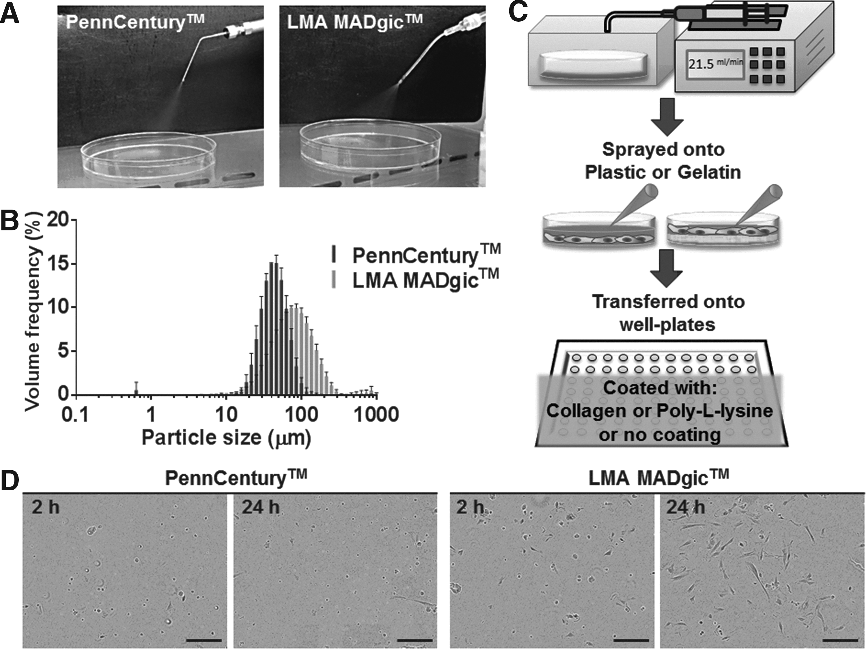
The LMA device yielded higher viability of cells after spraying, and the spraying process was better controlled by fitting the syringe of the device onto a syringe pump and using a preoptimized flow rate of 21.5 mL/min for aerosol production from the LMA device (Fig. 1C). This enabled the spraying process to be more consistent between users, as the flow rate is controlled and reproducible. Another reason for the exclusion of the PennCentury Microsprayer device was that Penn-Century, Inc., the only manufacturer of the patented device, has closed down. Although it is to be noted that the PennCentury devices are designed for animal use (and often considered the “gold standard”) and are no longer commercially available, our study aimed at demonstrating the superiority of the LMA device compared with the PennCentury IA-1C device for atomizing hAMSCs. The LMA device has other benefits for future research and clinical translation, including commercial availability and its current use in medical practice for humans. Hence, the LMA device was selected for the subsequent experiments in this study, and its new application for cell therapy was presented.
Substrates with different stiffness were designed to compare the mechanical stresses that cells are exposed to when “landing”
There has been no previous report on the analysis of mechanical stress related to the “landing” of the atomized MSCs on substrates with different stiffness. In the aforementioned studies that atomized fibroblasts,(36,40) the viability of cells measured was related to the mechanical stresses that the cells were exposed during the atomization process, without measuring the stiffness of substrates on which the cells landed. Kardia et al. atomized cells directly into a flask containing liquid medium,(36) whereas in the work of Sosnowski et al. “cell survival was analyzed solely in the liquid emitted as aerosol.”(40) These approaches do not mimic a potential environment of the lung and do not facilitate studying the effects of different levels of mechanical stress to which the cells are exposed, that is, the landing of cells on the substrate. This was considered in our study, as it may affect the viability and proliferation of the cells after being atomized. These findings may advance this field of research for future translation to in vivo studies.
The two major factors that determine the survival of cells after the spraying process were tested in this study—the stiffness of the substrate onto which the cells were sprayed and the cues available for cell attachment and proliferation. First, to examine an impact of mechanical stress when cells landed on the substrate, we considered two materials with distinctively different stiffness: “soft” that mimics healthy lungs (gelatin gel), and an extremely opposite “hard” surface that represents hard tissues (tissue culture plastic). The use of a different stiffness in this study was for the purpose of highlighting that the cells are able to survive after being sprayed onto surfaces of varying stiffness. Preliminary studies demonstrated that the distance from the nozzle of the sprayer to the recipient substrate had minimal impact; therefore, 5 cm distance was used consistently for all experiments. The gelatin gels used in this study were 10% (w/v) with ∼1 cm thickness and the stiffness of 8 kPa, which mimic the stiffness of healthy lung tissues. In contrast to the thin gelatin coatings used to help the attachment of cells on tissue culture plates (typically 0.1% [w/v]), our gelatin gel used as a model of healthy lung tissue was not designed for long-term culture of cells. The stiffness of gelatin gel and tissue culture plastic was averaged as 8 kPa and 1.2 GPa, respectively, as determined from nanoindentation using atomic force microscopy (AFM). This stiffness of the gelatin gel corresponds to the rigidity of healthy lung, 2–10 kPa,(43) whereas the stiffness of tissue culture plastic represents extreme conditions that cells may experience—“hard” substrate. The gelatin gel in this study was used solely for the purpose of mimicking the softer substrate at the time of spraying, not for enhancing cell attachment.
The atomization of the hAMSCs maintained a high viability of the cells
The initial cell viability was quantified at the time of atomization onto the substrates with different stiffness, immediately after the mechanical stresses experienced by the cells. Then subsequently, to observe whether the spraying process (mechanical stress) affected the long-term cell viability, it was necessary that the cells were collected immediately after spraying to be expanded in the same conditions on well plates for other long-term assays (Fig. 1C). This approach was required to reduce the effects of stiffness on cell growth.
When a live/dead cell staining assay was performed 4 hours after spraying onto the gelatin or plastic surfaces, the majority of the sprayed hAMSCs were found to be viable (Fig. 2A). Cells seeded directly onto the tissue culture plates without being sprayed were used as the control group. The assessment of the viability of cells sprayed onto gelatin and plastic showed that the cells did not have a compromised viability (81 ± 3.1% and 79 ± 11.6% for cells sprayed to plastic and gelatin, compared to 85 ± 4.8% for control/nonsprayed cells) confirmed with no statistically significant differences in viability compared with the control (Fig. 2B).
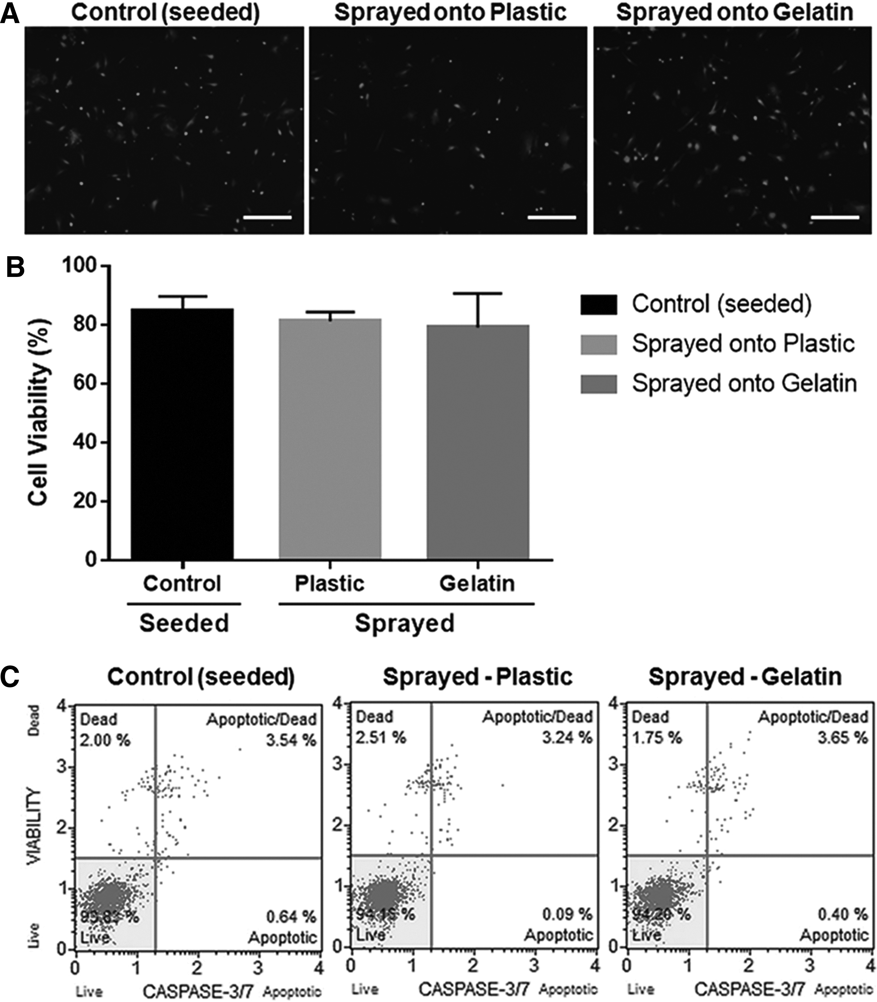
Before investigating the effects of the addition of co-factors such as collagen I or PLL, the impact of the spraying process was investigated using an apoptosis assay. The apoptotic profiles confirmed that the cells sprayed onto gelatin had a similar apoptotic profile compared with the nonsprayed control group (Fig. 2C). The cells sprayed onto plastic had a slightly higher percentage of dead cells but did not show a significantly different apoptotic profile compared with the other groups. The results confirmed that the mechanical stress caused by the atomization process, regardless of stiffness of substrate, was well tolerated by the hAMSCs. Through these analyses, the use of the LMA device, at these atomizing parameters, was validated for future application to lungs with varying stiffness, as may result from lung injuries.
In contrast to other pharmaceutical aerosol products, the conventional in vitro approaches (impactors) that are used to establish the distribution of particles cannot be used for studying cell delivery. The anticipated distribution of the hAMSCs in the lung cannot be precisely determined from the particle-size measurements, as the cells are viable and, thus, can migrate. Previous in vivo studies investigating the migration of MSCs in the lung after intratracheal instillation in various lung disease models demonstrated that MSCs have the capacity to migrate to different regions by responding to different chemoattractants and homing signals.(44–46) Therefore, to determine the actual distribution of the hAMSCs delivered via atomization, animal models are required, which was beyond the scope of this study. It was not the scope of this study to use animal models with injury since the objectives were in the fundamental science of ensuring the cell viability during the atomization process and of identifying the conditions in which the cells could survive after being atomized (using varied stiffness of substrate and functionalization).
However, we anticipate that the distribution of the cell suspension in lungs, as produced in this study, will be more uniform compared with the conventional intratracheal instillation without atomization. Similar, broader dispersion and more uniform distribution throughout the lung were previously reported for ink and insulin delivered to the lungs of rats using the PennCentury microsprayer device.(47,48) Likewise, an important benefit of using the LMA device to atomize hAMSCs is that it can improve the distribution of the cells in the airway so that the cells are more uniformly distributed and cover a larger area of the injured site. This creates ideal opportunities for cells to migrate to more distant sites of injury. These cells that have the capacity to migrate and home to sites of injury and inflammation(49) may yield significant improvements in cell-based therapies for lung diseases.
Sprayed cells cultured on collagen I-coated plate yielded higher proliferation of cells
After establishing that hAMSCs could be sprayed with high viability, the impact of the two commonly used coatings—collagen I and PLL—was investigated. This study tested whether the presence of PLL or collagen enhanced the attachment and proliferation of cells. There are no previous reports that measured proliferation of cells on substrates that were functionalized with different biomolecules after exposing the cells to the mechanical stress of atomization. Previous studies used noncoated plastic substrates that do not mimic cellular environments in any way. The purpose of functionalization with biomolecules was to assess whether there was an advantage to delivering them together with the cells, as they could overcome the mechanical stress of atomization and landing on a substrate with high stiffness. This part of the study was also necessary to determine whether the atomization of cells disrupts their ability to proliferate on both collagen I- and PLL-coated plates.
Preliminary studies comparing the growths of cells sprayed directly onto collagen I-coated plate and those sprayed onto noncoated plastic and then transferred to collagen I-coated plate found no statistical differences (Supplementary Fig. S2). The long-term cell quantification assays were utilized to investigate how the different coatings (collagen I/PLL/noncoated tissue culture plastic) affected migration and proliferation of the sprayed cells. Both coatings provide chemical cues for cells to attach and then proliferate. The disruption of the proliferation would suggest that cells were impacted by the mechanical stress during the atomization and landing on different substrates. Collagen I is an extracellular matrix protein that promotes cell adherence via interaction with integrins αvβ1 and α2β1.(50) PLL is a poly-cation that binds to DNA and enhances the electrostatic interaction with the negatively charged ions on the cell membrane.(51) Noncoated plastic was used as a control. We hypothesized that the presence of collagen I or PLL incorporated at the cell binding sites via pretreatment or co-delivery with the cells would be beneficial for cell adhesion and growth.
In addition, we have measured the stiffness of these functionalized dishes, which has not been previously demonstrated despite its substantial impact on cell growth. The average stiffness of the collagen I- and PLL-coated surfaces was 70.27 ± 23.37 and 170.36 ± 35.50 MPa, respectively, as determined by nanoindentation on the AFM. These were higher than gelatin gel and lower than noncoated tissue culture plastic (averaged 8 kPa and 1.2 GPa as mentioned earlier). It is important to highlight that both collagen and PLL coatings are thin (monolayers—a few nanometers); thus, nanoindentation results are affected by the substrate stiffness due to the indentation depth, which is greater than 10% of the coating thickness. As a rule of thumb, to reduce the influence of substrate on the measurement of the coating stiffness, the indentation depth should not exceed 10% of the coating thickness.(52) To measure the stiffness of only collagen I and PLL coatings, the depth of the indentations should be in the Å range, which coincides with the noise level of the AFM and, thus, the data would not be reliable. However, the experimental conditions used here are commonly accepted.
It was noted that when cells were sprayed, there were slightly (but not statistically significant) lower numbers of cells counted initially compared with the nonsprayed control group, possibly due to some cells being retained in the nozzle or not every cell being collected after being atomized. Nevertheless, the sprayed hAMSCs proliferated well despite the lower starting number of cells, especially when cultured on collagen I- or PLL-coated surfaces. The live/dead cell staining assay conducted shortly after the cells attached (4 hours after being sprayed/seeding) did not show that collagen I or PLL, onto which the cells were transferred and seeded after spraying, had a short-term impact on the percentage of live cells (data not shown). The effect of these coatings was more distinguished after 24 hours of culture.
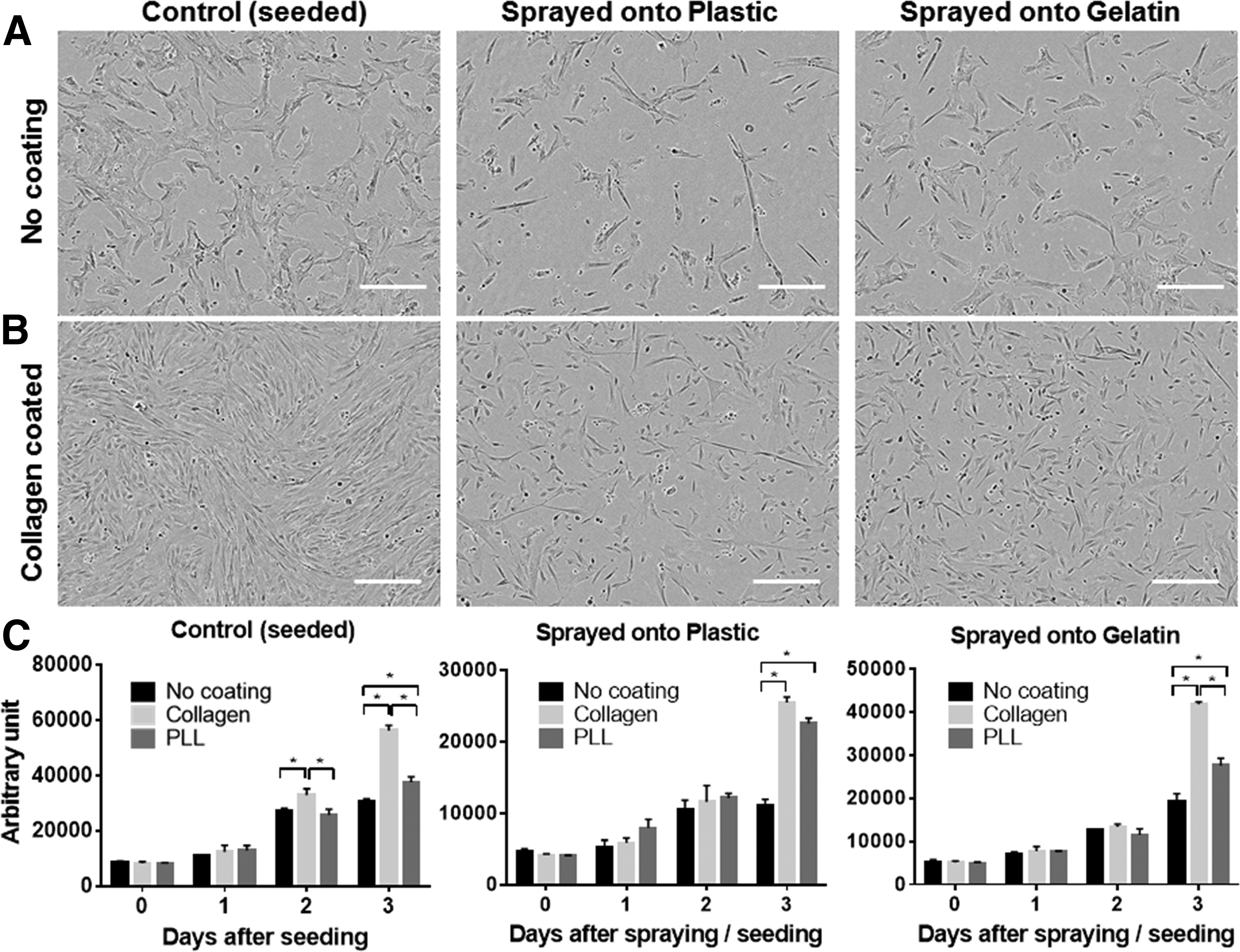
The morphology of sprayed cells seeded onto noncoated plastic was similar to that of the cells seeded without spraying (control group) (Fig. 3A). When the cells were seeded onto collagen I-coated plastic, the control group reached confluence by day 3 (Fig. 3B, left). The sprayed cells took longer to reach confluence compared with the control, as influenced by the lower initial cell number due to the spraying process. However, they proliferated and reached confluence after 4–5 days, demonstrating that the cells exposed to the mechanical stress of the spraying process were able to recover and proliferate normally. On comparing the cell numbers on day 3 (measured indirectly by DNA quantification using CyQUANT NF), the cells sprayed onto gelatin and then seeded onto collagen-coated plastic had the highest end total number of cells among the sprayed cells (Fig. 3C).
Phase images of hAMSCs 3 days after seeding onto noncoated plastic
Collagen I is known to facilitate cell attachment as well as proliferation.(50,53–55) Similarly, in our study, the effect of collagen I coating compared with noncoated plastic and PLL coating was investigated in each group (control, sprayed onto plastic, or sprayed onto gelatin). Collagen I coating yielded a higher number of cells, as evident from day 2 onward in the control group, and on day 3 for the sprayed groups (Fig. 3C). Collagen I is a fundamental component in the lung structure and function.(42) In our study, collagen I was demonstrated to enhance cell proliferation, regardless of whether the cells were sprayed or seeded without spraying (control). With the aid of the collagen I coating, the morphology of the sprayed cells (Fig. 4A, C) was similar to the morphology of the cells in the control group (Fig. 4B, D). The visualization of actin through phalloidin staining revealed that the sprayed cells had a well-developed network of actin fibers with filopodia stretched in all directions (nonpolarized).

Phase images of hAMSCs at 24 hours after being sprayed
It was evident that the cytoskeletal structure of the sprayed cells was not compromised, and it was similar to that of the control group. This confirmed that the cell viability was maintained even after spraying, and the cells were able to proliferate normally in the presence of collagen I. As determined from the CCK-8 assay conducted with optimized starting cell number for control and sprayed groups, the indirectly measured cell viability of the sprayed cells was similar to that of the control group, with no significant differences (Fig. 4E). These findings suggest that the presence of collagen I on the substrate is helpful for the sprayed cells to attach and proliferate, yet it requires further extensive investigation.
This novel and important part of the research has set the ground for future investigations, since the results suggest the need to co-deliver additional adhesive molecules in situations where the natural lung environment is compromised. After further assessment and optimization, the co-delivery of collagen I together with the hAMSCs using the LMA device is expected to promote cell localization and regeneration when delivered to the injured sites in the airways.
Conclusions
The feasibility to atomize hAMSCs was demonstrated in this study. Cell atomization was achieved using two commercial devices: PennCentury Microsprayer Aerosolizer, model IA-1C and LMA MADgic Airway MAD780. The LMA device was found to be superior in maintaining cell morphology and viability even after the mechanical stress of the spraying process and, therefore, was used for further assays. It was also demonstrated that the atomized hAMSCs maintained high viability on the substrates with varying stiffness, which mimic different lung conditions. In the light of cell therapy after lung injury, there were some important findings that required further extensive investigation, including that the sprayed cells had improved proliferation with the presence of collagen I on the substrate. The use of collagen I as an additive for the pretreatment of the substrate before cell delivery is suggested for the purpose of supporting cell adhesion and growth. Further studies of this cell atomization delivery system for the lung using animal models would show potential to enhance the effectiveness of the cell therapy for lung regeneration.
Footnotes
Acknowledgments
The authors are grateful to the Faculty of Pharmacy, the University of Sydney for the Innovation Challenge Funding and Teleflex Australia for providing LMA MADgic Airway MAD780 devices. J.K.B. was supported by an NHMRC Career Development Fellowship #1032695. The authors acknowledge the facilities and the scientific and technical assistance of the Australian Microscopy & Microanalysis Research Facility at the Australian Centre for Microscopy & Microanalysis at the University of Sydney.
Author Disclosure Statement
No competing financial interests exist.
References
Supplementary Material
Please find the following supplemental material available below.
For Open Access articles published under a Creative Commons License, all supplemental material carries the same license as the article it is associated with.
For non-Open Access articles published, all supplemental material carries a non-exclusive license, and permission requests for re-use of supplemental material or any part of supplemental material shall be sent directly to the copyright owner as specified in the copyright notice associated with the article.