
Abstract
Following a presentation with abdominal pain, a 22-year-old female was diagnosed with a massive primary liver immature teratoma with evidence of omental and pelvic metastases. Despite chemotherapy, the teratoma continued to rapidly increase in size. Significant treatment-associated myelosuppression was challenging as the patient did not want to receive any blood products (religious objections). The only feasible approach was surgical resection. Surgical resection of the primary tumor and abdominal metastases was undertaken despite unappealing perioperative risk with histological specimens demonstrating only mature teratoma. We report the first case of a liver teratoma suggestive of growing teratoma syndrome treated with myelosuppressive chemotherapy and major hepatectomy without the use of any blood products.
Case Presentation
A
Histopathology surprisingly demonstrated an immature teratoma comprising of cartilage, muscle, squamous, respiratory, and retina epithelium. Dedicated ovarian imaging showed no suspicious masses suggesting a diagnosis of a primary liver teratoma.
Chemotherapy was commenced in the form of Cisplatin/Etoposide (Cis/Etop). The complexity of the case was increased by the patient's religious beliefs, as a Jehovah's witness. Blood transfusion was not an option, even in the case of a life-threatening scenario. Myelosuppressive chemotherapy rendered the patient pancytopenic with significant anemia as low as 5.6 Hb/dL. In addition to this, the clinical scenario deteriorated with recurrent respiratory sepsis, pulmonary emboli, and pleural effusions requiring chest drain insertions. Three cycles of Cis/Etop were completed but further chemotherapy was held due to pancytopenia. The patient's tumor markers responded rapidly to the chemotherapy, however, the tumor continued to increase in size on repeat staging scans. This was most consistent with a diagnosis of “Growing Teratoma Syndrome.” This involves the “retroconversion” of an immature to a mature teratoma following the administration of effective chemotherapy. A surgical approach was deemed the most appropriate for the management of what was most likely the remaining benign tumor.
At the same time the patient was regularly seen by the Hematology team to optimize her hemoglobin without blood transfusions. Erythropoietin supplementation improved the situation but the patient remained anemic with a Hb around 7–8. In this scenario both the surgeons and anesthetists were hesitant to pursue surgical approaches in the absence of blood transfusion.
Finally, following many multidisciplinary meetings a decision was made to proceed with partial right hepatectomy with cava resection using a venovenous bypass approach with bilateral subphrenic, left paracolic gutter, and pelvic metastectomy. At the time of surgery, the primary tumor had increased in size to 31.4 × 25.4 × 42.1 cm (Figs. 1 and 2) and the metastatic deposits had increased proportionally. The left lobe of the liver was remaining (about 40% of the liver volume). Due to the high postoperative bleeding risk, the surgical plan included only partial abdominal closure and packing of the abdomen. This allowed adequate hemostasis to minimize blood loss. Additional measures included the use of cell-saver technology. Surgery was associated with a prolonged ICU admission and a complicated postoperative course, including anemia as low as 4 Hb/dL, [cytomegalovirus infection], and tracheostomy insertion. All surgical specimens (including the metastases) demonstrated mature teratoma supporting the hypothesis of retroconversion postchemotherapy. Further surgery was needed on a right mediastinal and diaphragmatic deposit 6 months later (1 year following initial presentation). Once again, these showed only mature teratoma and the patient has been well for the past 18 months (since her most recent surgery) without any disease recurrence on surveillance scans.

Primary liver tumor, weight 16 kg.
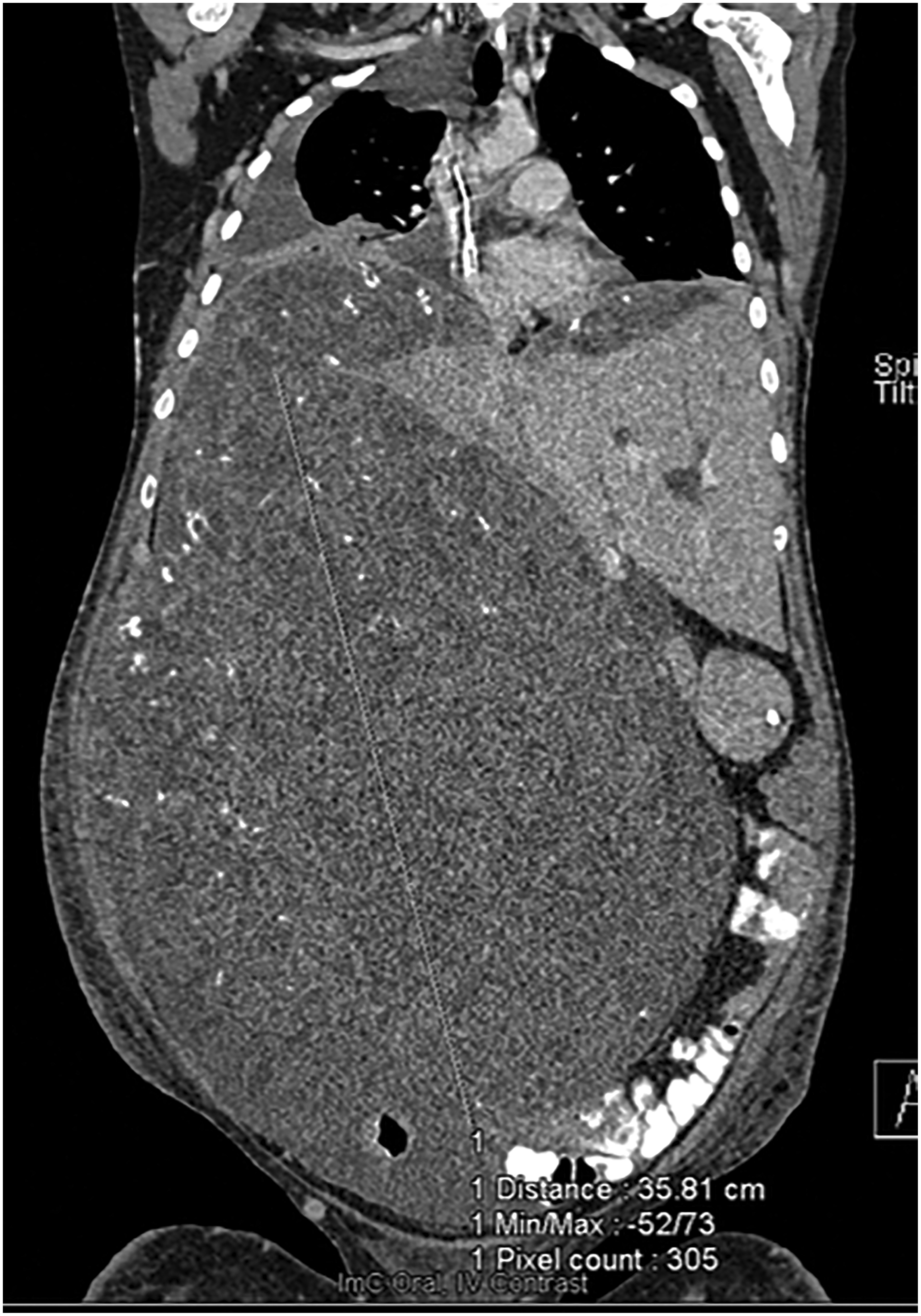
Preoperative CT scan with tumor involving most of abdomen. CT, computed tomography.
Case Discussion
The case reported has multiple remarkable clinical points, including (1) rare case of a liver teratoma (only nine previous reported cases in adults to the best of our knowledge, most of which were in the adolescent and young adult (AYA) population (2) first reported case of “Growing Teratoma Syndrome” of a primary liver tumor and finally (3) partial hepatectomy and abdominal metastectomy in a severely anemic patient without the use of blood products.
Liver Teratoma in the AYA Population
A search of PubMed using the keywords “Liver” AND “Teratoma” AND “Adult” yielded 332 results, of which 9 were identified as being case reports of liver teratomas in adults. Liver teratomas are much more common in the pediatric population, however, most of the case reports identified (8/9) include patients from the AYA population.1–9 Patients most commonly present with abdominal pain, but have been known to present more acutely with bleeding due to tumor rupture and cholangitis. In this cohort, teratomas have been reported as benign, malignant, both, or mixed with other types of germ cell tumors. Generally, management for immature teratomas involves chemotherapy to which they are generally extremely sensitive. Most previously reported cases were treated with surgery; however, Xu et al. did report a previous case treated with chemotherapy, although sadly the disease recurred and the patient died. 3
Growing Teratoma Syndrome
The apparent retroconversion of the immature teratoma to a tumor composed of only mature components is consistent with Growing Teratoma Syndrome. Although this is a relatively rare phenomenon, it is well described in the literature with regard to ovarian teratomas.10–16 It is sometimes described as “Chemotherapeutic Retroconversion.” This is highly relevant to the AYA population because although these cases are relatively rare, the median age found in a literature review of 101 cases, was 22 years. 14 However, our case represents the first report of this phenomenon in a primary liver teratoma. Logothesis first described the phenomenon in 1982 and he defined three criteria, which our case is consistent with: (1) normalization of serum tumor markers postchemotherapy (AFP in our case); (2) enlarging or new masses despite appropriate chemotherapy; and (3) exclusive presence of mature teratoma in the resected specimen. 17 Regarding the pathogenesis of this phenomenon, there are two predominant schools of thought: (1) chemotherapy treats the immature component effectively but the mature component is left untreated and thus thrives; (2) DNA-damaging chemotherapeutics result in altered cell kinetics resulting in transformation from a malignant immature teratoma to a benign mature teratoma. 7 The latter hypothesis is somewhat favored, however the debate is ongoing. This is supported by convincing changes in radiological features postchemotherapy more suggestive of a benign tumor. These radiological changes could include “increased density of mass lesions, whose margins became better circumscribed in relation to adjacent tissues, and the onset of internal calcification, with fatty areas and cystic change.” 17 The fact that all metastatic deposits in our case were identified as mature teratoma is highly convincing for the “Retroconversion” phenomenon.
Surgical Challenges
The surgical and anesthetic challenges posed by this case were remarkable. Careful preoperative planning included restoration of mineral reserves, and the use of erythropoietin. Intraoperatively, techniques included cell-saver, and venovenous liver bypass in conjunction with local perfusionists. The perioperative risk would have been extremely concerning if it was not for the age of the patient, and the limited alternative options available. There is, however, some small-scale evidence that major hepatectomy and liver transplantation can be completed without the need for blood transfusion if that blood transfusion is absolutely unacceptable to the patients. 18 Imperative is preoperative optimization and counseling in consultation with hematology. An important learning point for this case is the use of partial abdominal closure and packing to optimize hemostasis. This technique was shown to be technically feasible and successful in this case.
The prognosis for this condition is generally excellent with most cases resulting in cure through surgical resection. It has been estimated that the cure rate for those undergoing surgical resection is in the region of 90%. 17 For the unfortunate few with recurrence, or initially unresectable disease, there are very few alternatives with regard to treatment options with conventional chemotherapy and radiotherapy generally being seen as ineffective. However, there is some anecdotal evidence and Phase 2 data that cyclin-dependent kinase inhibitors (Palbociclib) may have some efficacy in this regard. 19
Conclusion
This case is an excellent example of the need for AYA medical oncologists. Liver teratomas are typically found in pediatrics and young adults. In this way, an AYA oncologist would be perfectly placed to coordinate care.
We present the world's first reported case, to the best of the author's knowledge, of growing teratoma syndrome in a liver primary in an AYA. In addition to that, we provide important evidence that major hepatectomy with abdominal metastectomy is possible without the use of blood transfusion, however, perioperative risk remains a grave concern.
Footnotes
Acknowledgments
The authors would like to thank all the other nursing and medical staff who were involved in this remarkable patient's care. Most importantly, they would like to thank the patient for allowing them to share her story to educate others.
Author Disclosure Statement
No competing financial interests exist.
Funding Information
No funding was received for this article.