
Abstract
Tumor suppressor melanoma differentiation-associated gene-7/interleukin-24 (mda-7/IL-24) has been extensively regarded as an anti-oncogene; however, that whether IL-24, as a member of IL-10 family, is involved in cancer pain was seldom reported before. In this study, we found that IL-24 mediated by adenovirus could significantly increase the plantar mechanical pain threshold in both operation side and contralateral side of the animal models, which were established by injecting 5×10³ Walker 256 rat breast cancer ascitic tumor cells into rats' tibia bone medullary canals; IL-24 could also suppress in vitro Walker 256 cells growth by inducing cell apoptosis. Pathologically, IL-24 could protect bone trabecula and substantia corticalis ossium from being completely destructed. Enzyme-linked immunosorbent assay (ELISA) showed that IL-24 treatment could increase the β-endorphin levels and decrease the IL-6 concentration in plasma of animals. Our study indicated that IL-24 has a potential treatment effect on cancers not only by inhibiting tumor proliferation, but also by the promotion of β-endorphin synthesis, inhibition of IL-6 secretion to relieve cancer pain.
Introduction
T
Our previous studies have shown the tumor inhibition property of IL-24 on lung carcinoma cell (Xie and others 2008; Zhu and others 2011), hepatocellular carcinoma (Wang and others 2007; Yu and others 2010), colon cancer (Chang and others 2011) by inhibiting tumor angiogenesis or suppressing tumor cell invasion and migration, or directly inducing apoptosis. Moreover, IL-24 can exert a potent immunostimulatory activity and enhance antitumor immunity (Caudell and others 2002), and exhibit a profound bystander antitumor activity and augment its antitumor therapeutic potential (Fisher 2005). However, whether there are some others mechanism for the application of IL-24 in cancer treatment remains largely unknown.
Pain is a common and devastating symptom of cancer affecting patients' lives sometimes more than the cancer with which they are diagnosed (Breivik and others 2009). Pain is the first symptom of cancer in 20%–50% of all cancer patients, and 75%–90% of advanced and terminal cancer patients must cope with chronic severe pain (Portenoy and Lesage 1999). Cancer pain can be managed with the various pharmacological and nonpharmacological methods currently available (Bruera and Kim 2003), but this is not always effective and many patients continue to suffer from pain. Fortunately, gene therapy for cancer pain is now being increasingly seriously considered (Lundstrom 2003). Therefore, based on our previous application of IL-24 on tumors gene therapy, we also try to explore some other mechanism for the treatment of cancer, for example, the probable inhibition of cancer pain.
In this study, we constructed tibia cancer models with Walker 256 rat ascitic tumor cells, and explored the possibility of pain inhibition by IL-24. We demonstrated that IL-24 had a role in inhibiting cancer pain, which was correlated with the up-regulation of β-endorphine (β-EP), as well as the down-regulation of IL-6. This would be a quite new mechanism for the application of IL-24 for cancer therapy.
Materials and Methods
Vectors, cell lines, reagents, apparatus and rats
The adenovirus-mediated IL-24 (Ad-IL-24) gene transfer vectors were constructed in the laboratory (Wang and others 2007). The Walker 256 rat breast cancer ascitic tumor cells was purchased from the American Type Culture Collection (Shanghai, China) and cultured in RPMI-1640 (Gibco, Shanghai, China) supplemented with 10% fetal bovine serum (Hyclone, Shanghai, China). The enzyme-linked immunosorbent assay (ELISA) kit for β-EP ELISA kit was purchased from BlueGene Company (Shanghai, China) and IL-6 was purchased from the Jingmei Company (Shanghai, China). The 3-(4, 5-dimethylthiazol-2-yl)-2, 5-diphenyltetrazolium bromide (MTT) kit was purchased from Sigma (Shanghai, China). Zoledronic acid was purchased from Chia Tai Tianqing Pharmaceutical CO., Ltd. (Jiangsu, China). The automated Dynamic Plantar Aesthesiometer was purchased from Ugo Basile (Milan, Italy). Female Sprague-Dawley rats were purchased from Shanghai Experimental Animal Center (Shanghai, China) and maintained in the animal facility at Soochow University with the approval from animal research ethics committee's of Soochow University.
MTT assay
The in vitro cytotoxic effect of Ad-IL-24 on Walker 256 cells was evaluated by MTT assay. Briefly, the walker 256 cells were dispensed into 96-well culture plates at 1×10⁴ cells/well. And after 24 h’ incubation at 37°C, the Walker 256 cells were infected with Ad-IL-24 at 100 MOI, Ad-GFP (used as a negtive adenovirus control at 100 MOI), zoledronic acid (25 pg/μL), or phosphate buffered solution (PBS), respectively, and cultured for the indicated time periods (0–5 days). Before treatment and at different time points after treatment, the viability of Walker 256 cells was analyzed using MTT kit according to company's protocol. The optical density of the plates was measured using the spectrophotometrical absorbance at 570 nm in Microplate Reader Model 550 (BIO-RAD, Shanghai, China).
Flow cytometric analysis
Walker 256 cells, after being treated with Ad-GFP, Ad-IL-24 at MOI of 100, zoledronic acid (25 pg/μL), and PBS, respectively for 72 h, were harvested and washed with cold PBS twice, and apoptosis was assessed by flow cytometric analysis using Annexin V-PE/7-AAD apoptosis detection kit following manufacturer's instruction. Briefly, the treated and untreated Walker 256 cells (5×10⁵) were incubated with 5 μL Annexin V-PE (early apoptotic marker) and 5 μL 7-AAD (late apoptotic marker) in 100 μL of 1× Annexin V binding buffer at room temperature. After 15 min's incubation, 400 μL of 1× binding buffer was added and the apoptotic cells were then analyzed by a flow cytometry.
Animal studies
Establishment of Tibia bone cancer pain model Female Sprague Dawley rats (except the normal control group), weighing 180–200 g, were anesthetized with 4% chloral hydrate (i.p. 10 mL/kg), the left leg was shaved and the skin was disinfected with 70% (V/V) ethanol. A 1 cm rostro-caudal incision was made in the shin over the proximal half of the tibia to expose the bone with minimal damage to the surrounding muscle or blood vessels. The tibia was pierced 5 mm below the knee joint distal to the epiphysial growth plate with a needle; after a breakthrough, the needle was pull out, and 5 μL of culture medium containing ∼5×10³ Walker 256 cells were injected into medullary canal with a 10 μL Hamilton syringe; the injection site was closed using surgical gumwater. Animals were placed in thermoregulated cages until they have regained consciousness and then returned to the home cage.
Grouping
Model animals were randomly divided into 5 groups with 8 rats per group, that is, the normal group, PBS group, Ad-GFP group, Ad-IL-24 group, and the zoledronic acid group. The normal group received no Walker 256 tumor cells inoculation; the remaining 4 groups received treatment of PBS, Ad-GFP, Ad-IL-24, or zoledronic acid, respectively.
Treatments upon animals
Eight days after the establishment of animal models, the hind limbs of the operation side were injected subcutaneously with 70 μL PBS, 70 μL Ad-GFP (1×10⁸ gene transfer unit, GTU), 70 μL Ad-IL-24 (1×10⁸ GTU) and zoledronic acid (30 μg/Kg weight) for each rat, respectively in PBS group, Ad-GFP group, Ad-IL-24 group, and zoledronic acid group. The injection was administered every other day for totally 3 times.
Mechanical allodynia measurement
Mechanical allodynia was assessed before injection of Walker 256 cells, and, respectively 3, 6, 8, 10, 12, and 14 days after injection of these cells. It was determined by measuring rat hind-paw withdrawal threshold (HWT) using an automated Dynamic Plantar Aesthesiometer at a daily fixed time, between 7:00 and 11:30 AM. Rats were put in a silent testing room for at least an acclimation period of 10 min. To prevent the rats from mechanical injury, the mechanical force, which was exerted on the middle of the hind paw, were kept less than 15 g. Force was recorded when the withdrawal reflex could be observed. Left and right hind paws were measured 3 times with a 2-min interval between stimuli, and the mean value was of HWT calculated. The baselines of HWT were obtained 1 day before the injection of Walker 256 cells.
Bone histology and detections of cytokine
The rats were anesthetized by 4% chloral hydrate (i.p. 10 mL/kg) and sacrificed 14 days after the injection of Walker 256 cells; their tibia bones were dipped in solution containing 4% neutral formalin for 7 days, followed by fixed in paraformaldehyde containing 10% methanoic acid for another 7 days, and then were decalcified for 24 h. The bones were rinsed, dehydrated, and then embedded in paraffin for haematoxylin and eosin staining to visualise the structure of the tumor infiltration and the bone destruction. Meanwhile, the rats' plasma were collected with EDTA and centrifuged for 15 min at 1,000 g within 30 min of plasma collection. These samples were stored at −20°C for the measurement of the concentrations of β-EP and IL-6 by ELISA using ELISA kits according to manufacturer's instruction.
Western blot
The tibia bones of these model rats in each group were collected, washed with cold PBS, grinded and lysed for western blot detection. Briefly, the total tissues lysates (100 mg per lane) were then resolved by 12% sulfate-polyacrylamide gel electrophoresis and subsequently transferred onto polyvinylidene difluoride membranes. The membranes were blocked with 5% (w/v) nonfat dry milk in Tris-buffered saline containing 0.05% Tween-20, incubated with primary antibodies specific for IL-24 and β-actin (an internal control) in the blocking solution for 1 h and incubated with peroxidase (horseradish peroxidase)-conjugated secondary antibody for 1 h. Subsequently, the membranes were washed and developed using the super-enhanced chemiluminescence detection kit (Jianglaibio, Shanghai, China), according to the manufacturer's protocol. The protein bands were visualized after exposing the membranes to X-ray film.
Statistical analysis
Results are represented by a mean±SD. Data were analyzed for statistical significance by 1-way analysis of variance followed by Newman-Keuls test or χ 2 test if necessary, using SPSS 10.0 statistical software. Values of P<0.05 were considered as indicative of significance.
Results
Time course of mechanical allodynia
During the first 3–8 days after the injection of Walker 256 tumor cells, plantar mechanical pain threshold of the operative side in each group at each time point decreased significantly (P<0.05). However, 2–6 days later, the mechanical pain thresholds in Ad-IL-24 group and the zoledronic acid group were significantly higher compared with PBS group and Ad-GFP group at each time point was (P<0.05). And there were no significant difference between PBS group and Ad-GFP group (P>0.05) (Fig. 1a); for the contralateral side, it was 12 and 14 days after the injection of tumor cells that there was a significant difference of the plantar mechanical pain threshold between intervention treatment groups and control groups (Fig. 1b).
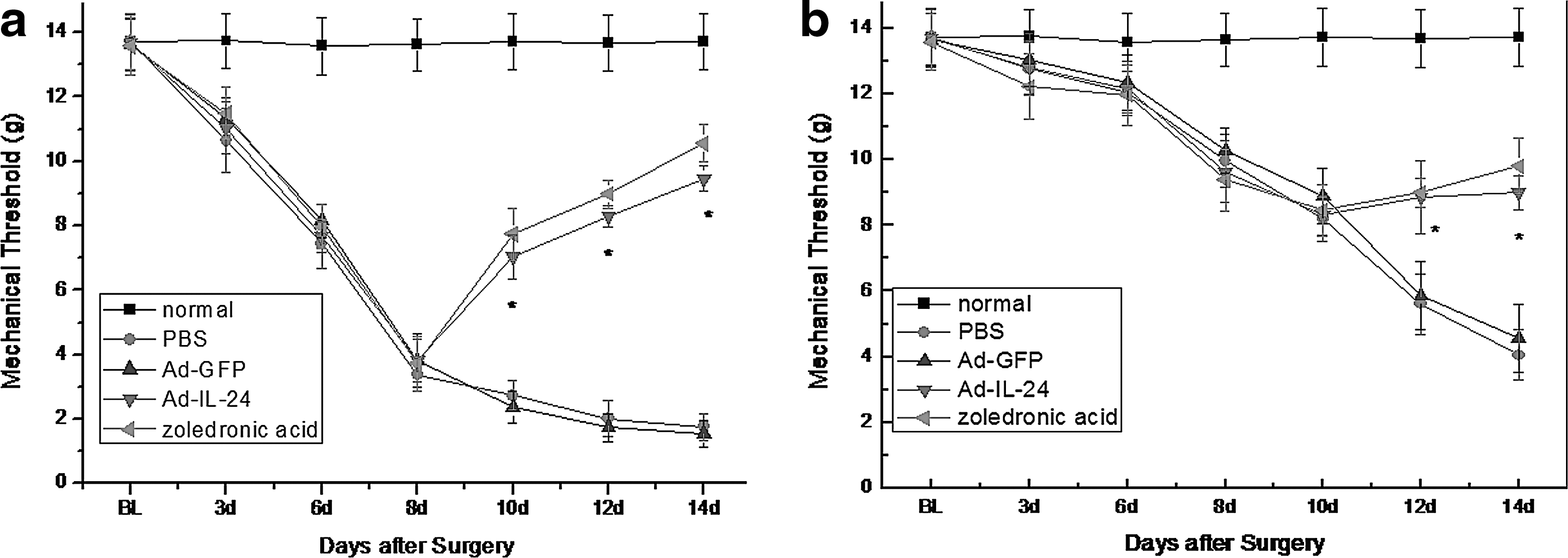
Effects of repeated treatment with Ad-IL-24 on the nociceptive pain behavior in rats of bone cancer model. We use zoledronic acid as the positive group and measure the mechanical thresholds of the ipsilateral
Ad-IL-24 induces in vitro tumor cell cytotoxicity
To explore the probable mechanisms for the cancer pain inhibition of IL-24, we first studied the cytotoxicity effect of IL-24 on Walker 256 cells in vitro. As shown in Figure 2a by MTT, compared with PBS and Ad-GFP control group, Ad-IL-24 expression significantly suppressed in vitro Walker 256 cells growth in a time-dependent manner (P<0.05). We also detected the apoptosis ratio in each group by a flow cytometry using Annexin–V-R-PE/7-AAD double staining. The results showed that Ad-IL-24 treatment induced 35.99% of Walker 256 cell apoptosis (P<0.001), whereas the apoptosis ratio of PBS or Ad-GFP group even zoledronic acid group were significantly lower (Fig. 2b, c), indicating that Ad-IL-24 induces Walker 256 carcinoma cell apoptosis.
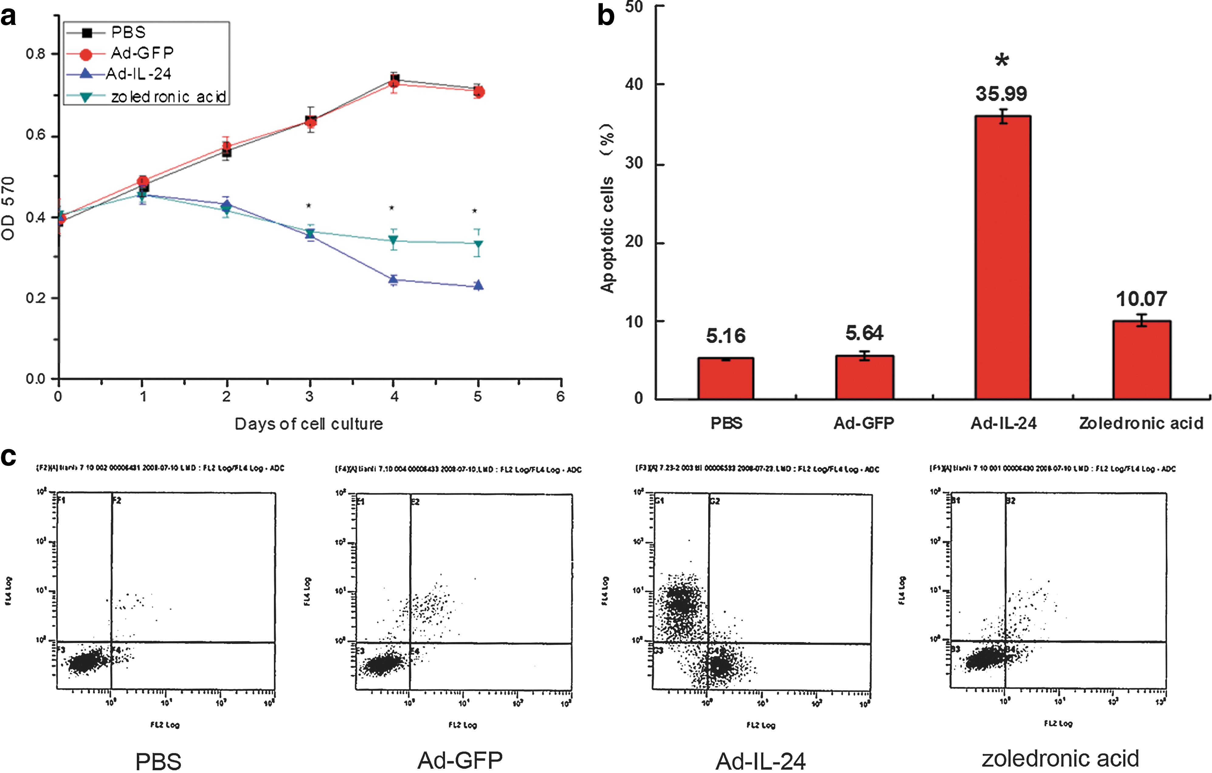
Ad-IL-24 induced tumor suppression on Walker 256 cells.
Pathology examination
Bones inoculated with Walker 256 cells showed infiltration of bone marrow cavity by malignant tumor 16 days after tumor cell injection. Western blot showed that IL-24 expression could only be detected in the Ad-IL-24 group (Fig. 3). Tumor growth and various degrees of bone destruction were observed in the animals received Walker 256 carcinoma cells (Fig. 4). Pathological sections with Hematoxylin-eosin staining showed that there were no abnormal changes in the left tibia bone marrow cavity of normal rats (Fig. 4a); in PBS group and Ad-GFP group, the tibia bone marrow cavities of the operation sides were completely filled with tumor cells, and even some tumor cells grew outside of marrow cavities, with bone trabecula and substantia corticalis ossium being completely destroyed (Fig. 4b, c); however, in Ad-IL-24 group and zoledronic acid group, less bone trabecula were destructed and substantia corticalis ossium also had a better retention (Fig. 4d, e). To quantify the extent of bone destruction by the tumor growth, we calculated the areal proportion of tumor tissues on the total area of the pathological section, and these results further confirmed that Ad-IL-24 could inhibit the growth of tumor in vivo (Fig. 4f).
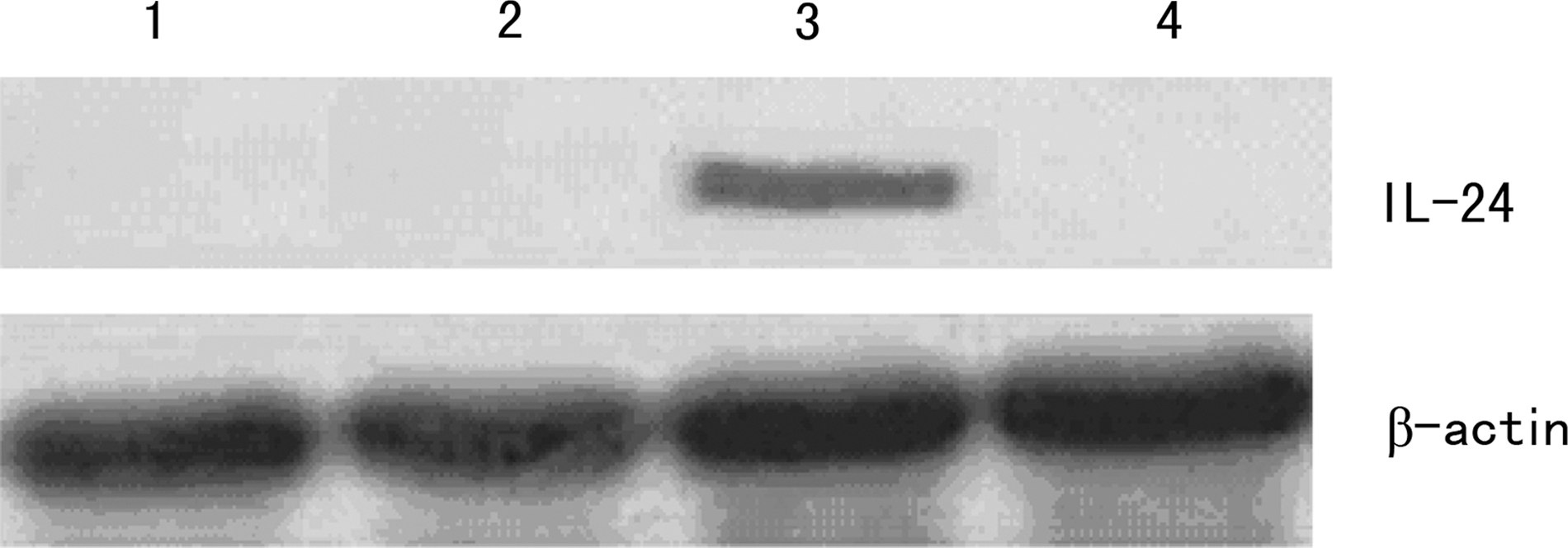
Western blot detection showed the expression of IL-24. 1, 2, 3, 4, respectively refer to PBS, Ad-GFP, Ad-IL-24, and zoledronic acid groups. IL-24 expression could be observed only in tumor tissues from the Ad-IL-24 groups.

Histology of tibial bone destruction (Hematoxylin-eosin staining). Longitudinal sections (7 μm) of the proximal tibia obtained from a control animal
Cytokine changes in vivo
The plasma β-EP concentration of rats treated with Ad-IL-24 was also higher compared with the PBS or Ad-GFP groups (P<0.05) (Fig. 5a); however, the plasma IL-6 concentration was lower compared with the PBS or Ad-GFP groups (P<0.05) (Fig. 5b).

Effects of Ad-IL-24 on the secretion of β-endorphine (β-EP) and IL-6.
Discussion
Interleukins are a group of cytokines that are expressed by a wide variety of body cells. Interleukins are extensively involved in immune function and differentiation and maturation of hematopoietic cells (Ymer and others 1985; Dorssers and others 1987) as well as inflammation process (Gabay 2006). However, a number of cytokines (IL-1b, IL-6, IL-8) released from a variety of immune cells can induce powerful hyperalgesia (Dray 1995). And some other kind of interleukin, for example, IL-10, could decrease algesthesia (Plunkett and others 2001). IL-24 is a cytokine belonging to the IL-10 family of cytokines. However, that whether IL-24 might also play a role in pains seemly has not been reported previously. In this study, we showed that IL-24 can alleviate cancer pain. Although the mechanism of bone cancer pain is not clear, at present, it is believed that the periosteum stimulation of tumor cell, reactive muscle spasm and inflammatory mediators and enzymes secreted by osteoclasts can lead to cancer pain of bone (Thürlimann and de Stoutz 1996; Mercadante 1997). Bone destruction caused by excessive activation of osteoclasts and inflammatory cytokines and prostaglandins secreted by tumor cells are also regarded as some other mechanism leading to bone cancer pain (Fulfaro and others 1998).
In our study, we used Zoledronic acid as a positive control, for Zoledronic acid is a potent inhibitor of osteoclastic activity (Fleisch 1991), cancer cell proliferation, and production of cytokines, such as IL-6 (Walker and others 2002). They have been shown to be efficacious in clinical conditions, such as tumor induced hypercalcaemia, metastatic bone disease (Body 2000), and they are even effective against pain associated with bone cancer (Fulfaro and others 1998; Bonabello and others 2001). According to our results, it could be concluded that the cancer pain inhibition of IL-24 was related to the following mechanisms: (1) IL-24 can palliate cancer pain by inhibiting tumor growth and reducing tumor volume. Our studies in vitro have shown that IL-24 can inhibit Waker 256 cell proliferation and promote apoptosis (Fig. 2); animal experiment manifested that IL-24 could also inhibit tumor growth in vivo (Fig. 4). (2) Tumor mass was generally limited to bone medullary substance due to inhibition of tumor proliferation by IL-24, and bone cortex can be better retained (Fig. 4), which leads to the reduction of periosteum stimulation and palliated pain. (3) IL-24 can alleviate the cancer pain by adjusting synthesis or releasing of inflammatory mediators and neurotransmitters. A lot of evidence suggested that an increase in plasma IL-6 had a hyperalgesic effect (Cunha and others 1992; De Jongh and others 2003); meanwhile, the inhibition of IL-6 production might play a neuroprotective role in the treatment of neuropathic pain (Zanjani and others 2006). In our study, we found that the application of exogenous IL-24 lead to a decrease of IL-6, which in turn result in the pain suppression. Moreover, Beta- EP is a part of the endogenous opioid system and functions in providing analgesia and assisting in dealing with stressors. β-EP is released into the plasmafollowing exposure to a painful stimulus (Rasmussen and Farr 2003; Bruehl and others 2007; Mechlin and others 2007). In this study, IL-24 could promote the expression and release of β-EP into plasma, and consequently leading to hypoalgesia in both the operative side and the contralateral paw, which were probably the results of the increase of β-EP and decrease of IL-6 in plasma. Meanwhile, the transduction of IL-24 could also lead to the enhanced secretion of β-EP from Waker 256 cells in vitro, which also leads to a local or general hypoalgesia.
Pain leads to great suffering for patients and results in a heavy burden for society. The most commonly used therapy for chronic pain is the application of opioid analgesics and nonsteroidal anti-inflammatory drugs, but these drugs can lead to addiction and may cause side effects. Further studies of the mechanisms of pain have opened the way for development of new treatment strategies, one of which is gene therapy (Huang and others 2011). For example, administration of a virus encoding endogenous opioid β-endorphin or prepro-β-endorphin into the cerebrospinal fluid surrounding the spinal cord attenuated hyperalgesia (Finegold and others 1999; Storek and others 2008). So, it was regarded that, in the not-too-distant future, gene therapy may provide one or more novel options for physicians who struggle to treat patients suffering from intractable pain (Pohl and Fink 2008). IL-24 displays ubiquitous antitumor property and tumor-specific killing activity in a broad spectrum of cancer cells. In our study, we found that adenovirus-vectored IL-24 not only could inhibit the proliferation of Waker 256 cell in vitro and in vivo, but also attenuate the pain in the animal model of bone cancer. So, Ad-IL-24 genetherapy in the treatment of cancer has multiple advantages.
Footnotes
Acknowledgment
The current research was supported by National Natural Science Foundation of China (No. 81001016; 81101909).
Author Disclosure Statement
The authors declare no conflict of interest.