
Abstract
Short palate, lung, and nasal epithelium clone 1 (SPLUNC1) is a kind of secretory protein, and gets expressed abundantly in normal respiratory epithelium of humans. As a natural immune molecule, SPLUNC1 is proved to be involved in inflammatory response and airway host defense. This review focuses on summarizing and discussing the role of SPLUNC1 in regulating airway surface liquid (ASL) and participating in airway host defense. PubMed and MEDLINE were used for searching and identifying the data in this review. The domain of bactericidal/permeability-increasing protein in SPLUNC1 and the α-helix, α4, are essential for SPLUNC1 to exert biological activities. As a natural innate immune molecule, SPLUNC1 plays a significant role in inflammatory response and airway host defense. Its special expression patterns are not only observed in physiological conditions, but also in some respiratory diseases. The mechanisms of SPLUNC1 in airway host defense include modulating ASL volume, acting as a surfactant protein, inhibiting biofilm formation, as well as regulating ASL compositions, such as LL-37, mucins, Neutrophil elastase, and inflammatory cytokines. Besides, potential correlations are found among these different mechanisms, especially among different ASL compositions, which should be further explored in more systematical frameworks. In this review, we summarize the structural characteristics and expression patterns of SPLUNC1 briefly, and mainly discuss the mechanisms of SPLUNC1 exerted in host defense, aiming to provide a theoretical basis and a novel target for future studies and clinical treatments.
Introduction
The airway is a vital part of our respiratory system, which sustains our life through inspired air. Owing to connecting directly with external environment, our respiratory tract is continually exposed to some unwanted or even detrimental constituents, such as bacteria, virus, and other particulates. Simultaneously, airway host defense system, including the specific natural defense system and the antigen recognition adaptive immune response system, is activated to protect and keep the organismic homeostasis. Modulation of airway surface liquid (ASL) volume and secretion of antimicrobial peptides (AMPs), defensins, mucins, and related cytokines are the first line of host defense that airway epithelial cells play roles in.
However, the self-regulation of our body is restrained, which results in a number of airway diseases from inflammation to cancer. Therefore, molecular targeted therapy or immunological therapy has become increasingly significant in clinical treatment.
Short palate, lung, and nasal epithelium clone 1 (SPLUNC1), also known as palate, lung, and nasal epithelium clone (PLUNC), lung-specific protein-X (LUNX), nasopharyngeal carcinoma (NPC)-related protein, and secreted protein from upper respiratory tract, is a protein mainly expressed in normal respiratory epithelium (Weston and others 1999; Bingle and Bingle 2000). As a natural immune molecule, SPLUNC1 possesses its uniqueness in structural characteristics and expression patterns, and is proved to participate in host protection (Akram and others 2018; De Smet and others 2018) and correlate with the progression of tumors (Zhang and others 2014a; Wang and others 2017).
This review introduces the structural characteristics and expression patterns of SPLUNC1 briefly, and exhibits current researches about the possible mechanisms of airway host defense related to SPLUNC1 to provide a new sight for clinical therapies.
The Structural Characteristics of SPLUNC1
Plunc gene was first identified to be expressed in the palate and nasal epithelium of embryo in murine, and also in the lung of adult murine (Weston and others 1999), which is the origin of the name of PLUNC. Subsequently, human plunc gene has been detected in upper airway and nasopharygneal regions (Bingle and Bingle 2000). Plunc located in chromosome 20q11.2 is a protein containing 256 amino acids (Bingle and Bingle 2000). At N-terminal domain, there is 19 amino acid signal peptides, which indicates that PLUNC is a secretory protein (Weston and others 1999; Bingle and Bingle 2000). The members of human PLUNC family are divided into 2 groups based on their sizes (Bingle and others 2004). One group was known as the SPLUNC, and another group was designated as the long PLUNC. Among them, SPLUNC1, renamed by the original plunc gene, was investigated in most PLUNC-related studies.
Bactericidal/permeability-increasing protein (BPI) and lipopolysaccharide (LPS) binding protein can specifically be interacted with LPS that exists in the outer membrane of Gram-negative bacteria, and this interaction was supposed to participate in modulating LPS-induced inflammation (Weiss 2003). Studies have manifested that SPLUNC1 has a domain of BPI in N terminus and can bind to LPS (Bingle and Craven 2002; Ghafouri and others 2004; Zhou and others 2008). Owing to such structural homology, SPLUNC1 is also known as a member of BPI fold-containing family A, named BPIFA1 (Bingle and others 2011; Tarran and Redinbo 2014). However, based on the studies of Campos, Ning, and their colleagues, no interaction has been observed between LPS and SPLUNC1 in vitro lipid-binding experiments (Campos and others 2004; Ning and others 2014). Recently, Walton and others (2016) have assessed the influence of SPLUNC1 variants on LPS binding. They discovered that the variants of SPLUNC1 with surfactant activity could bind to LPS and discussed the possibility of an overlap between surfactant-regulating and LPS-binding regions. Besides, they also found that a unique α-helix, α4, is essential for SPLUNC1 to exert surfactant activity, bacteriostatic function, and LPS-binding activity (Walton and others 2016).
Moreover, Garland and others (2013) first reported the crystal structure of SPLUNC1 in humans (hSPLUNC1), and revealed the 2 pH-sensitive salt bridges that were involved in the regulation of ASL volume. Subsequently, in their latest research, the crystal structure of SPLUNC1 was also observed in mice, and its orientations of the α4 structural motifs were confirmed to be distinct with those in the hSPLUNC1 (Little and Redinbo 2018). Further differences were also found in the surface electrostatics of some regions (Little and Redinbo 2018), which indicated the important role of SPLUNC1 in the cross-species researches.
The Specific Expression Features of SPLUNC1
SPLUNC1 has been found to present strong expression in nasal epithelium of the embryonic mouse and lung of the adult mouse (Weston and others 1999) and similar expression profiles were also observed in humans. In accordance with the RNA blot analysis, the expression of human SPLUNC1 was restricted to the salivary gland, nasopharyngeal epithelium, upper airway, and trachea (Bingle and Bingle 2000). Our group used suppression subtractive hybridization combined with cDNA microarray technology to isolate and identify specific expression genes in human nasopharyngeal epithelial tissue, and splunc1 gene was observed to express highly in the trachea and lower in salivary gland and lung (Zhang and others 2003). Consistently, similar expression patterns were identified by Yao and others based on the results of in situ hybridization, and they further confirmed that the expression was gradually downregulated from the upper to lower end of the respiratory tract (Wang and others 2016). Furthermore, our laboratory detected the distribution of SPLUNC1 in the human fetus, and demonstrated that SPLUNC1 was not only expressed in nasopharyngeal epithelium and trachea, but also in stomach mucosa, conjuctiva, and subcutaneous adipose tissue, which indicated that the distribution of SPLUNC1 was more widespread in the human fetus than in the human adults (Zhou and others 2006). Recently, Schicht and others (2015) detected the SPLUNC1 expression of the ocular surface from human adults, and proposed that the SPLUNC1 was indeed expressed in tear system through the results of polymerase chain reaction, Western blot, and immunohistochemistry. Besides, the expression of SPLUNC1 in early fetal salivary gland and middle ear of humans was also well established by the latest studies (Alves and others 2017; Hadzhiev and others 2017).
In some pathological conditions, such as inflammatory or infectious diseases, SPLUNC1 also displays its special expression patterns (Table 1). For instance, patients with chronic rhinosinusitis demonstrated lower expression of SPLUNC1 in nasal polyp relative to the corresponding controls (Seshadri and others 2012; Ding and others 2017). In cystic fibrosis (CF), Saferali and others reported that single-nucleotide polymorphisms were associated with decreased SPLUNC1 (Saferali and others 2015). Moreover, Sesma and others demonstrated that the expression of SPLUNC1 was apparently reduced and promptly degraded in sputum of CF group than in healthy ones (Sesma and others 2019). As for chronic obstructive pulmonary disease (COPD), SPLUNC1 was increased and correlated with severity of the disease (De Smet and others 2018). SPLUNC1 expression also has been increased after the infection of airway pathogens, like Mycoplasma pneumoniae (Chu and others 2007), but with a significantly reduced trend after the infection of Pseudomonas aeruginosa (Jiang and others 2013a; Tsou and others 2013). Schicht's group examined the SPLUNC1 expression of ocular surface not only under healthy conditions, but also from dry eye diseases (Schicht and others 2015). They found the level of SPLUNC1 expression was distinctly increased in dry eye diseases. Even though the level of SPLUNC1 in saliva is relatively high, researchers discovered the decrease of salivary SPLUNC1 in the type 2 diabetes mellitus patients and considered SPLUNC1 as a potential biomarker of such group (Guo and others 2017). Hadzhiev and Mulay have found the expression of SPLUNC1 was relevant to chronic otitis medium, and the loss of SPLUNC1 might result in more severe otitis medium (Hadzhiev and others 2017; Mulay and others 2018).
The Expression Features of Short Palate, Lung, and Nasal Epithelium Clone 1 in Pathological Conditions
BAL, bronchoalveolar lavage; CF, cystic fibrosis; COPD, chronic obstructive pulmonary disease; CR, chronic rhinosinusitis; DED, dry eye disease; MP, Mycoplasma pneumoniae; NF-κB, nuclear factor-kappa B; NLF, nasal lavage fluid; OM, otitis medium; SNPs, single-nucleotide polymorphisms; SPLUNC1, short palate, lung and nasal epithelium clone 1; T2DM, type 2 diabetes mellitus.
Multifunctional Roles of SPLUNC1
As a natural innate immune molecule, SPLUNC1 is considered to participate in host defense. In recent years, researchers have concentrated on investigating the roles that SPLUNC1 played in inflammatory response and airway host defense (Akram and others 2018; Britto and others 2019; Schaefer and others 2019). Even though some progress has been achieved, a majority of studies are still unable to form systematic networks in exploring signaling pathways and just stay in presenting results with the alteration of individual factors. Therefore, in this section, we introduce the biological properties and regulatory function of SPLUNC1 (Fig. 1), and propose potential mechanisms wherein the SPLUNC1 may work in immune defense.
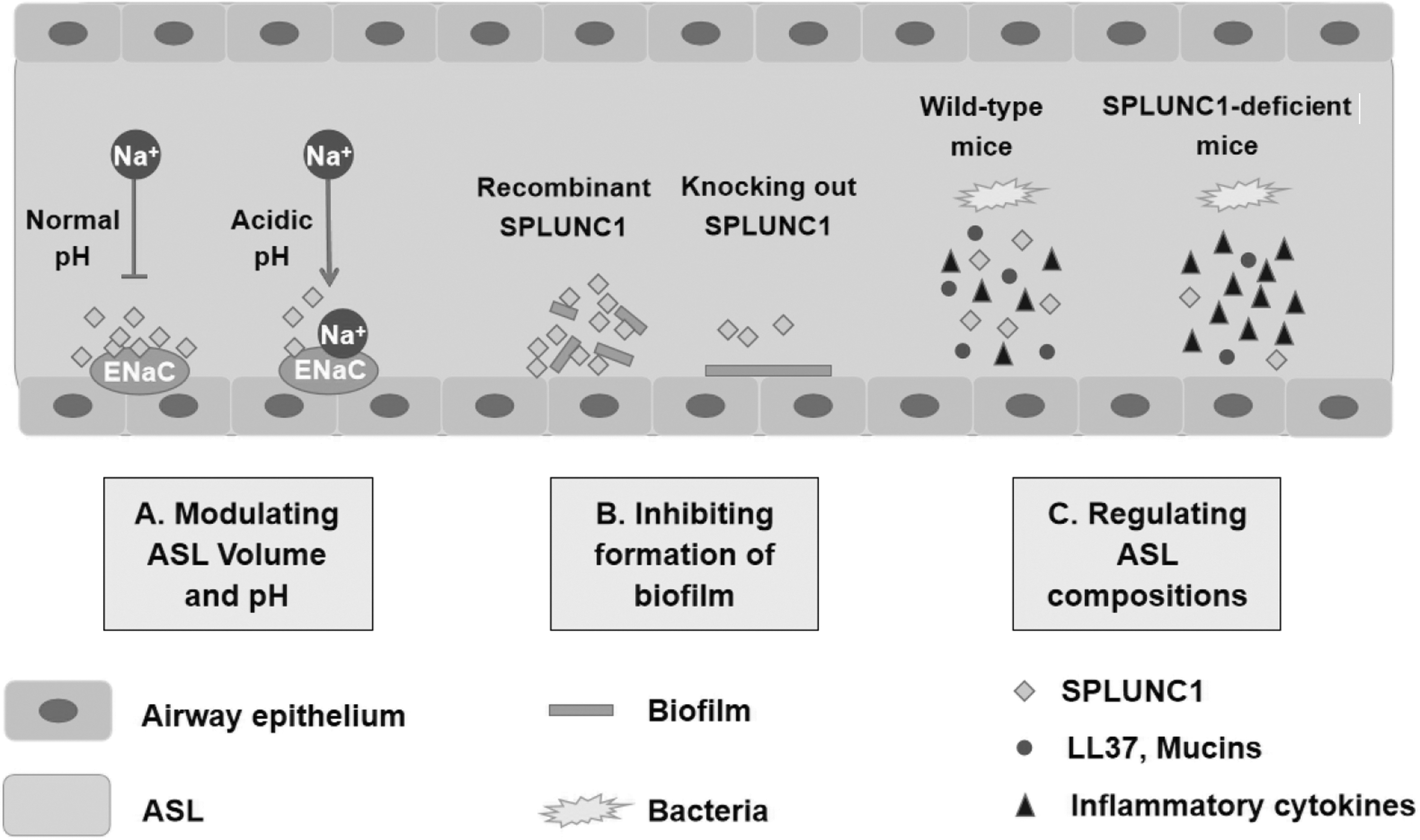
Multifunctional roles of SPLUNC1 in ASL.
Modulating ASL volume
ASL, a thin layer in mammalian airways covering and lubricating airway epithelium surface, is imperative for mucus clearance and for avoidance of infection. In recent researches, ASL has been studied mostly in CF (Berkebile and others 2019; Simonin and others 2019), a lung disease caused by the mutation of CF transmembrane conductance regulator, which was considered to affect the transport of bicarbonate ions and induce the reduction of pH (Coakley and others 2003). A multitude of investigators found that the pH of ASL decreased and airway dehydrated significantly in CF (Tang and others 2016; Simonin and others 2019). Therefore, restoration of ASL height and pH is targeted on therapies of CF (Delpiano and others 2018; Mroz and Harvey 2019). SPLUNC1 compromises up to 10% of the total soluble proteins in ASL (Campos and others 2004). Garland and others observed that SPLUNC1 was with an epithelial Na+ channel (ENaC) inhibitory domain, S18 region. This region, specifically, contained pH-sensitive salt bridges that are essential in restraining the binding between SPLUNC1 and ENaC at acidic pH, and the interaction between SPLUNC1 and ENaC has been shown to be in a pH-dependent manner (Garland and others 2013). They also found that the SPLUNC1 function in the inhibition of ENaC could be recovered after the ASL pH in CF was increased to a normal level (Garland and others 2013).
Furthermore, SPLUNC1 could serve as an allosteric modulator of ENaC and form a SPLUNC1-β-ENaC complex at the plasma membrane to restrain ENaC, maintaining airway fluid homeostasis (Kim and others 2018). However, in CF patients, SPLUNC1 was degenerated rapidly in CF secretions, and recombinant SPLUNC1 was unable to restore the process, resulting in ENaC activation and ASL dehydration (Webster and others 2018). To solve this intractable problem, SPX-101 and S18, belonging to SPLUNC1-derived peptides, have emerged (Scott and others 2017; Terryah and others 2018). They could remain active and intact in CF because of the resistance to the degradation by protease, which might shed new lights on the application of clinical trials to the CF treatments.
Functioning as a surfactant protein and inhibiting formation of biofilm
ASL is covering respiratory tract epithelium and is implicated in preventing alveolar collapse. The surfactant proteins in airway are essential for reducing the surface tension of ASL, which is indispensable for us during breathing and host defensive responses (Pilecki and others 2018; Choi and others 2019; Ordonez and others 2019). Recent studies observed the direct interaction between surfactant protein A (SP-A) and surfactant protein B (SP-B) by coimmunoprecipitation, which implied the potential mechanisms of functional cooperation in the pulmonary surfactant protein system (Martinez-Calle and others 2019).
Based on its marked hydrophobicity (Campos and others 2004), SPLUNC1 was hypothesized to possess potent surfactant properties and later studies have verified the conjecture (Gakhar and others 2010; Liu and others 2013a). As already mentioned, Walton's group found that α4 of SPLUNC1 was conducive to exerting surfactant activities and considered there may exist a link between surfactant regulating and LPS binding (Walton and others 2016). Besides, previous studies also discovered that the surface tension of air–liquid interface was pronouncedly reduced by adding recombinant human PLUNC (Gakhar and others 2010). To sum up, SPLUNC1 can act as a surfactant protein to participate in host defense.
The formation of biofilm is closely associated with respiratory infection. Therefore, increasing attention has been poured into exploring the mechanisms of the biofilm formation and other inducible factors (Kiedrowski and others 2018; Vargas and others 2018), and proposing some methods to attenuate or prevent biofilm formation (Bedi and others 2017; Martin and others 2019). Researches found that the absorption of crystal violet staining biofilm was decreased followed by the treatment with PLUNC, comparing with that of nontreated and buffer-treated biofilms (Gakhar and others 2010). According to the measurements of biofilm biomass and colony forming units assay, it was observed that SPLUNC1 as a surfactant protein indeed inhibited the formation of biofilm by Gram-negative bacteria such as Klebsiella pneumonia (Liu and others 2013a) and P. aeruginosa. Furthermore, the growth and biofilm formation of Burkholderia cepacia complex, a pathogenic bacteria in CF, were disrupted by SPLUNC1, with the α4 helix playing an important role in this process (Ahmad and others 2016). In a recent study, apart from Gram-negative bacteria, SPLUNC1 also affected the biofilm formation of Gram-positive bacteria, like Staphylococcus aureus (Yu and others 2018). It should be noted that the α4 region is also crucial for biofilm inhibition activity and a new AMP, α4-short as the derivative of SPLUNC1, has emerged, which could be used to effectively kill broad-spectrum bacteria and inhibit biofilm formation (Jiang and others 2019). However, it is still contradicted whether the inhibition of biofilm formation is due to the surfactant activity of SPLUNC1 or its direct binding with bacterial LPS.
Regulating ASL compositions
LL37
LL37 is a cationic peptide and belongs to the AMPs family. A study showed that LL37 was associated with the seasonal allergic rhinitis, and decreased the level of some cytokines in tonsil monocytes (Bogefors and others 2014). In some infectious or inflammatory diseases, such as CF, LL37 can exert antibacterial or bactericidal activities against P. aeruginosa (Wnorowska and others 2015). Hosoda and others found that LL37 could improve the survival rate of mice with cecum ligation and puncture sepsis model, and clarified the protective mechanism of LL37 by illustrating its effects on the release of neutrophil extracellular traps (Hosoda and others 2017). Currently, Luo and others (2019) found that LL37 was directly combined with fungus to restrain excessive inflammation caused by Aspergillus fumigatus. According to a study of Weng's group, LL37 was contributed to restoring the function of glucocorticoid, which was widely used in the COPD treatment, and might be involved in suppression of PI3K/Akt signaling pathway (Weng and others 2019a).
In the mice model after P. aeruginosa infection, the SPLUNC1 knockout mice were observed with a significant reduction in the level of LL37 (Liu and others 2013b), which highlighted the potential effect of SPLUNC1 on the expression of LL37 to exert antimicrobial activity. Interestingly, as another member in the AMPs family, the β-defensins 2, demonstrated no similar alteration as the LL37 in the SPLUNC1 knockout mice (Liu and others 2013b). This result showed that the SPLUNC1 may be associated in the selective regulation of some molecules. Therefore, we believe that the SPLUNC1 and LL37 may exert their effects in a synergistic manner against pathogens, and more explorations are needed to prove it.
Mucins
Mucins are glycoproteins and the homeostasis of mucins secretion is vital for trapping and clearing inhaled pathogens. MUC5AC and MUC5B are predominant and localized dissimilarly in the human airways (Okuda and others 2019). Abnormal expression of MUC5AC or MUC5B can induce some respiratory diseases and may be involved in potential signaling pathways (Liu and others 2019; Na and others 2019; Xu and others 2019). Recently, Choi and others (2018) found that tussilagone, an anti-inflammatory medicine, can suppress MUC5AC production through nuclear factor-kappa B (NF-κB) signaling pathway and, therefore, proposed the possibility of tussilagone in treating inflammatory airway diseases. In SPLUNC1 knocked mice, both MUC5AC and MUC5B were downregulated compared with those in wild-type mice (Liu and others 2013b). Furthermore, the decreased expression of MUC5AC and MUC5B still persisted after the P. aeruginosa infection, which indicated that SPLUNC1 exerted antibacterial infection in host defense by influencing the production of mucins. Hence, it should be noted that SPLUNC1 and mucins exert synergistic effect in airway host defense through similar inflammation-relative signaling pathways.
Neutrophil elastase
Human neutrophil elastase (NE), secreted by neutrophils, belongs to the chymotrypsin-like family of the serine proteinase (Yang and others 2016), and its importance in the inflammatory response and host defense has been well established. For example, NE was related to the enhancement of proinflammatory sphingolipid signaling in CF sputum, especially in women with methicillin-resistant Staphylococcus aureus (Karandashova and others 2018). Besides, a latest research demonstrated that the activity of NE in patients' sputum was associated with the severity of diseases (Shoemark and others 2019), and Thulborn and others (2019) also considered NE as a biomarker in COPD with bacteria infection. Hence, therapies targeting on NE are examined or applied to some pulmonary disorders (Keir and others 2019; Watz and others 2019). In hSPLUNC1 transgenic mice, NE activity was significantly increased, comparing with in the wild-type mice (Gally and others 2011). Moreover, due to the critical role the NE played in directly inhibiting the growth of Mycoplasma pneumoniae, the SPLUNC1, as a factor to influence the activity of NE, might be implicated in the antibacterial effect (Gally and others 2011). Ahmad and others also found that SPLUNC1 disrupted the formation of biofilm after the NE pretreatment (Ahmad and others 2016). However, NE can degrade SPLUNC1(Hobbs and others 2012; Jiang and others 2013b) and elevated Haemophilus influenzae level in human airway epithelium cell. According to these results, it is formidable to define the role of NE in the host defense, and more efforts were needed to explore the possible antagonistic or synergistic effect between SPLUNC1 and NE.
Inflammatory cytokines
Cytokines are small molecular proteins with broad biological activity, and their combination with relative receptors is supposed to play a role in immunity response. It was reported that inhibition of interleukin (IL)-19 or its receptor can alleviate allergic airway inflammation, which was expected to be a new therapeutic strategy in treating asthma (Weng and others 2019b). Besides, a new antagonist of IL-33, IL-33trap, also showed anti-inflammatory properties (Holgado and others 2019). Multiple studies have found the interaction effect between SPLUNC1 and cytokines. IFN-γ was helped to inhibit the expression of SPLUNC1 mRNA (Britto and others 2013). In nasal polyp epithelial cells, SPLUNC1 was reduced followed by IL-13 incubation (Yeh and others 2010). Tsou and others (2015) also presented that IL-13 significantly suppressed SPLUNC1 expression in LPS-treated nasal epithelial cells. Liu and others (2013a) established SPLUNC1-deficient mice model by genetic ablation to analyze cell profile and quantify various cytokines levels in bronchoalveolar lavage fluid. Results showed that the number of inflammatory cells was increased more significantly in SPLUNC1 knockout mice than in wild-type mice after Klebsiella pneumoniae infection. Cytokines, such as tumor necrosis factor-α (TNF-α), IL-1α, and IL-6, were elevated distinctly in SPLUNC1-deficient mice at 48 h postinfection. Another group also demonstrated that proinflammatory cytokines IL-1β was increased significantly in SPLUNC1 knockout mice after P. aeruginosa infection (Liu and others 2013b). In Epstein–Barr virus-associated NPC, IL-6, IL-8, IL-1β, and TNF-α were reduced in the SPLUNC1 transfection group, which was involved in the Toll-like receptor 9 (TLR9)/NF-κB signaling pathway (Ou and others 2015). Currently, Zhang and others reported that IL-6 and some chemokine ligands were elevated in SPLUNC1 knockdown mice injected with LPS (Zhang and others 2018). In summary, further investigations should be focused more on these 2 debated points: (1) SPLUNC1 was directly employed as antagonist of inflammatory cytokines to ameliorate airway inflammation and (2) SPLUNC1 indirectly decreased the production of inflammatory factors through acting on other molecules.
Conclusions and Perspectives
The pathogenesis of respiratory diseases is various and complicated, and limitations exist potentially in clinical treatment. Accordingly, there is an imperative demand for seeking a novel target on airway-related diseases. As a natural immune molecule, SPLUNC1 was described to be engaged in host defense, and a surging number of investigations have demonstrated the broad function of it in modulating ASL volume, acting as a surfactant protein, inhibiting biofilm formation, as well as regulating ASL compositions.
A unique α-helix, α4, has been manifested to be paramount for SPLUNC1 to exert surfactant, LPS-binding, and bactericidal activities. In addition, researchers harbored the idea of a potential crosslink between LPS binding and surfactant regulating. Hence, the diverse functions of SPLUNC1 were closely associated with its specific structure. The homeostasis of ASL compositions is indispensable for inflammatory response and host defense in airway, and the expression of some ASL compositions tend to be altered after the infection of bacteria or the invasion of other external stimulus, including the LL-37, mucins, NE, and inflammatory cytokines. It is noted that these ASL compositions were supposed to interact with each other. For example, the production of MUC5AC was induced by LL-37 through TACE-TGF-α-EGFR pathway (Zhang and others 2014b). Besides, NE was also considered as a possible inducer of MUC5AC (Luo and others 2016; Komiya and others 2018; Xu and others 2018). However, a Germany group reported mucin can be degraded by NE in CF sputum, and an inhibitor of NE named KRP-109 was emerged to rescue this process by reversing airway obstruction (Chillappagari and others 2016). These data revealed that the regulation of ASL was particularly complicated and the molecules might exert different effects in different conditions. Surprisingly, similar complexity and diversity were also retained in the regulation of SPLUNC1, with the SPLUNC1 reduced or elevated in distinct inflammatory or infectious disease, which made it difficult to conclude the absolute alteration. For instance, SPLUNC1 was increased in the mice followed by Mycoplasma pneumoniae infection, and this significant enhancement was only observed with infection at 4 h, rather than at 72 h. It was probable that the host was able to resist a temporary infection, with SPLUNC1 mRNA expression increased. As for long-term infection, our body might not be capable of sustaining resistance to pathogens and the level of SPLUNC1 was gradually downregulated. Moreover, the mechanisms of how SPLUNC1 expression is induced during infections are not completely clear, and more studies are needed to clarify it.
Further investigations are supposed to form a systematical network among SPLUNC1 and ASL compositions, or other facets. The network is conducive to better researching or understanding the potential mechanisms of SPLUNC1 in inflammatory response and host defense. We hold the belief that the inhibition of biofilm formation may result from the surfactant activity of SPLUNC1 or its direct binding with bacterial LPS. For that reason, the development of SPLUNC1-derived surfactant protein is an effective approach to limit biofilm formation. Furthermore, a multitude of data have implied the correlations among ASL compositions, which suggests the possibility that SPLUNC1 influences the properties of 1 molecule by acting on another 1. Further directions are recommended to shed light on the potential interaction between SPLUNC1 and some ASL compositions to construct a feasible network for future treatment in airway diseases.
Footnotes
Ethical Statement
No humans or animals were involved in the study, so no ethical approval was required for this article.
Data Availability Statement
The data that support the findings of this study are available in public domain resources including PubMed and MEDLINE.
Author Disclosure Statement
No competing financial interests exist.
Funding Information
This study was supported by grants from the National Natural Science Foundation of China (Grant Nos. 81672688, 81372907, 81472531, 81472595, 81672683, and 81772928).