




Abstract
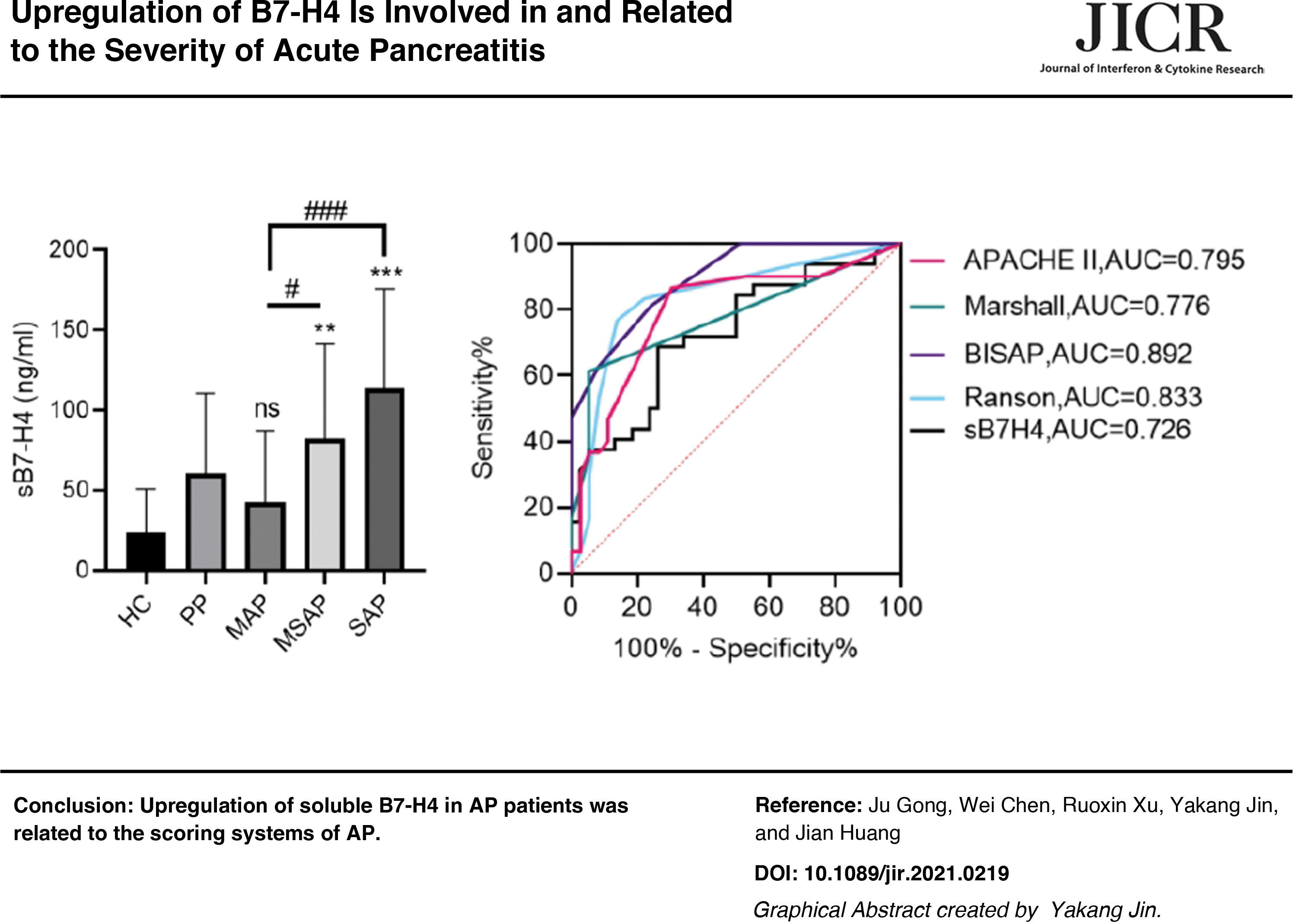
The expression and clinical significance of co-stimulator B7-H4 in acute pancreatitis (AP) is still unclear. In vitro study showed that the expression of soluble B7-H4 (sB7-H4) and proportions of membrane B7-H4-positive CD14+ cells in the peripheral blood mononuclear cells were upregulated in response to stimulation with plasma from AP patients, lipopolysaccharides, or tumor necrosis factor α (TNF-α). sB7-H4 in the plasma of AP patients were positively correlated with interleukin (IL)-6, IL-10, IL-17A, TNF-α, and interferon-γ The areas under the curves (AUCs) of receiver operating characteristic (ROC) curves of plasma sB7-H4 to distinguish the AP patients from healthy donors, the mild AP (MAP) from the moderately severe acute pancreatitis (MSAP)+severe acute pancreatitis (SAP) or the SAP from the MAP+MSAP were 0.78 (P < 0.001) or 0.773 (P < 0.001) or 0.764 (P < 0.001). sB7-H4 in the plasma of patients were positively correlated with the RANSON scores, Bedside Index of Severity of Acute Pancreatitis scores, Marshall scores, and Acute Physiology And Chronic Health Evaluation II scores; and the AUCs of ROC curves of plasma sB7-H4 in the prediction of local complications was 0.726 (P = 0.001). In conclusion, the co-stimulator B7-H4 is involved in the immune response in AP.
Color images are available online.
Introduction
Acute pancreatitis (AP) is an inflammation response of the pancreas caused by local pancreatic damage that may lead to multiorgan failure or death. According to the Revised Atlanta Classification, AP could be classified as mild AP (MAP), moderately severe acute pancreatitis (MSAP), or severe acute pancreatitis (SAP), and the prognosis of AP depends on its severity. MAP is a common, self-limited acute abdominal disease with a good prognosis, whereas SAP has a high mortality rate owing to pancreatic necrosis, systemic inflammation, and persistent multiple organs dysfunction associated with concurrent infections (Valverde-López and others 2018; Lee and Papachristou 2019; Petrov and Yadav 2019).
Many efforts have been contributed to understand the pathogenesis of pancreatitis, but the mechanism is still unclear (Watanabe and others 2017). Recent studies reveal that immune cells and inflammatory cytokines play an important role in the pathogenesis of pancreatitis (Kim and others 2017; Sendler and others 2018). Uncontrolled or dysregulated activation of the immune system may result in systemic complications of AP. Pancreatic acinar cells are considered to be the common target of intrinsic (genetic) and extrinsic factors (such as biliary stones, alcohol, drugs, and metabolic disorders). Necrotic cells activate innate immune cells and recruit them to sites of inflammation to propagate the severity of AP by releasing damage-associated molecular patterns, cytokines, chemokines, and other molecules, which further activate the adaptive immune system.
As a member of the B7 family, B7-H4 plays a role as a negative regulator in T cell-mediated immune responses in various cancers, such as pancreatic cancer, ovarian cancer, breast cancer, bladder cancer, glioma, and prostate cancer (Ye and others 2018; Qi and others 2019; Fang and others 2020). Expression of B7-H4 inhibits the proliferation of T cells and the production of cytokines in tumor microenvironment (Chen and others 2018). Knockdown of B7-H4 not only significantly inhibits the proliferation, invasion, and migration of tumor cells, but also promotes the apoptosis of tumor cells (Zhang and others 2017). High expression of B7-H4 is a poor prognostic factor for pancreatic cancer (Shen and others 2017). However, there are few studies on the expression of B7-H4 and its role in pancreatitis.
In this study, we first determined that the expression pattern of B7-H4 in cultured human peripheral blood mononuclear cells (PBMCs) could be changed by the stimulation of plasma from AP patients, lipopolysaccharides (LPS), or tumor necrosis factor α (TNF-α). Then, we analyzed the relationship between expression levels of soluble B7-H4 (sB7-H4) and cytokines in AP patients. Finally, the relationships between the expression levels of B7-H4 and clinical features were analyzed. This study reveals that the expression of B7-H4 is involved in AP.
Materials and Methods
Patients
This study was approved by the Ethics Committee of the First Affiliated Hospital of Suzhou University (Protocol No. 2021-008). Seventy AP patients (30 MAP patients, 21 MSAP patients, and 19 SAP patients), 20 abdominal pain patients without AP, and 20 healthy donors were recruited for this study. All participants provided written informed consent before study inclusion. The patients were diagnosed according to the revised Atlanta Classification for AP in 2012. Venous blood samples of patients were collected on admission. The plasma was separated by centrifugation and stored at −80°C until further analysis.
Preparation of PBMCs
Human PBMCs were isolated with gradient centrifugation. Four milliliters of heparin anticoagulant peripheral blood was mixed with 4 mL of Lymphoprep solutions (Axis Shield, Abbott Diagnostics Technologies AS, Oslo, Norway) and centrifuged at 2000 rpm for 20 min. The white blood cells were carefully transferred to new tubes and centrifuged at 1500 rpm for 10 min. Following washing, cells were resuspended in RPMI-1640 at a concentration of 5 × 10⁶/mL. Cell viability was detected by Trypan blue staining.
PBMCs stimulation
Isolated PBMCs were cultured in 24-well plates. PBMCs were co-incubated with 200 μL of plasma from AP patients or healthy volunteers for 24, 48, and 72 h. PBMCs were also stimulated with TNF-α (PeproTech, Inc., Rocky Hill, NJ) (100 ng/mL) or LPS (Biosharp Life Sciences, Hefei, Anhui, China) (1 μg/mL) for 24, 48, and 72 h. Then, cells were collected to detect the expression of B7-H4 on the cell membrane by flow cytometry or sB7-H4 by enzyme-linked immunosorbent assay (ELISA).
Flow cytometry
First, red blood cells in the blood needed to be lysed with 1 mL of ACK lysis buffer (Beyotime Biotechnology, Shanghai, China). For the expression of B7-H4 on CD14+ cells, the monocytes in the blood of AP patients were stained with FITC-conjugated anti-CD14 (Biolegend Coulter, San Diego, CA) and PE-conjugated anti-B7-H4 (Suzhou Bright Scistar Biotechnology Company Ltd., Suzhou, Jiangsu, China) at 4°C for 30 min in dark. After washing with 1 mL of phosphate-buffered saline (PBS), cells were collected with a centrifuge at 1200 rpm for 5 min. After being resuspended with 400 μL of PBS, cells were analyzed using BD FACSCanto II (BD Bioscience, Franklin Lakes, NJ).
Enzyme-linked immunosorbent assay
The levels of sB7-H4 in plasma were measured using human sB7-H4 ELISA kits from Shanghai Tongwei Industrial Co., Ltd. (Shanghai, China) according to the manufacturer's instructions. Data were analyzed using the standard curve plotted to calculate the amount of sB7-H4.
Statistical analysis
Data analyses were performed using IBM SPSS Statistics 22.0. All data values were presented as mean ± standard error of the mean. The difference between the 2 groups was evaluated using the Student's t-test; differences among 3 or more groups were evaluated by analysis of variance (ANOVA) and followed by an unpaired t-test or Mann–Whitney test. The relationships between sB7-H4 with cytokines were analyzed using the Pearson correlation analysis. The receiver operating characteristic (ROC) curves were calculated by GraphPad software. A value of P < 0.05 was considered as statistical significance.
Results
Changes in B7-H4 were induced by stimulation with plasma from AP patients
To determine whether B7-H4 is involved in AP, we first stimulated PBMCs with plasma from AP patients, sB7-H4 in the supernatants and proportions of membrane B7-H4-positive CD14+ cells were determined by ELISA and flow cytometry. When compared with healthy donors' plasma, stimulation with AP plasma significantly increased both the sB7-H4 and proportions of mB7-H4-positive CD14+ cells (Fig. 1A–D). The AP patients' plasma induced changes in the expression pattern of B7-H4 in PBMCs, indicating that B7-H4 was involved in AP.
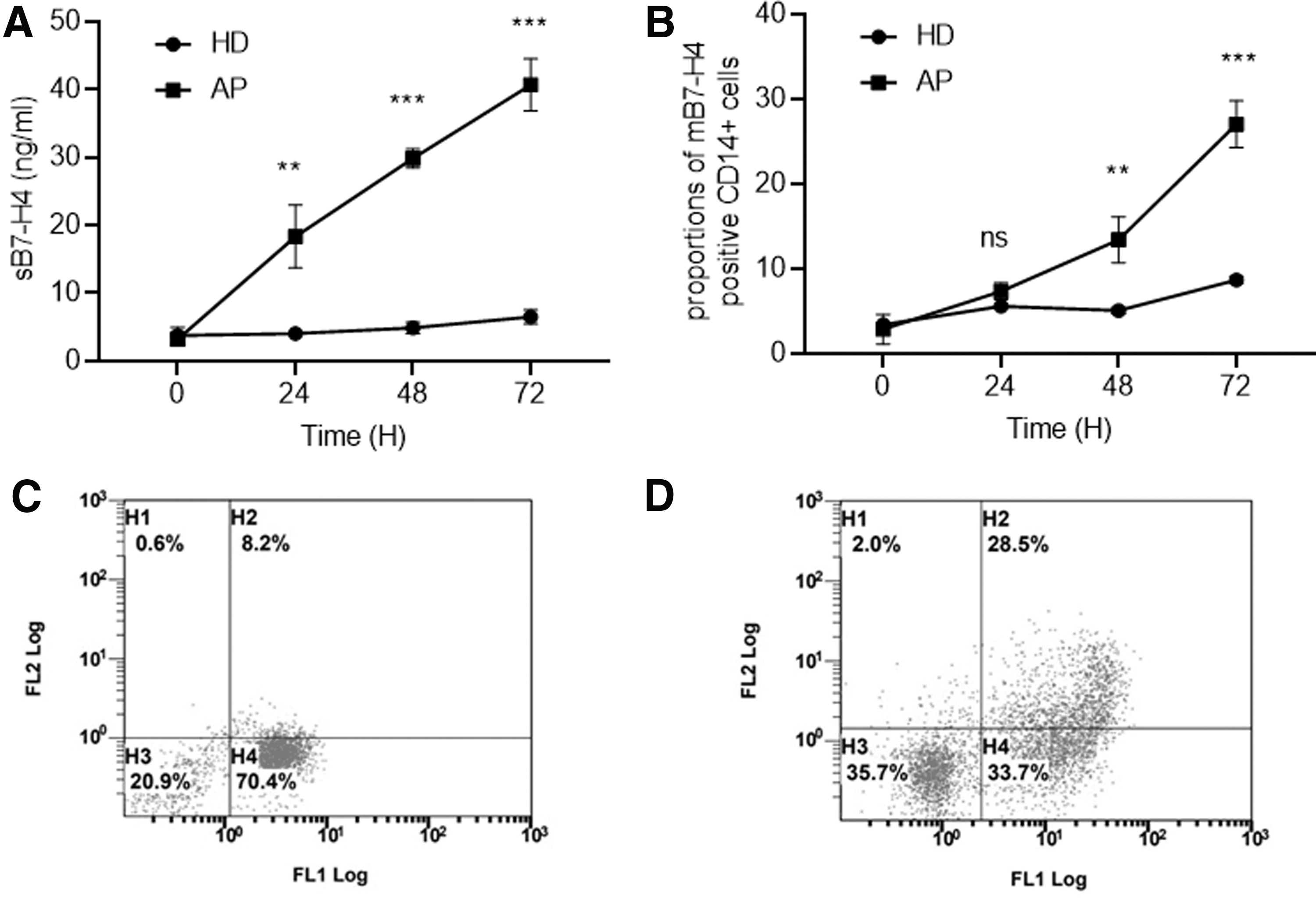
Upregulation of sB7-H4 and membrane B7-H4 in PBMCs by serum from AP patients.
Changes in B7-H4 were induced by stimulation with LPS or TNF-α
The changes in B7-H4 induced by serum from AP patients suggested that the activation of immunity responses was involved. Thus, LPS and TNF-α were used to stimulate the isolated PBMCs. Both LPS and TNF-α were found to significantly increase the expression of sB7-H4 and mB7-H4 in PBMCs (Fig. 2A–D). These results indicated that activation of innate inflammatory responses by LPS or TNF-α induced the changes in the expression of B7-H4.
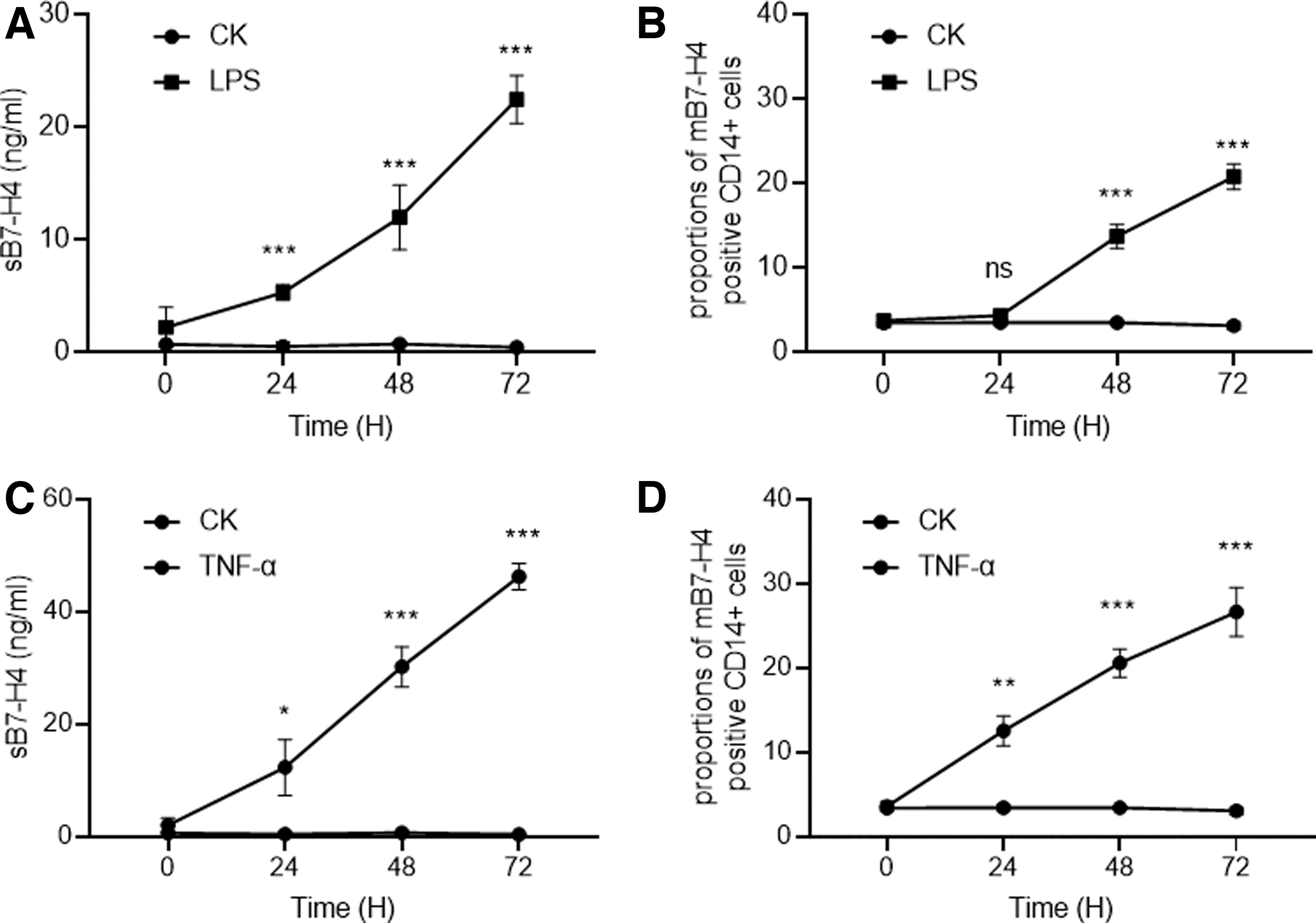
Upregulation of sB7-H4 and membrane B7-H4 in PBMCs by LPS or TNF-α.
Levels of sB7-H4 were correlated with interleukin (IL)-6, IL-10, IL-17A, TNF-α as well as interferon-γ in AP patients
Because B7-H4 could be induced by innate immunity responses in vitro, we wondered whether the expression levels of B7-H4 were related to the cytokine expression levels in AP patients. Because sB7-H4 was much easier to measure, we mainly focused on sB7-H4.
First, the patients were divided into 2 groups according to the median expression levels of sB7-H4 and the expression levels of cytokines were compared. The expression levels of IL-6, IL-10, IL-17A, TNF-α, and interferon-γ (IFN-γ) were significantly higher in the patients with high sB7-H4 expression levels than those in patients with low sB7-H4 expression levels, whereas no difference was found in thrombopoietin (TPO) and IL-33 (Fig. 3A, B). Then, the correlations between expression levels of sB7-H4 and these cytokines were analyzed. sB7-H4 expression levels were significantly positively correlated with IL-6, IL-10, IL-17A, TNF-α, TPO, and IFN-γ levels (Table 1). The correlation between B7-H4 and cytokines indicated that B7-H4 is involved in AP progress in vivo.

Differential expression of cytokines in AP patients with low or high sB7-H4 expression levels.
The Relationship Between Expression Levels of Soluble B7-H4 and Clinical Features
When P < 0.05, it is shown in bold.
APACH II, Acute Physiology And Chronic Health Evaluation; BISAP, Bedside Index of Severity of Acute Pancreatitis; IFN-γ, interferon γ; IL, interleukin; RANSON; TNF-α, tumor necrosis factor α; TPO, thrombopoietin.
Levels of sB7-H4 were related to the severity of AP
Because B7-H4 regulates the adaptive immunity response through the second signaling pathway, which plays an important role in SAP development, we speculated whether the expression levels of sB7-H4 are related to the severity of AP.
As given in Fig. 4A, sB7-H4 gradually increased in the healthy donors, MAP, MSAP, and SAP patients, whereas no significant difference was found between AP patients and abdominal pain without AP patients. Besides, the expression levels of sB7-H4 in MSAP and SAP were significantly higher than those in healthy donors or MAP (Fig. 4). Of interest, although the expression levels of sB7-H4 could barely distinguish between abdominal pain without AP patients and AP patients (Fig. 4B); the areas under the ROC curves (AUCs) were 0.78 in differentiating AP patients from healthy donors (Fig. 4C). Moreover, the AUCs were 0.773 and 0.764 in differentiating the MAP from the MSAP+SAP or the SAP from the MAP+MSAP (Fig. 4D, E). The results indicated that levels of sB7-H4 could differentiate the severity of AP patients efficiently.
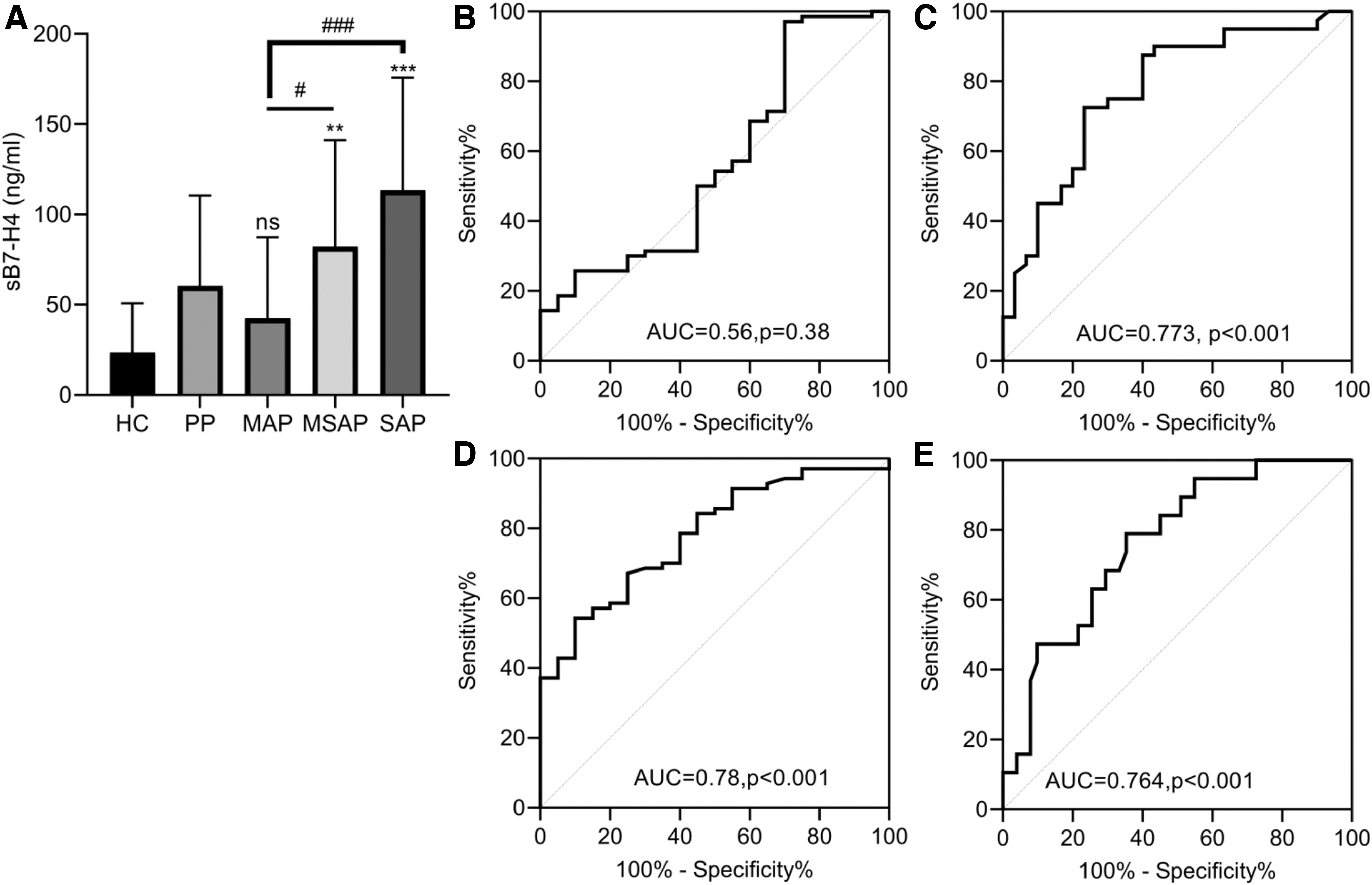
sB7-H4 was related to the severity of AP.
Levels of sB7-H4 were related to the scoring systems of AP
Considering the positive correlation between levels of sB7-H4 and cytokines, the relationship between sB7-H4 and the scoring systems was analyzed. First, we compared the scores of the scoring systems in the high and low sB7-H4 expression groups. The RANSON scores, Bedside Index of Severity of Acute Pancreatitis (BISAP) scores, Marshall scores, and Acute Physiology And Chronic Health Evaluation II (APACH II) scores were significantly higher in patients with high sB7-H4 expression levels than in patients with low sB7-H4 expression levels (Fig. 5A). Moreover, the expression levels of sB7-H4 were significantly positively correlated with the RANSON scores, BISAP scores, and APACH II scores (Table 1).
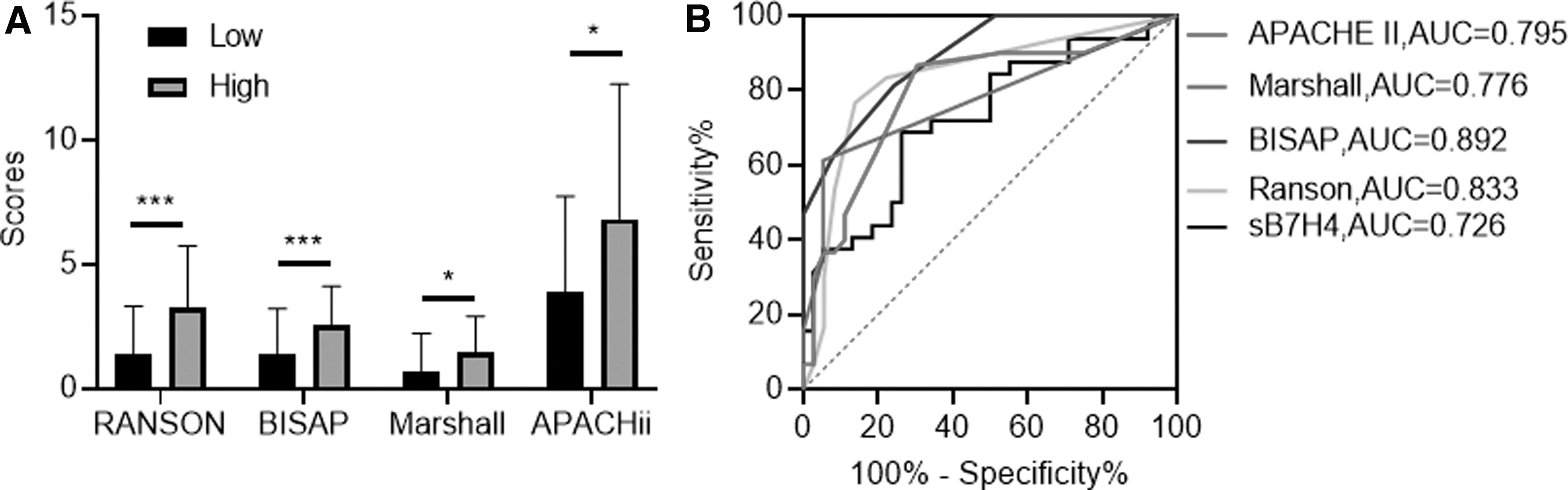
sB7-H4 was related to the scoring systems of AP.
And then, the ROCs of sB7-H4 and the scoring systems in the prediction of local complications were plotted. The AUCs of sB7-H4, RANSON score, BISAP score, Marshall score, APACH II score were 0.726 (P = 0.001), 0.833 (P < 0.001), 0.892 (P < 0.001), 0.776 (P < 0.001), and 0.795 (P < 0.001), respectively (Fig. 5B). The results indicated that the levels of sB7-H4 might be correlated with the outcomes of AP patients.
Discussion
AP is initially a local protective inflammation secondary to the necrotic acinar cell, gradually progressing to an overwhelming systemic immune reaction, including systemic inflammatory response syndrome (SIRS) and compensatory anti-inflammatory response syndrome (CARS), ultimately leading to persistent multiorgan failure, which seems to be responsible for the majority of mortality (Lee and Papachristou 2019). The coexistence of SIRS and CARS is known as “mixed antagonist response syndrome” (MARS), and it causes the dysregulation of pro- and anti-inflammatory cytokine production as well as more serious damage to the body. The imbalance in the host immune response is considered relevant to the severity and prognosis of AP (Ge and others 2020).
The systemic activation of the adaptive immune system in AP is mediated by macrophages and T cells. Co-signaling molecules, which are divided into co-stimulatory and co-inhibitory molecules, are expressed on the cell surface of activated antigen-presenting cells such as T cells, B cells, monocytes, and dendritic cells (Ni and Dong 2017). Previous studies have shown that the co-signaling molecules, as the second signals, play a pivotal role in regulating the intensity of T cell immune responses, determining the outcome of T cells, and directing the activation or inhibition of lymphocytes and the fate of the immune response (de Ramon and others 2018).
B7-H4, an important member of the B7 family, is a co-inhibitory molecule and negatively regulates T cell immune response and promotes immune escape by inhibiting the proliferation, cytokine secretion, and cell cycle of T cells (MacGregor and Ohashi 2017). With the exacerbation of AP, the gradually increased expression level of sB7-H4 was considered an index for the severity assessment of AP. However, the expression characteristics of this molecule in AP patients are not clear.
Despite B7-H4 mRNA being widely expressed, B7-H4 protein has not been detected in most normal tissues in lymphoid and nonlymphoid organs. The B7-H4 protein possesses 282 amino acid residues, comprising an amino terminal extracellular domain, a large hydrophobic transmembrane domain, and a very short intracellular domain, aside from B7-H4 being anchored to the cell membrane through a GPI linkage, which can be cleaved to produce sB7-H4 from the cell surface through metalloproteinase (Radichev and others 2014). In this article, it is observed that both the sB7-H4 and mB7-H4 were induced by plasma from AP patients, LPS, or TNF-α, suggesting that the inflammatory response may promote B7-H4 expression on monocytes and accelerate the activity of metalloproteinase to release sB7-H4.
The pancreatic innate immune activation and rapid systemic inflammatory response are usually followed by macrophages activation. As a result, TNF-α, IL-6, and IFN-γ are produced in greater amounts, promoting tissue damage and disease progression. In the process of inflammation, apoptosis of activated immune cells is a timely switch of the immune response to shutdown process of excess immune responses, avoiding tissue damage and maintaining body homeostasis. Our in vitro studies found that the expression of B7-H4 was upregulated in response to inflammatory stimulation. As a co-inhibitory molecule, the upregulation of B7-H4 seems to have a protective role in the initial stage of AP.
However, we further found that the gradually increasing levels of sB7-H4 had a significant correlation with scoring systems. These results demonstrated that sB7-H4 was related to the severity of AP patients, and may even be related to their prognosis. In AP patients with SIRS, the body also mounts an anti-inflammatory response to reduce tissue damage. Overcompensation of CARS suppresses the immune system and induces susceptibility to infection, which is usually more common in patients with SAP. Thus, the relationship between high expression levels of B7-H4 and the severity of AP indicates an important role of B7-H4 in CARS.
Conclusion
Collectively, sB7-H4 and mB7-H4 were upregulated in AP, and upregulation of sB7-H4 was related to the severity and the scoring systems of AP. However, we found no relationships between expression levels of sB7-H4 and hospital stay or mortality, which was found by another group (Cheng and others 2020). This is probably owing to the relatively small number of patients enrolled in our study. Further studies with larger samples and better experimental design may strengthen the value of sB7-H4 in AP diagnostics, severity, and prognosis.
Footnotes
Authors' Contributions
J.G. performed experimental work, data analysis, obtained funding, and wrote the article. W.C. performed experimental work, data analysis, assisted with data acquisition and analysis. X.R.X. assisted with data acquisition and analysis. J.H. conceived the idea, designed the study, and obtained funding. All authors were involved in the final approval of the article. J.Y.K. assisted in designing the study, revised the article.
Author Disclosure Statement
No competing financial interests exist.
Funding Information
This work was supported by Science and Technology Plan Project of Changshu Commission of Health (csws202012), Science and Technology of Tongren City (No. 2018-1-5), Suzhou University (Nos. P112200519 and P112200719).