




Abstract
Oral lichen planus (OLP) is a T cell-mediated chronic inflammatory mucocutaneous disease affected by the interaction between keratinocytes and T cells. Recent evidence indicates that vascular cell adhesion molecule-1 (VCAM1) plays a vital role in mediating immune and inflammatory responses. In this study, the expression of VCAM1 in OLP was detected by immunohistochemical staining and its correlations with clinical features were analyzed. The disease severity of OLP was assessed by the reticular, atrophic, and erosive scoring system. We found that VCAM1 was generally localized in the cytoplasm of epithelial cells, and in nucleus, cytoplasm, and extracellular matrix of subepithelial infiltrated cells in superficial layer of lamina propria. Moreover, VCAM1 levels in epithelium and lamina propria of OLP were significantly higher than that in controls, respectively. In addition, VCAM1 level in epithelium was increased compared with that of lamina propria. There were no significant differences for VCAM1 expression between nonerosive and erosive forms of OLP. The expression of VCAM1 in OLP was not associated with the severity of disease, gender, and age. Thus, we speculated that spatial expression differences of VCAM1 in local lesions of OLP may involve the pathogenesis of OLP.
Introduction
Oral lichen planus (OLP) is a T cell-mediated inflammatory mucocutaneous disease with a prevalence of 0.5%–3% (El-Howati et al, 2022; Gonzalez-Moles et al, 2021). OLP has been defined as an oral potentially malignant disorder by the World Health Organization (WHO) (Warnakulasuriya et al, 2021). The histological observation of OLP showed that CD4+ T cells predominantly reside within the lamina propria, whereas CD8+ T cells are most abundant within the epithelium and adjacent to damaged basal keratinocytes (Sugerman et al, 2002). Its immunopathogenesis is supposed to comprise antigen presentation, T cell activation and migration, and keratinocyte apoptosis (Alrashdan et al, 2016). T cells, in response to antigen, are activated and recruited to oral mucosa, and then direct a deleterious immune response against oral keratinocytes (Roopashree et al, 2010). The migration of T cells and their interactions with keratinocytes are mediated by specific cell surface adhesion molecules (El-Howati et al, 2022; Krummel et al, 2016).
Vascular cell adhesion molecule-1 (VCAM1) is a membrane protein belonging to the superfamily of immunoglobulins and as a key player with multiple functionalities (Cook-Mills et al, 2011; Kong et al, 2018). In resting cells, VCAM1 is usually expressed at a very low level, but it can be upregulated on the surface of activated cells, including keratinocytes, T cells, particularly under high levels of inflammation and chronic conditions in some diseases (Cook-Mills et al, 2011; Diaz et al, 2005; Groves et al, 1993; Ishiyama et al, 1998; Kong et al, 2018; Maarof et al, 2016). Studies have revealed that the protein levels in local lesions of VCAM1 is elevated in several human autoimmune diseases, including multiple sclerosis and Crohn's disease (Allavena et al, 2010; Danese et al, 2005). However, the expression of VCAM1 in OLP patients is still unknown.
In this study, we analyzed the expression of VCAM1 on epithelial cells and subepithelial infiltrated cells in OLP lesions. Besides, the difference of VCAM1 expression between epithelium and lamina propria was further explored to investigate the potential roles of VCAM1 in local environment of OLP. In addition, the association between VCAM1 expression and clinicopathological factors was also measured.
Materials and Methods
Participants and tissue specimens
The experiments were approved by the Ethics Committee Board of School and Hospital of Stomatology, Wuhan University (No. 2019A17, and followed the principles of the Declaration of Helsinki in the use of human samples. All tissue specimens used in this study were obtained from the Department of Oral Medicine, School and Hospital of Stomatology, Wuhan University. Twenty-five OLP patients were clinically diagnosed and histopathologically confirmed as OLP according to the diagnostic criteria of OLP made by WHO (van der Meij and van der Waal, 2003), and they were simplified into the nonerosive group (NEOLP) and erosive group (EOLP). Eleven age- and gender-matched healthy volunteers were recruited as the controls (CON). All participating subjects gave their informed consents, and neither of them had any systemic disorders (such as cardiovascular disease, diabetes mellitus, etc.) nor any soft tissue lesions in the oral mucosa.
Smokers and severe alcoholics were excluded. Besides, patients on immunotherapy or receiving any medical treatment of OLP within 3 months were not included either. The clinical characteristics of subjects are listed in Table 1. Reticular, atrophic, and erosive (RAE) scoring system recommended by our previous study was used to assess the severity of OLP in different clinical forms (Zhou et al, 2012).
Clinical Characteristic of Oral Lichen Planus Patients and the Controls
Immunohistochemical staining
The tissue samples were fixed in 4% formalin at room temperature for 24 h. Sections were routinely deparaffinized in xylene, rehydrated in an alcohol gradient, and embedded in epoxy resin, followed by blocking the activity of endogenous peroxidase and antigen retrieval with ethylenediaminetetraacetic acid (1 mM, pH = 8.0) buffer. After treatment with hydrogen peroxide and blocking with normal goat serum, the sections were incubated with the primary rabbit antibody against VCAM1 (1:500; Abcam, ab134047, Cambridge, United Kingdom) overnight at 4°C, and followed by HRP polymer-conjugated anti-rabbit secondary antibody for 10 min at 37°C. Immunoreactivity was visualized by incubating with diaminobenzidine (Detection Kit, GK600505; Gene Tech, Shanghai, China). Hematoxylin was then used to stain the nucleus.
VCAM1 level in OLP and control tissues was assessed semiquantitatively. Images were captured using CaseViewer (3D HISTECH Ltd., Budapest, Hungary). The expression level was measured by mean optical density value using computer-assisted image analysis software (Image-Pro Plus 6.0 version). On microscopic examination, the oral mucosa was divided into 2 layers as epithelium and lamina propria. Each layer was evaluated for their staining intensity. The evaluation for all the staining images of immunohistochemistry was performed on at least 5 randomly selected at 200 × .
Statistical analyses
All statistical graphs were generated using Graph Prism 8 (GraphPad Software, San Diego, CA). Anderson–Darling test, Kolmogorov–Smirnov test, and Shapiro–Wilk test were used to measure the normal distribution of data. Student's unpaired 2-tailed t test was performed when comparing 2 groups to each other, and Spearman's test was used to analyze the correlations. Representative data from separate experiments were shown as mean values ± standard deviations (SD). A value of P < 0.05 was considered as statistically significant.
Results
VCAM1 were overexpressed on epithelial cells and subepithelial infiltrated cells in OLP lesions
The expression of VCAM1 was mainly found in the cells of epithelium and superficial layer of lamina propria in OLP (Fig. 1A). Especially, VCAM1 was generally localized at the cytoplasm of epithelial cells. As for subepithelial infiltrated cells in superficial layer of lamina propria, the distribution of VCAM1 was restricted to nucleus, cytoplasm, and extracellular matrix (Fig. 1B). VCAM1 expression was upregulated in general field, epithelium, and lamina propria of OLP when compared with that in controls (P < 0.05, Fig. 1C–E). We further analyzed the expression of VCAM1 in epithelium and lamina propria of OLP, respectively, for exploring the distinct characteristics of VCAM1 spatial distribution. The results showed that the epithelium of OLP had a demonstrated overexpression of VCAM1 compared with that in lamina propria (P < 0.05, Fig. 1F).
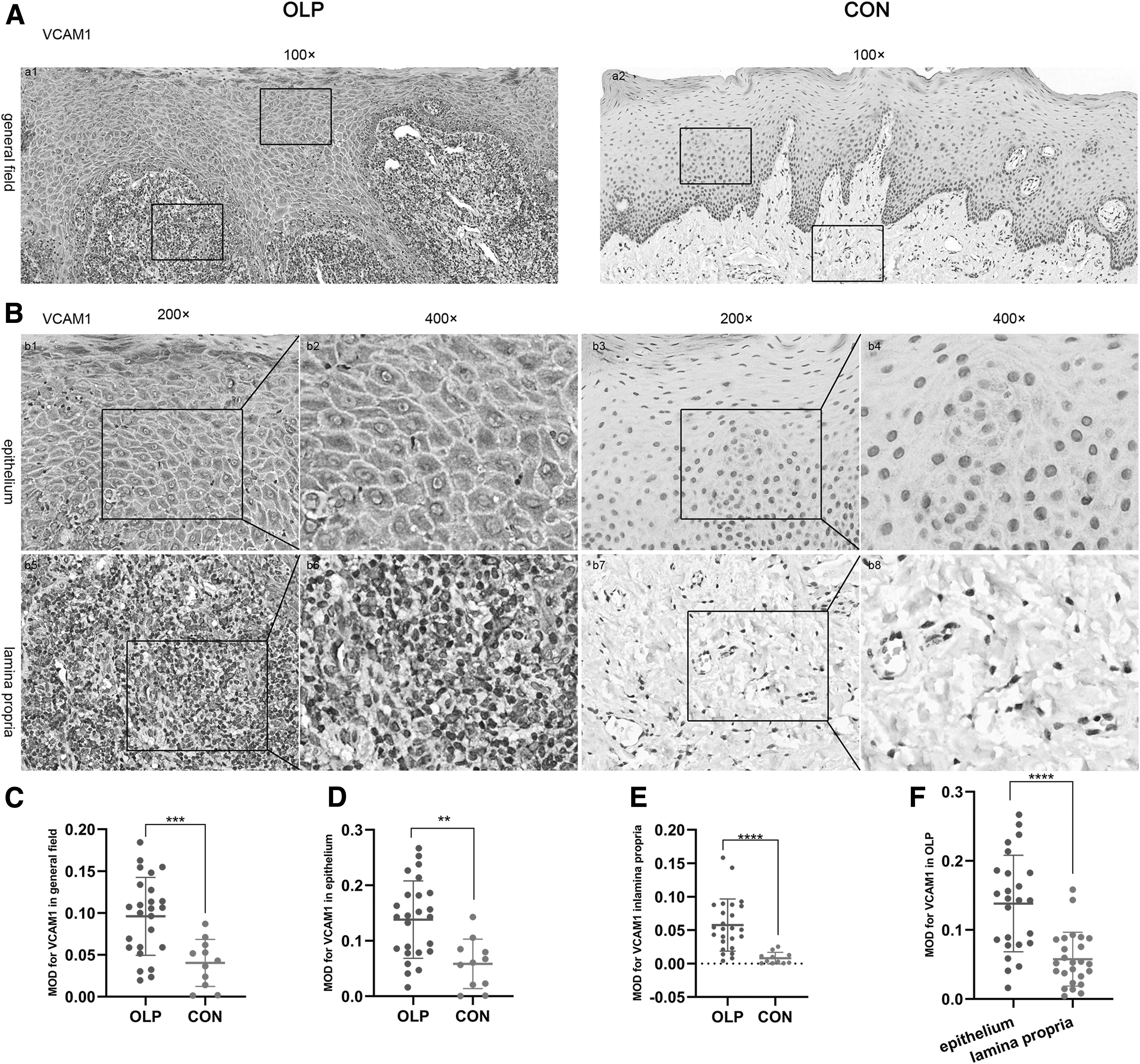
VCAM1 were overexpressed on epithelial cells and subepithelial infiltrated cells in OLP lesions. The expression of VCAM1 in mucosal tissues of OLP (n = 25) and controls (n = 11) was identified by immunohistochemistry and semiquantitatively assessed by MOD value. Magnification: 100 × , scale bar, 50 μm; 200 × , scale bar, 100 μm; 400 × , scale bar, 25 μm. Positive staining was analyzed using MOD of at least 3 randomly captured fields at × 200 magnification. VCAM1 levels of general field
Expression of VCAM1 in different clinical forms of OLP
The expressions of VCAM1 of general field, epithelium, and lamina propria showed no significant difference between different clinical types of OLP (P > 0.05, Fig. 2A–C). To further investigate the clinical significances of VCAM1 in OLP, we also examined correlations of VCAM1 expression in OLP patients with RAE scores. However, no correlation was observed between the expression of VCAM1 and RAE scores (P > 0.05, Fig. 2D–F).
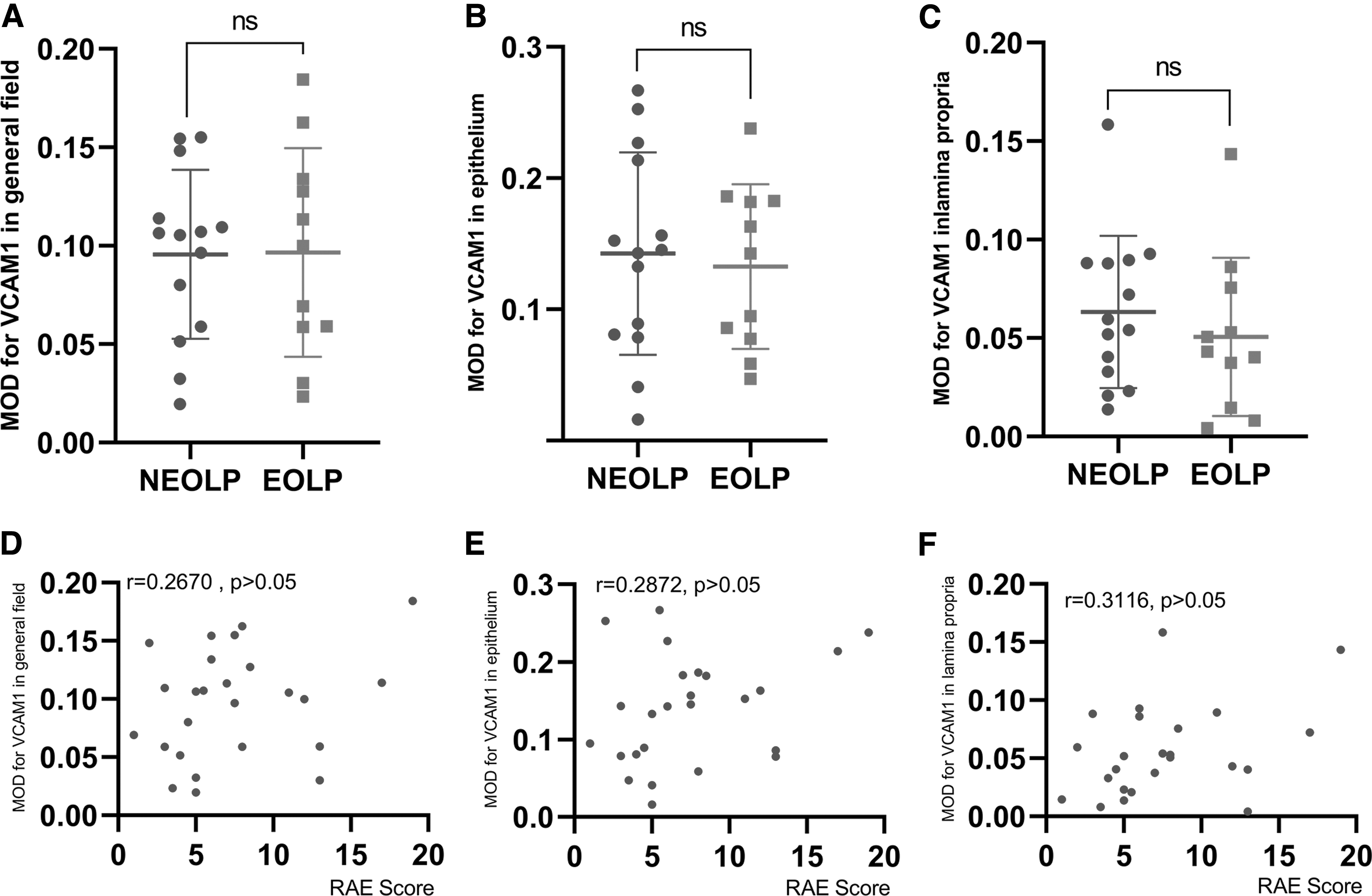
Expression of VCAM1 in different clinical forms of OLP. Expressions of VCAM1 in the general field, epithelium, and lamina propria of different clinical forms of OLP
VCAM1 expression in different genders of OLP patients
The higher VCAM1 levels were observed in the general field, epithelium, and lamina propria both of males and females in OLP patients, compared with the control group (P < 0.05, Fig. 3A–B, E, F, I, J). VCAM1 levels had no statistical difference between males and females either in OLP or controls (P > 0.05, Fig. 3C, D, G, H, K, L).
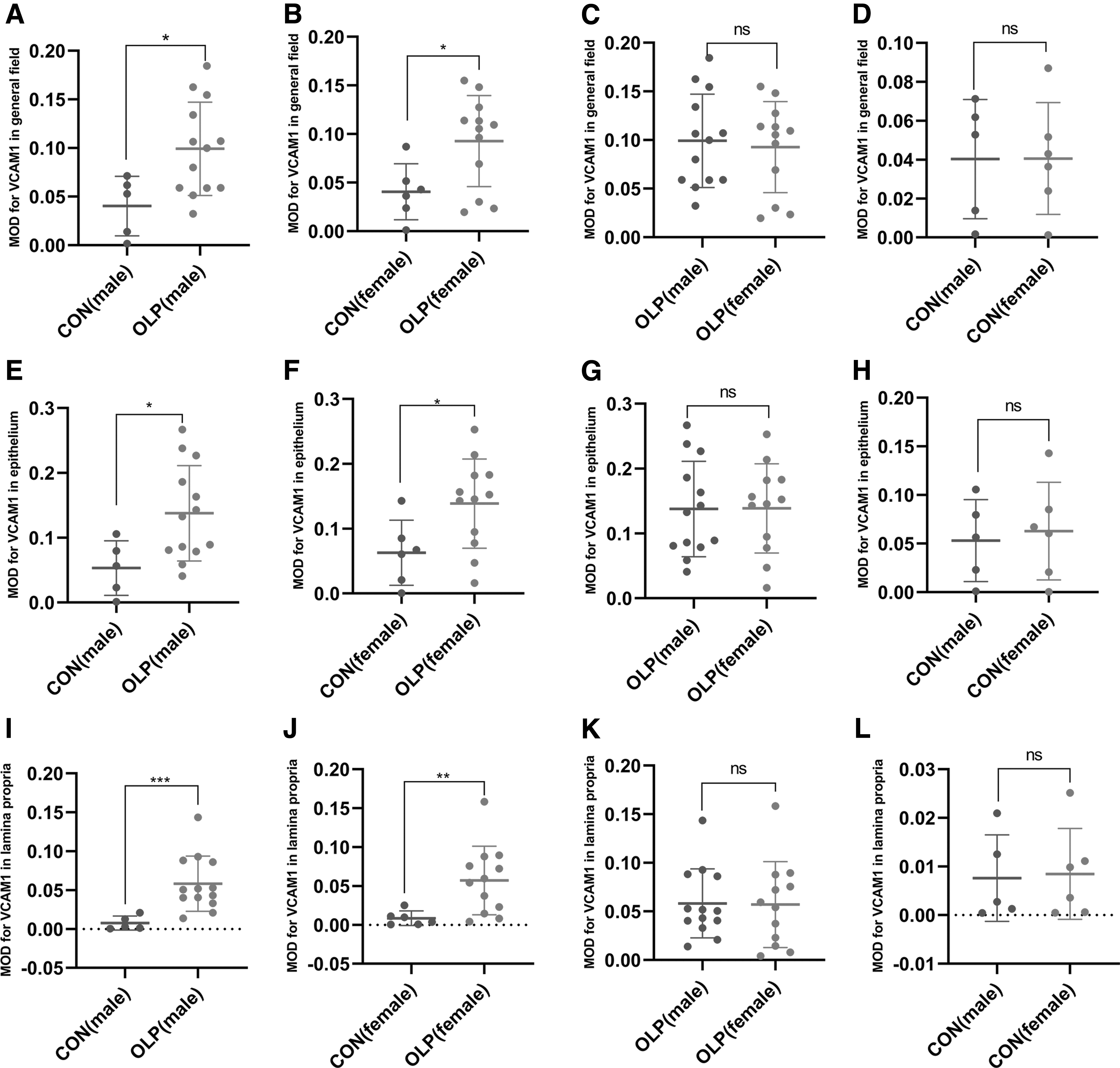
VCAM1 expression in different genders of OLP patients. VCAM1 expression of the general field
VCAM1 expression in different ages of OLP patients
When the subjects were divided into age groups, no statistical difference in VCAM1 levels was found between OLP subjects with the age beyond 50 years and the age equal to or less than 50 years (P > 0.05; Fig. 4A). The correlations of VCAM1 levels with ages in OLP were then examined. The results showed no correlation between VCAM1 expression with the age of OLP patients (r = 0.1131, P > 0.05; Fig. 4B).
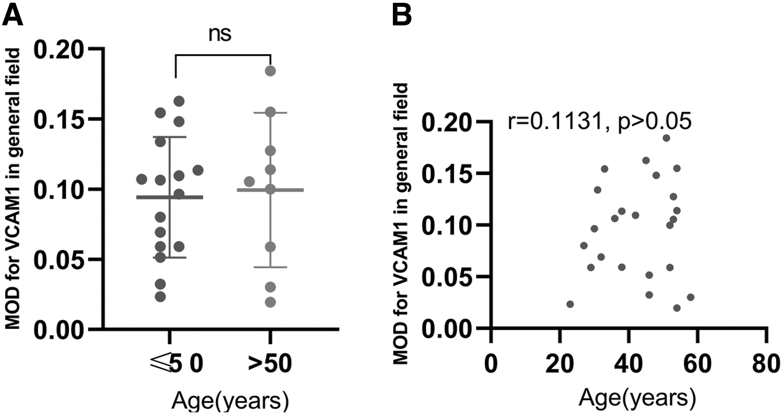
VCAM1 expression in different ages of OLP patients.
Discussion
Studies have demonstrated that VCAM1 expression can be induced by cytokines in the tissue, including NF-kB, TNF-α, IFN-γ, and IL-1β (Cook-Mills et al, 2011). For example, TNF-α upregulates VCAM1 expression on fibroblast-like synoviocytes through NF-kB in rheumatoid arthritis, and VCAM1 activates signaling pathways within the activated fibroblast-like synoviocytes that allow the recruitment and retention of leukocytes through interaction with α4β1 integrin expressed on leukocytes (Cook-Mills et al, 2011; Li et al, 2000). As reported in our previous studies, NF-kB, TNF-α, IFN-γ, and IL-1β were abnormally expressed in OLP lesions (Hu et al, 2015; Hu et al, 2013; Lu et al, 2015; Ma et al, 2019; Peng et al, 2019; Zhou et al, 2012; Zhou et al, 2009). Recruitment and retention of lymphocytes is a requisite event for OLP. Activated antigen-specific T cells enter the bloodstream and are recruited to the OLP lesion site by chemokines and adhesion molecules (El-Howati et al, 2022). The present study demonstrated that the expression of VCAM1 in OLP was significantly higher than healthy individuals.
Furthermore, our results also showed that the epithelium of the OLP had a demonstrated overexpression of VCAM1 compared with that in lamina propria. In addition, according to the immunohistochemical assessment of Seyedmajidi et al (2013), VCAM1 protein was significantly overexpressed compared with the normal mucosa samples taken from around the lesions. The histological observation of OLP showed that the majority of lymphocytes in the lamina propria are CD4+ T cells, while most lymphocytes within the epithelium and adjacent to damaged basal keratinocytes are activated CD8+ T cells (Sugerman et al, 2002). It reported that T cell migration from the endothelial surface into lesional tissue was directed by chemokine gradients and VCAM1 concentration (Dominguez et al, 2015; Gómez et al, 1997; Little et al, 2003).
Lalor et al (1997) demonstrated that lymphocytes rolled on lower concentrations of VCAM1, while they were stationary at high concentrations. Thus, we speculated that the increased VCAM1 may be upregulated by various inflammatory mediators and the unique expression pattern of VCAM1 in epithelium and lamina propria may help form a specialized local microenvironment, facilitating regional localization and migration of T cells into the superficial layer of lamina propria in OLP lesions.
VCAM1 was generally localized in epithelial cells and subepithelial infiltrated cells in superficial layer of lamina propria in OLP patients. VCAM1 is a major regulator of leukocyte adhesion and migration through selective interaction with VLA4 integrin (Lu et al, 2015). VLA4 integrin expressed on leukocytes adheres to VCAM1, and activates signaling pathways within the activated cells that promotes the adhesion, posterior rolling, and migration of leukocytes in the inflammatory scenario of different tissues (Anderson et al, 2019; Dong et al, 2011). Previous studies support the theory that VLA4/VCAM1 interactions do not only have adhesive functions in inflammatory cell recruitment, but also signaling and regulatory components (Hyun et al, 2009; Jones et al, 1994). A body of evidence indicates that the antiadhesion molecules' therapeutic strategy may have effects in lymphocyte costimulation, polarization, and survival, in addition to blocking lymphocyte trafficking (Gorfu et al, 2009; Jones and Walker, 1999; Steeber and Tedder, 2000).
Besides, the high level of VCAM1 may increase the various defects of proliferation and apoptosis of T cells (Afford et al, 2014; Neurath et al, 2001; Sturm et al, 2008). Leussink et al suggested that blockage of signaling through VCAM1 caused increased T cell apoptosis in experimental autoimmune neuritis. Specifically, they observed a clear reduction of mRNA coding for proinflammatory cytokines (such as IFN-γ) as mediators of inflammatory processes in anti-VCAM1 monoclonal antibody-treated rats (Neurath et al, 2001). Therefore, we presumed the upregulated VCAM1 expressed in T cells may play an important role in the reduction of T cell apoptosis and regulation of proinflammatory cytokines.
Recent evidence suggests that VCAM1 is closely associated with the progression of various immunological disorders, including rheumatoid arthritis, asthma, and transplant rejection (Chen et al, 2010; Cook-Mills et al, 2011; Salem and Zahran, 2021). Ilgner and Stiehl (2002) reported that VCAM1 levels of local lesions in rheumatoid arthritis might be related to the disease condition (McMurray, 1996; Veale and Maple, 1996). Nevertheless, no correlation between VCAM1 expressions with RAE score was established in OLP and there were no significant differences for VCAM1 expression between nonerosive and erosive forms of OLP, implicating VCAM1 in oral lesions may not reflect the severity of OLP.
Generally, OLP affects females more frequently than males and occurs predominantly in middle-aged adults (Roopashree et al, 2010). In the present study, we tried to investigate the potential association between gender and VCAM1 expression patterns in OLP patients. In both males and females, VCAM1 expression was significantly increased in OLP patients than in healthy controls. However, there was no significant difference between males and females in OLP. Studies have reported that humans had significantly increased levels of plasma soluble VCAM1 with advanced age (Chow et al, 2019). VCAM1 molecules on the surface of endothelial cells are usually shed into the blood to form soluble proteins (Garton et al, 2006). In this study, we compared the VCAM1 expression patterns in different age groups of OLP patients with control subjects. However, neither in the age ≤50 nor the age >50 showed statistical differences in VCAM1 levels in OLP. Likewise, no correlation between VCAM1 expressions with age was established in OLP. Thus, the expression of VCAM1 in OLP was not associated with gender or age.
Conclusion
This preliminary study revealed that VCAM1 levels in OLP were significantly higher than that in controls. Meanwhile, in OLP lesions, VCAM1 level in epithelium was increased compared with that of lamina propria. We proposed the spatial expression differences of VCAM1 in local lesions of OLP may affect the pathogenesis of OLP. In future studies, deeper investigations are needed to explore functional significance and regulatory mechanisms of VCAM1 in OLP; it might be of interest to elucidate whether the VCAM1 pathway plays a role in migration, proliferation, and apoptosis of T cells and keratinocytes in OLP.
Authors' Contribution
X.-L.Z.: Conceptualization; Data curation; Formal analysis; Investigation; Methodology; Software; and Writing—original draft. F.W.: Conceptualization; Formal analysis; Investigation; Methodology; Software; and Writing—review and editing. G.Z.: Funding acquisition; Supervision; and Writing—review and editing.
Data Availability Statement
The data that support the current findings are available from the corresponding author upon reasonable request.
Footnotes
Author Disclosure Statement
All authors declare that they have no conflicts of interest.
Funding Information
This work was supported by a grant from the National Natural Science Foundation of China (Nos. 81970949, 82270983) to Gang Zhou.