




Abstract
Angiogenesis, retinal neuropathy, and inflammation are the main molecular features of diabetic retinopathy (DR) and should be taken into consideration for potential treatment approaches. Retinal pigmented epithelial (RPE) cells play a major role in DR progression. This study evaluated the in vitro effect of interferon (IFN) α-2b on the expression of genes involved in apoptosis, inflammation, neuroprotection, and angiogenesis in RPE cells. RPE cells were cocultured with IFN α-2b at 2 doses (500 and 1,000 IU) and treatment periods (24 and 48 h). The quantitative relative expression of genes (BCL-2, BAX, BDNF, VEGF, and IL-1b) was evaluated in the treated versus control cells through real-time polymerase chain reaction (PCR). The result of this study demonstrated that IFN treatment at 1,000 IU (48 h) led to significant upregulation of BCL-2, BAX, BDNF, and IL-1b; however, the BCL-2/BAX ratio was not statistically altered from 1:1, in any of the treatment patterns. We also showed that VEGF expression was downregulated in RPE cells treated with 500 IU for 24 h. It can be concluded that IFN α-2b was safe (BCL-2/BAX ∼1:1) and enhanced neuroprotection at 1,000 IU (48 h); however—at the same time—IFN α-2b induced inflammation in RPE cells. Moreover, the antiangiogenic effect of IFN α-2b was solely observed in RPE cells treated with 500 IU (24 h). It seems that IFN α-2b in lower doses and short duration exerts antiangiogenic effects and in higher doses and longer duration has neuroprotective and inflammatory effects. Hence, appropriate concentration and duration of treatment, according to the type and stage of the disease, should be considered to achieve success in IFN therapy.
Introduction
Diabetic retinopathy (DR) is the most common cause of vision loss worldwide and is known as a diabetic associated microvascular and neuroretinal complication (Yau et al., 2012). Pathological factors identified in DR include the disruption of retinal blood vessels and damaged neural retina due to hyperglycemia (Schwartz et al., 2017). The vascular and neuroretinal injury caused by hyperglycemia, in turn, trigger inflammatory responses that exacerbate the condition during the disease (Semeraro et al., 2015). Hence, it can be said that angiogenesis, neurodegeneration, and inflammation are the critical features of DR and should be taken into mind to address the potential therapeutic approaches (Sinclair and Schwartz, 2019).
The damaged retinal blood vessels, followed by excessive and abnormal growth of the choroidal blood vessels, promote choroidal neovascularization (CNV), leading to retinal pigmented epithelial (RPE)/photoreceptors dysfunction and irreversible blindness (Krzystolik et al., 2002; Singh et al., 2021). Based on recent studies, RPE cells have a pivotal role in the development and pathogenesis of DR (Tonade and Kern, 2021). RPE cells, in response to mechanical and oxidative stress, produce large amounts of angiogenic agents and promote angiogenesis (Marmorstein et al., 1998).
The invasion of choroidal blood vessels and fluid accumulation below RPE are the potential sources of mechanical stress. Retinal neovascularization causes the retina to swell and bleed, which in turn makes RPE cells undergo more changes in molecular and cellular levels (Notomi et al., 2013; Yehoshua et al., 2011). Hence, investigating the RPE response to different drugs seems to be of great help in the control and treatment of DR.
Interferons (IFNs) are a group of glycoprotein cytokines featured with antiviral effects, inhibiting cell proliferation, regulating immunity, and treating malignant tumors (Dunn et al., 2006; Teijaro, 2016). IFN-α is routinely applied for the treatment of several ocular disorders including uveitis (Sobacı et al., 2010), vernal keratoconjunctivitis (Zanjani et al., 2017), refractory diabetic macular edema (Maleki et al., 2020), and ocular squamous neoplasia (Kim and Salvi, 2018). Recent studies suggest that IFN-α may be associated with DR in the pathogenesis of inflammation and treatment (Afarid et al., 2020; Cheung et al., 2012; Li et al., 2021).
However, the mechanisms by which RPE cells specifically respond to IFN-α have not been fully elucidated. This study was conducted to investigate the expression of genes involved in apoptosis (BCL-2, BAX), inflammation (IL-1B), neuroprotection (BDNF), and angiogenesis (VEGF) in IFN α-2b-treated RPE cells at 2 different concentrations and treatment periods. To the best of our knowledge, this is the first in vitro study on the effect of IFN α-2b on RPE cells to investigate the expression level of IFN safety and efficacy-related genes.
Materials and Methods
This study was reviewed and approved by the ethics committee of Shiraz University of Medical Sciences (
RPE cell culture
Human eye globes (n = 2), obtained from cadaver candidates for organ donation (Central Eye Bank of Iran), were used for RPE cell culture. The explant culture of RPE cells was established as explained in detail previously (Sanie-Jahromi et al., 2021). In brief, the adnexal tissues were removed, the globe was incised, and the vitreous was drained. The remaining tissue was rinsed 3 times using sterile phosphate buffered saline. The neural retina tissue was then removed from the globe. The outermost layer of the retina, which is made up of a sheet of RPE cells, was detached and gently sliced.
The small pieces that were cut from this layer were placed on a plate and left there for 10–15 min at room temperature. Then, a culture medium consisting of Dulbecco's modified Eagle medium (DMEM)/F12 (Shellmax), 10% fetal bovine serum (FBS; Gibco), and 1% penicillin/streptomycin (Shellmax) was added to the plate. After ∼10 days, RPE cells began to grow and multiply from the edge of the small pieces that were placed on the plate. Immunocytochemistry (ICC) was performed to evaluate the expression of the RPE65 marker in RPE cells (rabbit antihuman RPE65 polyclonal antibody; Santa Cruz) (Sanie-Jahromi et al., 2012). The cells from passages 5–7 were used for IFN α-2b treatment.
Experimental design
In this study, we applied IFN α-2b (PDferon B®; Pooyesh Darou CO.) in 2 doses (500 and 1,000 IU) and 2 periods of time (24 and 48 h) for the treatment of RPE cells. RPE cells were seeded in the concentration of 106 cells/mL and treated with IFN α-2b as given in Table 1. For each time point, the controls were the RPE cells that did not receive IFN α-2b. Relative gene expression was quantified in the treated RPE cells versus the control cells.
The Pattern of Interferon α-2b Treatment on the Retinal Pigmented Epithelial Cells
RNA isolation, complementary DNA synthesis, and real-time polymerase chain reaction
The total RNA pool was extracted from the treated versus control RPE cells by using the RNeasy kit (Parstous) considering the manufacturer's protocol. Complementary DNA (cDNA) synthesis kit (Parstous) was applied for the synthesis of cDNA regarding the manufacturer's instruction. We used AllelID software (v.7.5; Premier Biosoft International, Palo Alto, CA) to design the primers for BCL-2, BAX, BDNF, VEGF, IL-1b, and β-actin (Table 2). β-actin was applied as the reference gene for relative expression analysis. For quantitative gene expression analysis, the volume of each PCR was 15 μL (7.5 μL RealQ Plus Master Mix Green; [Ampliqon], 1.5 μL F and R primer mix, and 6 μL [100 ng] of template DNA).
The Primer Sequences of the Genes Under Study
The reactions were run in the StepOne™ Real-Time PCR system (Applied Biosystems). The adjusted thermal profile included 45 cycles of 95°C hold phase (15 min), 95°C denaturation phase (10 s), and 61°C annealing and extension phase (45 s). Finally, the relative expression of RNA was calculated by using the 2−ΔΔCt method.
Statistical analysis
The experiments were performed in triplicates. Data are represented as means ± standard error. Gene expression analysis was evaluated by 1-way analysis of variance following an Fisher's least significant difference (LSD) post hoc test using the SPSS statistical software package (v. 20; SPSS, Inc., Chicago, IL). P < 0.05 was considered the level of significance.
Results
RPE cell morphology and characterization
RPE cells began to proliferate from the margin of the retinal explants ∼1 week after the first day of culture. The cells represented the typic morphology of highly pigmented RPE cells in the early passages (Fig. 1A). Although the pigmentation disappeared over time and after repeated cultures, the cells represented a spindle-shaped morphology (Fig. 1B). RPE65 marker expression was confirmed in the primary cultures of RPE cells using the ICC test (Fig. 1C).
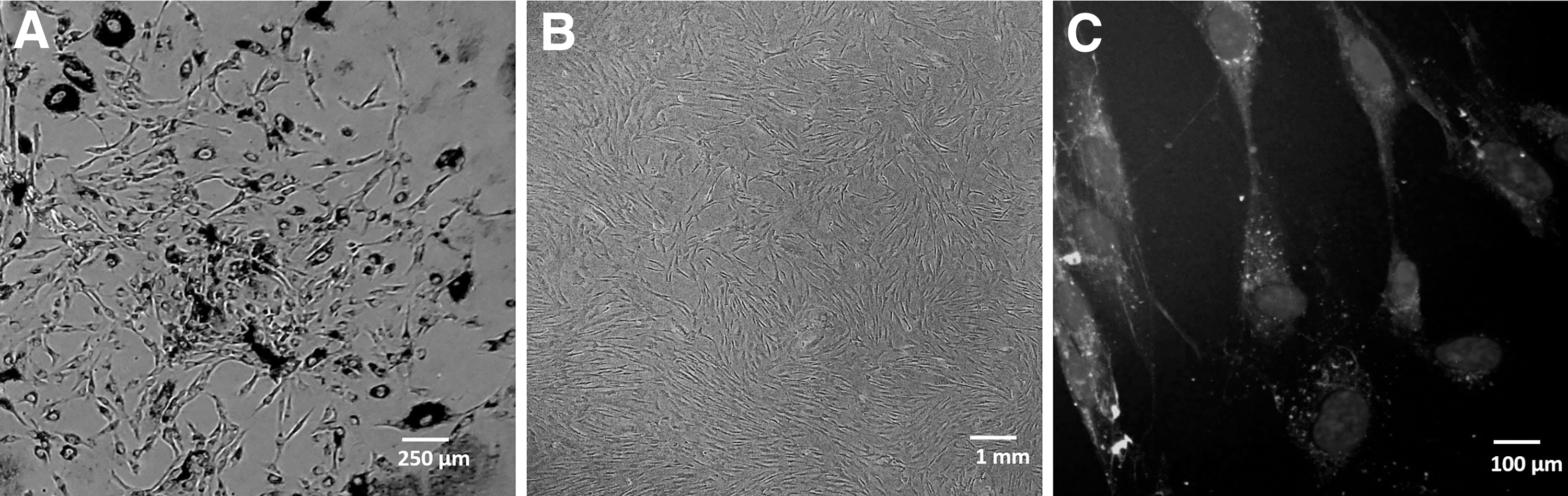
Morphological characteristics of retinal pigmented epithelial (RPE) cells.
RPE gene expression
BCL-2 and BAX relative gene expression
The results of this study showed that BCL-2 gene expression was significantly increased in RPE cells treated with IFN at the dose of 1,000 IU for 48 h (2.12 ± 0.20, P = 0.0001); however, the expression of this gene was not significantly changed in the other treatment patterns. The BAX gene expression showed the same pattern and increased in RPE cells treated with IFN 1,000 IU for 48 h (2.12 ± 0.58, P = 0.025). No statistically significant alteration of BAX expression was observed in any of the treatment patterns (Fig. 2). We also analyzed the ratio of BCL-2/BAX, as the apoptosis index, in the treated RPE cells versus controls; our data showed no significant alteration of BCL-2/BAX ratio from 1:1 in any of the treatment patterns, indicating the in vitro safety of IFN α-2b in RPE cells (Table 3).
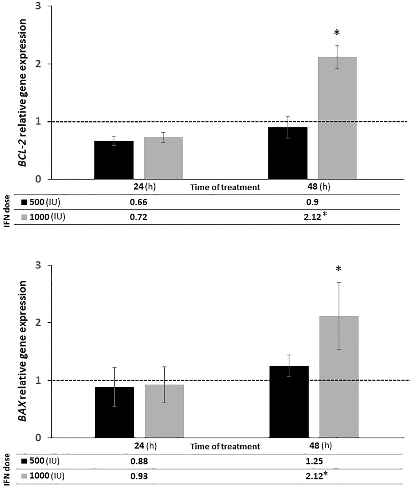
Relative gene expression of BCL-2 and BAX genes in IFN α-2b-treated RPE cells (dark bars) in comparison with controls (light bars) in the 24 and 48 h treatment periods. The mean value of relative expression ± standard deviation is represented in the graph. The asterisk signs (*) bold the statistically significant differences (P < 0.05) by using the LSD post hoc test. IFN, interferon; LSD, Fisher's least significant difference.
BCL-2/BAX Ratio in the Different Dose and Time Treated Retinal Pigmented Epithelial Cells
The mean value of relative expression ± standard error of mean is represented. LSD post hoc test was performed to analyze the statistical significance considering P < 0.05. LSD, Fishe's Least Significant Difference.
BDNF relative gene expression
Our data demonstrated that BDNF gene expression was significantly increased in RPE cells treated for 48 h with 1,000 IU of IFN (5.44 ± 1.34, P = 0.0001). No significant alteration was found in BDNF gene expression of the BDNF gene in RPE cells treated with other patterns (Fig. 3).
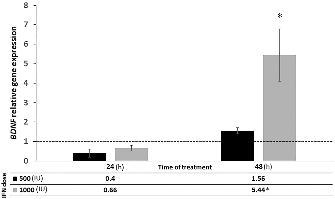
Relative gene expression of BDNF genes in IFN α-2b-treated RPE cells (dark bars) in comparison with controls (light bars) in the 24 and 48 h treatment periods. The mean value of relative expression ± standard deviation is represented in the graph. The asterisks signs (*) bold the statistically significant differences (P < 0.05) by using the LSD post hoc test.
IL-1b relative gene expression
IL-1b expression was significantly upregulated in RPE cells treated with IFN at 1,000 IU (48 h) (3.98 ± 1.21, P = 0.003). The level of the IL-1b gene expression was not significantly changed in RPE cells treated with IFN in the patterns of 500 IU (24 h), 1,000 IU (24 h), and 500 IU (48 h) (Fig. 4).
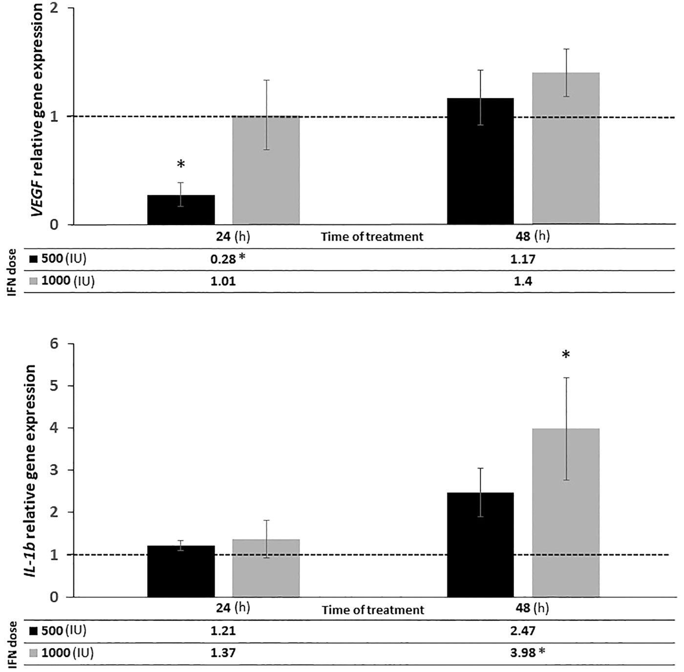
Relative gene expression of VEGF and IL-1b genes in IFN-α-2b-treated RPE cells (dark bars) in comparison with controls (light bars) in the 24 and 48 h treatment periods. The mean value of relative expression ± standard deviation is represented in the graph. The asterisk signs (*) bold the statistically significant differences (P < 0.05) by using the LSD post hoc test.
VEGF relative gene expression
The results of this study showed that RPE cells treated with IFN at the dose of 500 IU for 24 h showed a significant downregulation in the expression level of the VEGF (0.28 ± 0.12, P = 0.023). However, VEGF expression in RPE cells treated with IFN in the patterns of 500 IU (48 h), 1,000 IU (24 h), and 1,000 IU (48 h) was not altered significantly (Fig. 4).
Discussion
IFNs are a group of cytokines with immune regulatory properties (Negishi et al., 2018). Previous studies have shown that IFNs exert their effect at the cellular level by modifying the expression of different genes (Baechler et al., 2003; Moore et al., 2008). Of course, the other regulatory molecules and chemical products may also impress the function of IFNs. Although in vitro studies may not be able to reflect the exact effect of the drug on the body (for reasons such as drug clearance and interaction with other active chemicals in the body), the molecular level in vitro studies could provide a clue as to the mechanism of action of the drug and enabled the design of new treatment strategies.
In experimental animal studies, IFNs are commonly used to treat inflammatory diseases (Crow and Ronnblom, 2019; Prencipe et al., 2018). The antiangiogenic effect of IFNs has also been confirmed in previous studies (Indraccolo, 2010; Yıldırım et al., 2015). Our study showed that the effect of IFN on the expression of genes involved in apoptosis, inflammation, vascularization, and neuroprotection might be significantly affected in a time- and dose-dependent manner. The represented data on the expression of apoptotic genes showed that the ratio of BCL-2/BAX was not significantly changed from 1:1 in any of the doses and timelines; confirming the in vitro safety of IFN α-2b treatment on RPE cells.
BCL-2 and BAX are the main role players in the process of intrinsic apoptosis, which, although closely related, have opposite effects on cell survival and apoptosis. BCL-2, as an antiapoptotic factor, leads to cell survival, however, BAX acts as a proapoptotic protein and promotes programmed cell death (Bagci et al., 2006). Previous studies have shown that the alteration of the BCL-2/BAX ratio from 1:1 can determine the cellular fate of whether or not to go through the process of intrinsic mitochondrial apoptosis (Korsmeyer et al., 1993; Raisova et al., 2001). Our observation confirmed the safety of IFN α-2b on RPE cells, which is in agreement with previous studies (Ghembaza and Lounici, 2018; Qian et al., 2021).
This study also showed that IFN α-2b at the dose of 1,000 IU (48 h) enhanced the neuroprotective effect in RPE cells by increasing the BDNF gene expression. In line with this observation, recently it has been reported that IFN-β increases the level of BDNF both in the peripheral blood mononuclear cells and in the serum of multiple sclerosis patients (Lalive et al., 2008; Shajarian et al., 2021). Therefore, it seems that this drug can be a potential candidate for the management of retinal neurodegenerative diseases.
Most studies, examining the neuroprotective effect of IFNs, have focused on the β and γ IFNs (Ibrahim et al., 2020; Lin et al., 2012). Our study investigated the effect of IFN α-2b on BDNF gene expression and revealed that IFN α-2b treatment leads to overexpression of BDNF in RPE cells treated with 1,000 IU IFN α-2b for 48 h.
The represented study also demonstrated that IFN α-2b treatment, at the dose of 1,000 IU (48 h), could significantly upregulate the expression of the IL-1b gene (coding for an inflammatory cytokine). Despite numerous reports of the inhibitory effect of IFN on IL-1b (Afarid et al., 2020; Afarid et al., 2016; Butler et al., 2012; Celiker et al., 2019; Gillies and Su, 1995; Kertes et al., 1995; Maleki et al., 2018; Reznikov et al., 1998), our study showed that IFN α-2b treatment could stimulate the expression of IL-1b in RPE cells. However, there is also evidence, in agreement with our study, that type I IFN can upregulate IL-1b expression in some bacterial and viral infections (Fernandes-Alnemri et al., 2010; Jones et al., 2010; Mayer-Barber and Yan, 2017; Rathinam et al., 2010; Sauer et al., 2010).
The impact of IFN on immune system is complex and can vary depending on the context and cell type involved (Moll et al., 2011). Although it can suppress the inflammation through upregulation of the expression of interleukin-1 (IL-1) receptor antagonist, which is a natural inhibitor of IL-1 signaling, it is also reported to be able to upregulate the expression of IL-1b, which is a proinflammatory cytokine involved in the initiation and maintenance of immune responses (Mayer-Barber and Yan, 2017). The mechanism by which IFN α-2b upregulates IL-1b gene expression is not completely understood.
However, IFN α-2b may upregulate the expression of IL-1b gene through multiple potential mechanisms, including the activation of transcription factor NF-κB, which can bind to the promoter region of the IL-1b gene and enhance its transcription; the induction of Toll-like receptor 4 (TLR4) expression, which can activate the transcription factor AP-1 and increase IL-1b transcription; and the stimulation of other proinflammatory cytokines, such as TNF-α and IL-6, which can indirectly promote IL-1b production (Georgel, 2021; Schroder et al., 2004).
VEGF is another key role player in the progression of DR. VEGF is markedly increased in the process of inflammation and ischemia (Al-Kharashi, 2018; Rossino et al., 2019). The antiangiogenic feature of IFNs has been previously reported (Indraccolo, 2010; Yıldırım et al., 2015). In confirmation with previous studies (Cao et al., 2008; Maleki et al., 2018; Wu et al., 2005), this study showed that IFN α-2b treatment reduced VEGF expression in RPE cells treated with 500 IU IFN α-2b for 24 h.
A previous study has shown that IFN α-2b can improve the function of retinal microvascular endothelium in vitro and promotes tissue homeostasis, indicating the IFN α-2b potential for the treatment of ocular diseases associated with vascular endothelium leakage (Gillies and Su, 1995). These findings were also approved by recent clinical reports (Faghihi et al., 2022). Our study also confirmed the previous reports and showed that IFN α-2b could exert anti-VEGF effect in RPE cells. However, it should be noted that according to the present data, the antiangiogenesis of IFN is transient and disappears over time.
Considering the increased expression of the IL-1b gene in the long-term treatment, it seems that the long-term treatment pattern might upregulate inflammation. Therefore, it is suggested that the antiangiogenesis property of IFN applies to a short-time treatment, and increasing the duration of treatment may exacerbate the disease, particularly in patients with advanced inflammation.
Conclusion
In conclusion, the data of this study hypothesize that IFN α-2b is a safe drug for the treatment of RPE complications. IFN α-2b in lower doses and short duration may have antiangiogenic effects, and in higher doses with longer duration exerts neuroprotective and inflammatory effects. Hence, appropriate concentration and duration of treatment, according to the type and stage of the disease, should be considered to achieve success in IFN therapy. These findings might have valuable applications in different clinical conditions.
Footnotes
Acknowledgment
The authors thank the vice-chancellor of Shiraz University of Medical Sciences for supporting this research (
Authors' Contributions
F.S.-J. and M.A. conceived and designed the study; F.S.-J. performed the experimental analysis; F.S.-J. and M.A. analyzed the data; and F.S.-J., H.B., and M.A. wrote the main article and revised the final version. All authors reviewed the article.
Author Disclosure Statement
The authors report no commercial or proprietary interest in any product or concept discussed in this article.
Funding Information
This study was supported by Shiraz University of Medical Science (Grant No. 20767), Shiraz, Iran.