
Abstract
The present study evaluated the efficacy of fennel seed methanolic extract (FSME) for its antioxidant, cytotoxic, and antitumor activities and for its capacity to serve as a nontoxic radioprotector in Swiss albino mice. We also assessed the natural antioxidant compounds of FSME for use in industrial application. Cytotoxic activity of FSME was evaluated in a mouse model of Ehrlich ascites carcinoma (EAC) and on different types of human cell lines in vitro. The safety and optimum dose of FSME were determined. FSME, 100 mg/kg, was injected intraperitoneally into mice bearing EAC before the mice were exposed to three 2-Gy doses of gamma irradiation. After 30 days, mice were fasted for 18 hours and then sacrificed to observe the lifespan of EAC-bearing hosts. Malondialdehyde (MDA), catalase activity, glutathione content, and total protein in serum, liver tissue, and ascitic fluid were determined. Iron, total iron-binding capacity, transferrin, and ferritin were also evaluated in serum. The data showed the presence of different types of compounds in FSME, such as flavonoids, terpenoids, alkaloids, phenols, and sterols; estragole (71.099%) was the predominant alcohol, gallic acid was the phenolic compound (18.895%), and L-limonene was the most prevalent monoterpene hydrocarbon (11.967%). The mean±standard deviation 50% inhibitory concentrations were 50±0.03 μg/mL for the MCF7 breast cancer cell line and 48±022 μg/mL for the Hepg-2 liver cancer cell line. The significant increase in MDA levels and the significant decrease in catalase activity and glutathione content in liver and tumor tissue in mice bearing EAC were ameliorated after FSME administration. In contrast, total protein content was increased in ascitic fluid. Serum iron was inversely proportional to the levels of ferritin and transferrin and total iron-binding capacity. Administration of FSME before irradiation exerted a cytoprotective effect against gamma irradiation, as manifested by a restoration of the MDA level, catalase activity, and GSH content to near-normal levels. In conclusion, FSME may have remarkable anticancer potential against a breast cancer cell line (MCF7) and liver cancer cell line (Hepg-2). It also showed strong free radical–scavenging activity (100%). Thus, FSME may reduce oxidative stress and protect mouse cells from damage caused by reactive oxygen species. In addition, it could be used as a safe, effective, and easily accessible source of natural antioxidants to improve the oxidative stability of fatty foods during storage. FSME also exhibited an antitumor effect by modulating lipid peroxidation and augmenting the antioxidant defense system in EAC-bearing mice with or without exposure to radiation.
Introduction
F
Drugs from natural origins have recently been studied as chemoprevention agents to inhibit tumor genesis; as a result, the natural extracts or purified compounds have become well established means for studying various aspects of cancer. 9 Fennel essential oil has a potent hepatic-protective action against carbon tetrachloride–induced hepatic damage in rats. 10 Volatile components of fennel seed extracts by chromatographic analysis include trans-anethole, fenchone, methyl chavicol, limonene, α-pinene, camphen, β-pinene, β-myrcene, α-phellandrene, 3-carene, camphor, and cis anethole. 10
Bharat and Shishir 11 reported that anethole is the active component in anise, camphor, and fennel and that the molecular targets modulated may be the basis for how these dietary agents not only prevent but also treat cancer and other diseases. Moreover, Al-Harbi et al. 12 studied the anticarcinogenic potential of anethole in Ehrlich ascites tumor (EATs) in the paws of Swiss albino mice. This study revealed that anethol increases survival time and reduces tumor weight and volume and body weight of the EAT-bearing mice. Anethol had a significant cytotoxic effect on EAT cells in the paw, reduced the levels of nucleic acids and malondialdehyde (MDA), and increased nonprotein sulfhydryl concentrations. 12 In addition, Singh et al. 13 described 7 phenolic acids in fennel extract, all with antitumor activity: tannic, gallic, caffeic, cinnamic, chlorogenic, ferulic, and vanillic acids. Gallic acid also exhibits antimicrobial activity. Fatima et al. 14 reported that fennel seeds extract protects against diethyldithiocarbamate-induced liver toxicity in rats.
The present study evaluated the efficacy of fennel methanolic extract as an agent with antiantioxidant, cytotoxic, and antitumor activity and as a nontoxic radioprotector in Swiss albino mice. We also assessed the natural antioxidant compounds of fennel seed methanol extract (FMSE) for industrial application
Materials and Methods
Drug and chemicals
Fennel seeds (Foeniculum vulgare) were provided by an herbal orchard in Sohag Governorate, Egypt. Refined sunflower oil (antioxidant free) was purchased from Misr Oil and Soap Co., El Mansoura, Egypt. Butylated hydroxy toluene and Tween 80 and 5-fluorouracil (5-FU) were obtained from Sigma Chemicals Co., Steinheim, Germany. Ehrlich ascites carcinoma (EAC) and human tumor cell lines were obtained from National Cancer Institute, Cairo University, Cairo, Egypt.
Diets
The experimental diet (g/kg) was prepared according to the formula described by Kim et al. 15 It included the normal diet for mice (fat 5%, carbohydrates 60%, protein 20.3%, L-cysteine 2.5%, cellulose 5%, mineral mixture 3.7%, vitamin mixture 1%, and choline bitartrate 2.5%) and was purchased from El-Gomhoria Co., Cairo, Egypt.
Diet consumption was calculated at the same time each day by subtracting the amount of diet left over in each cage barrier from the amount of diet provided on the previous day. The average diet consumptions were represented as g/day/mouse which were calculated according to the following equation:
Experimental animals
One hundred white albino mice weighing a mean±standard deviation of 24±6 g were studied to determine the acute toxicity of FSME at 0, 50, 100, 250, 500, 1,000, and 2,000 g/kg body weight.
Eighty adult female Swiss albino mice weighing 18–20 g were studied used to determine the optimum radiation dose with FSME. We used radiation doses of 6, 10, and 12 Gy alone as well as with FSME, 100 mg/kg body weight.
Three hundred female Swiss albino mice weighing 18–20 gram were studied to determine the radioprotective and antitumor effects. All mice were supplied by the National Cancer Institute, Cairo, Egypt. They were housed in polypropylene boxes in a controlled environment (temperature 23°C±2°C and 12-hour/12-hour dark/light cycles) with standard laboratory diet and water ad libitum. These conditions were in accordance with the guidelines of the Institutional Ethical Committe at the National Cancer Institute, Cairo, Egypt. The mice were housed 2 weeks before being used for experiments. After treatments, mice were kept in similar conditions for 30 days. Food consumption per group, as well as the body weight of each mouse, was monitored daily at the beginning of the experiment (day 1) and sequentially on days 5, 9, 14, and 30 (the last day of the experiment).
Experimental cell lines
All cell lines under investigation were supplied from the National Cancer Institute in Cairo. The following cell lines were used: MCF-7 (breast carcinoma cells), Hepg-2 (liver carcinoma cell line), HT-29 (adenocarcinoma of human colon cell line), Hela (cervical carcinoma cell line), H460 (lung carcinoma cell line), and U251 (brain tumor cell line).
Radiation facility
Whole-body gamma irradiation was performed by using a gamma cell Co-60 unit installed at the Middle Eastern Regional Radioisotopes Center for the Arab Countries, Giza, Egypt.
Extraction of volatile oil
One hundred grams of ground seeds were suspended in 1.5 L of distilled water. Continuous steam distillation extraction was performed for 3 hours, and the oil was collected and stored at 4°C until used.
Identification of volatile compounds
Volatile compounds contained in the extracted fennel oil were identified by using gas chromatography and gas chromatography–mass spectroscopy (GC-MS) at the Central Laboratory of Kato Aromatic Co., Cairo, Egypt. GC was performed with a Hewlett-Packard gas GC (Model 5890 Series II) equipped with a flame ionization detector and coupled to an electronic integrator. The chromatograph was fitted with a methyl silicone column (20 m×0.2 mm; 0.33-mm film thickness). GC analytical conditions were as follows: carrier gas, helium; flow rate, 1 mL/min; injector and detector temperatures, 200°C and 250°C, respectively; and oven temperature, programmed to increase from 60°C to 200°C at a rate of 3°C/min. Quantitative data were obtained by electronic integration of flame ionization detector area data without the use of response factor correction. GC-MS was performed by using a Hewlett-Packard MS 5970 equipped with a total ion chromatogram mass detector. The operation conditions were as follows: carrier gas, helium; ionization voltage, 70 eV; scanning speed, 1 second over an amu range of 20–550; and electron multiplier voltage, 1800 eV. The column and conditions were the same as described for GC except that the injector temperature was 150°C. Separated peaks were identified by matching with data from the library of mass spectra and compared with those of standard compounds and published data. 16 The quantitative determination was performed according to peak area integration.
Preparation of methanolic fennel extract
FSME was prepared by adding 300 mL of methanol water (1:1) to 50 g of fennel seed powder for 10–12 hours. The extract was filtered by using a 0.22-μm membrane, and the solvent was evaporated to near dryness under vacuum with a rotary evaporator. The resultant extract was pooled, dehydrated, and lyophilized in an oven at 50°C for 24 hours; it was then stored at 4°C until assessment of its antioxidant activity. The extract was emulsified in Tween-80 and diluted with ethanol to the appropriate concentration. It was intraperitoneally injected at different concentrations 24 hours before irradiation to allow enough time for accumulation in the bone marrow.
Extraction, determination, and identification of phenolic compounds
Phenolic compounds from fennel seeds were extracted according to the method reported by Marie et al., 17 with methanol used as a solvent
Total phenolic compounds of FSME was determined by using the Folin–Ciocalteu reagent, with gallic acid as a standard, according to the method described by Miliauskas et al. 18 The phenolic compounds were estimated as mg gallic acid/100 g of dry weight material.
Phenolic compounds of FSME were identified by using high-performance liquid chromatography (HPLC) with Hewlett-Packard Model 1100 according to the method described by Gertz. 19
Assessments of antioxidant activity
The stable free radical species content of fennel seed powder was measured by using electron spin resonance (ESR) at the Central Laboratory of the National Research Center, Dokki, Egypt. The free radical content of the seeds was measured by double integration area.
1,1-diphenyl-2-picrylhydrazyl (DPPH) radical scavenging assay
Radical scavenging activity of FSME against stable DPPH was measured according to the method described by Ramadan et al. 20 by using a Brucker Elexsys-500 operated at X-band frequency. The following parameters are generalized to all samples: microwave frequency: 9.79821 GHz; receiver gain: 60; microwave power: 0.00202637 W; sweep width: 6000, center at 3480. Briefly, the sample was measured at room temperature in an ultra-pure silica tube.
DPPH solution, 10−3 μg/mL in methanol, was used as a standard. One milliliter of DPPH solution plus 1 mL of methanol was used as a standard for comparison with a mixture of 1 mL of methanolic extract plus 1 mL of DPPH solution. Double integration area signal of free radical was measured before and after addition of DPPH to the sample, and the difference between the two values over the original DPPH value was taken as a measure of antioxidant activity.
Oxidative stability of sunflower oil
The influence of dried FSME on the oxidative stability of sunflower oil was evaluated by the rancimat method using 679 Rancimat (Metrohm), as described by Hasenhuttle et al. 21 One gram of dried FSME was mixed well with 100 mL of sunflower oil and filtered through filter paper, then added at levels of 0.01%, 0.02%, 0.03%, 0.05%, and 0.07% (v/v). It was mixed well with sunflower oil using a magnetic stirrer to complete dispersion in the oil.
BHT (0.02% w/v) and sunflower oil sample without any addition were used as control. The induction period was conducted with Rancimat at 100°C and calculated at 25°C; the temperature coefficient of 2.2 was used for the induction period according to the method reported by Hadorn et al., 22 and 2.5 was used for the expired period according to the method reported by Pardun et al. 23 Increasing index was the difference between the induction period of the addition of methanolic extract and the induction period of the control.
Measurements cytotoxicity by sulphodiamine-B assay
The cytotoxic assay was performed at the Pharmacology Unit, Cancer Biology Deptartment, National Cancer Institute, using the sulforhodamine B assay described by Skehan et al. 24 The stock cells maintained in 75 cm2 polystyrene flasks (Falcon) with minimal essential medium containing 10% fetal bovine serum, 1 mM sodium pyruvate and 1.5 g/L sodium bicarbonate, penicillin 100 IU/mL, streptomycin 100 μg/mL, and amphotericin B 5 μg/mL in a humidified atmosphere of 5% CO2 at 37°C. The cells were dissociated with 0.2% trypsin in phosphate-buffered saline (PBS) solution. The stock cultures were grown in 25-cm2 tissue culture flasks, and all cytotoxicity experiments were carried out in 6 well plates. Cell lines in exponential growth phase were washed with PBS solution and trypsinized and resuspended in complete culture media. Cells were equal to 3×10 4 cells/well and were plated in 96-well plates for 24 hours before treatment with FSME to allow attachment of the cells to the wall of the plate. The cells were incubated for 24 hours, during which time a partial monolayer formed. After incubation, the cells were exposed to various concentrations (25, 50, 125, 150, 250, 500, and 1,000 μg/mL) of FSME. The control well received only maintenance of medium. Triplicate wells were prepared for each individual dose. The monolayer cells in the plates were incubated at 37°C in a humidified incubator with 5% CO2 for 24 hours. Morphologic changes in the drug-treated cells were examined by using an inverted microscope and compared with the control cells. After 48 hours, the cells were fixed, washed, and stained with sulforhodamine B stain according to methods described by Freshney. 25 Excess stain was washed off with acetic acid, and the attached stain was recovered with Tris-EDTA buffer (pH, 9.0). Color intensity was measured in an enzyme-linked immunosorbent assay reader. The relationship between the surviving fraction and the drug concentration was plotted to determine the survival curve for each tumor cell line. The 50% inibitory concentrations (IC50) were derived from a nonlinear regression model (curve fit) based on sigmoidal dose-response curve (variable) and were computed by using GraphPad Prism software, version 4.00. Data were expressed as mean±standard error. All tests and analyses were run in triplicate, and mean values were recorded.
Measurements antitumor activity in vitro
The antitumor activity of FSME was determined by using EAC in vitro in accordance with the method described by McLimans et al.
26
Use of EAC cells was based on the finding that it is an excellent tool for studying the biological behavior of malignant tumors and drug action within cells.
27
Sterile test tubes were used, and 2.5×10⁵ tumor cells per mL were suspended in 2 mL PBS. The following amounts of FSME were added to the suspension and kept at 37°C for 2 hours: 50,100, 200, 400, 800, 1,000, and 2,000 μg/mL. The trypan blue dye exclusion test was then performed to calculate the percentage of nonviable cells. The antitumor activity of FSME in vitro was determined to calculate the percentage of nonviable cells. The following equation was used:
Determination of acute toxicity of FSME
A group of male albino mice (10 mice/group) were injected intraperitoneally with increasing doses of FSME (50–1,000 mg/kg body weight). Mortality, hypoactivity or hyperactivity, lethargy, and food intake were recorded daily. The dose–mortality curve was constructed and the 50% lethal dose (LD50) was graphically determined by using the Lorke 28 method.
Survival dose-response curves for radiation
Studies of survival in both control and FSME-treated mice (30 mice in each group) used whole-body gamma radiation in different doses: 6, 10, and 12 Gy. Mice were checked for mortality daily for 30 days. Regression analysis was performed to obtain the lethal dose required to kill 50% of mice in 30 days (LD50/30) and to determine dose reduction factor.
In vivo assays
Radio-protective and antitumor studies
Experimental design
Three hundred fifty female Swiss albino mice weighing 18–20 g were divided into 7 groups (30 mice in each).
Group I: Animals received 0.2 mL emulsified 1% Tween80 in ethanol (intraperitoneally); this mice served as the vehicle group (negative control).
Group II: Animals were inoculated intraperitoneally with 2.5×10⁶ EAC tumor cells. These mice made up the positive control group.
Group III: Mice were intraperitoneally inoculated with 2.5×10⁶ EAC cells; 24 hours after inoculation, they were intraperitoneally injected on days 1, 5, and 9 with 100 mg/kg FSME.
Group IV: Mice were exposed to 2 Gy of fractionated gamma irradiation once a day on days 2, 6, and 10, up to a cumulative dose of 6 Gy. They received an equal volume of Tween-80 intrapretanially.
Group V: On days 1, 5, and 9, mice received an intraperitoneal injection of FSME (100 mg/kg). Twenty-four hours after each treatment with FSME, the animals were irradiated at a dose of 2 Gy/d (days 2, 6, and 10). The total irradiation dose was 6 Gy.
Group VI: Mice were inoculated with 2.5×10⁶ EAC cells. Starting 24 hours after inoculation, mice were intraperitoneally injected with FSME (100 mg/kg) on days 1, 5, and 9. Twenty-four hours after each injection of FSME, the animals were irradiated at a dose of 2 Gy/d (days 2, 6, and 10). The total irradiation dose was 6 Gy.
Group VII: Mice were inoculated with 2.5×10⁶ EAC cells and received 5-FU intraperitoneally (20 mg/kg) on days 1, 5, and 9. After 24 hours mice were irradiated at adose of 2 Gy at days 2, 6, and 10. This group served as the positive control.
Mice were exposed to fractionated gamma irradiation delivered as 2 Gy once a day on days 2, 6, and 10, to a cumulative dose of 6 Gy. Irradiation was delivered over a 15-day period in perforated Plexiglass chamber (25×25 cm) with a distance of 77.5 cm from the source. Irradiation was delivered at a dose-rate of 1.33 Gy/min. To overcome the resistance of hypoxic cells present in tumors to radiation therapy, fractionated doses of gamma irrradiation were used. All groups were observed daily for up to 30 days, and changes in body weight were recorded on days 1, 5, 9, 14, and 30. Animals were monitored for mortality and any sign of sickness and behavioral toxicity. The animals were killed and dissected 30 days after the first irradiation dose.
Sampling collection
At the end of the experimental period, mice were fasted overnight, anesthetized under diethyl ether (Sigma Co.), and then killed. After growth of the EAC tumor cells, ascitic fluid was collected. Blood was collected in dry clean tubes by using the retro orbital plexus method. The blood percolates along the wall of the centrifuge tube to prevent risk for hemolysis. The blood tubes were left for 30 minutes and then centrifuged at 3000 rpm for 30 minutes. After centrifugation, the serum was separated at once, divided into aliquots, and stored at −80°C until used in the biochemical analysis. The liver tissues were excized and homogenized in bi-distilled water using a Potter–Elvehjem homogenizer under cooling and sterilized conditions and preserved at −80°C until used.
Biochemical assays
Sampling of fluid exudate
Ascitic fluid was collected from the peritoneal cavity of normal and treated mice by needle aspiration under aseptic conditions. The ascitic fluid was diluted with sterile saline so that 0.1 mL contained 2.5×10⁶ cells. Fluid was then left at −80°C until used.
Liver tissues and fluid exudate analysis
In liver tissue homogenates and ascitic fluid, glutathione (GSH) content was determined according to the method of Beutler et al. 29 Lipid peroxide content was determined by quantifying MDA according to the method of Yoshioka et al. 30 Catalase activity and total protein in tissue samples were determined according to the methods described by Chance et al. 27 and Lowery et al., 31 respectively.
Serum analysis
Blood was drawn from each mouse by using the retro orbital plexus method. Serum iron and iron-binding capacity (TIBC) were determined according to the colorimetric methods described by Haper et al.
32
Ferritin was assayed by radioimmunoassay techniques using commercial kits that depended on solid-phase radioimmunoassay (Coat-A-Count, Diagnostic Product Corp). Transferrin was assessed according to measurements of TIBC by using the following formula designed by Kalantar-Zadeh:
33
This formula assumes that 1 mg of transferrin may bind 1.25 mg of iron as a maximum.
Antitumor studies
The antitumor effect of FSME was assessed by estimating the ascitic tumor volume and packed cell volume (pcv) of the ascitic fluid. The fluid was collected from the peritoneal cavity of normal and treated mice by needle aspiration under aseptic conditions, pcv was measure in a graduated centrifuge tube after centrifugation at 1000 rpm for 5 minutes. Part of the ascitic fluid was diluted with sterile saline so that 0.1 mL contained 2.5×10⁶ cells, and this solution was used to determine the viable and nonviable tumor cell count.
Viable/nonviable tumor cell count
To count the tumor cells in ascitic fluid, we used a white blood cell pipette and diluted the sample 100 times. A drop of the diluted cell suspension was placed in a Neubauer counting chamber, and the number of cells in the 64 small squares was counted. The cells were then stained with trypan blue (0.4% in normal saline) dye. The cells that did not take up the dye were viable and those that took the stain were nonviable. These viable and nonviable cells were counted according to the following formula:
The antitumor effect of FSME was also assessed by evaluating the changes in the weight of the solid tumor and the tumor growth inhibition ratio, calculated according to the following formula:
34
Statistical analysis
We used SPSS software, version 12, for statistical analyses. Quantitative variables were summarized by using means and standard deviation. Comparisons between values before and after treatment within each group were analyzed with a paired t-test and 1-way analysis of variance for multiple comparisons. Differences were considered significant when the P value was less then .05 and highly significant when the P value was less than .01. 35
Results
Separation and identification of the chemical compounds of FSME
The components of volatile oil fractions of fennel obtained by traditional steam distillation were separated by GC, and the produced peaks were identified by using GC-MS. Table 1 lists the concentrations of the fennel volatile oil components.
More than 44 components were separated, and about 19 components were identified in volatile fennel oil. Alcohols were the major group identified, followed by monoterpene hydrocarbons (79.278% and 16.473% of peak area, respectively). The predominant alcohol was estragol, which was also the major component of volatile fractions. It had the highest peak area percentage, 71.099 % (Table 1 and Fig. 1). Bicyclo[3,3,1]non-2-en-9-ol,9-methyl and P-menth-3-en-1-ol were the major monoterpene alcohols, with percentages of 1.944% and 0.726%, respectively. Hydrocarbons were the second major group identified. The oxygenated derivatives commonly named terpenoids are considered an important flavor compound. L-limonene was the most common monoterpene hydrocarbon, representing 11.967% of the peak area. Other substances were detected as follows: benzaldede, 1.944%; benzene,1-methyl-4-(1-methylethyl), 1.6781%; α-pinene, 0.8202%; E-citral, 0.313%; anethole, 0.2718%; l-fenchone, 0.1378%; β-pinene, 0.1257%. Fennel volatile oil was rich in oxygenated compounds and poor in terpene hydrocarbons.

Gas chromatography–mass spectrometry for fennel seed volatile oil constituents.
Antioxidant content of FSME
The total volatile oil and phenolic compound contents of FSME are shown in Table 2. Volatile oil was the most important component of fennel seeds and represented about 1.825%±0.189%, with a specific gravity of 0.9222±0.132 gm/cm3. Table 2 also shows that FSME had a total phenolic compound content of 29.64 mg/g dry matter (gallic acid equivalent), and the total dried methanolic extract was 7.02%. These results indicated that FSME contains a high level of total phenolic compounds and thus may contribute to retardation of lipid peroxidation as a natural antioxidant.
Data expressed as mean±SD.
Radical scavenging activity of FSME
Fennel seed dried powder (as solid) was measured by ESR to determine the content of the stable free radical (Fig. 2). Figure 2 shows that fennel seed powder had double integration area of 8.095 arbitrary units. This factor indicates the quantity of stable free radical in fennel seed powder and can be considered a fast measure of antioxidant activity. In addition, the radical scavenging activity of FSME was measured by ESR using DPPH assay (Fig. 3). FSME almost completely inhibited DPPH absorption, and the radical scavenging activity was found to be 100% (Table 2) at a concentration of 29.64 mg/g total phenolic compounds dry matter.

Stable free radical species content of fennel seeds.
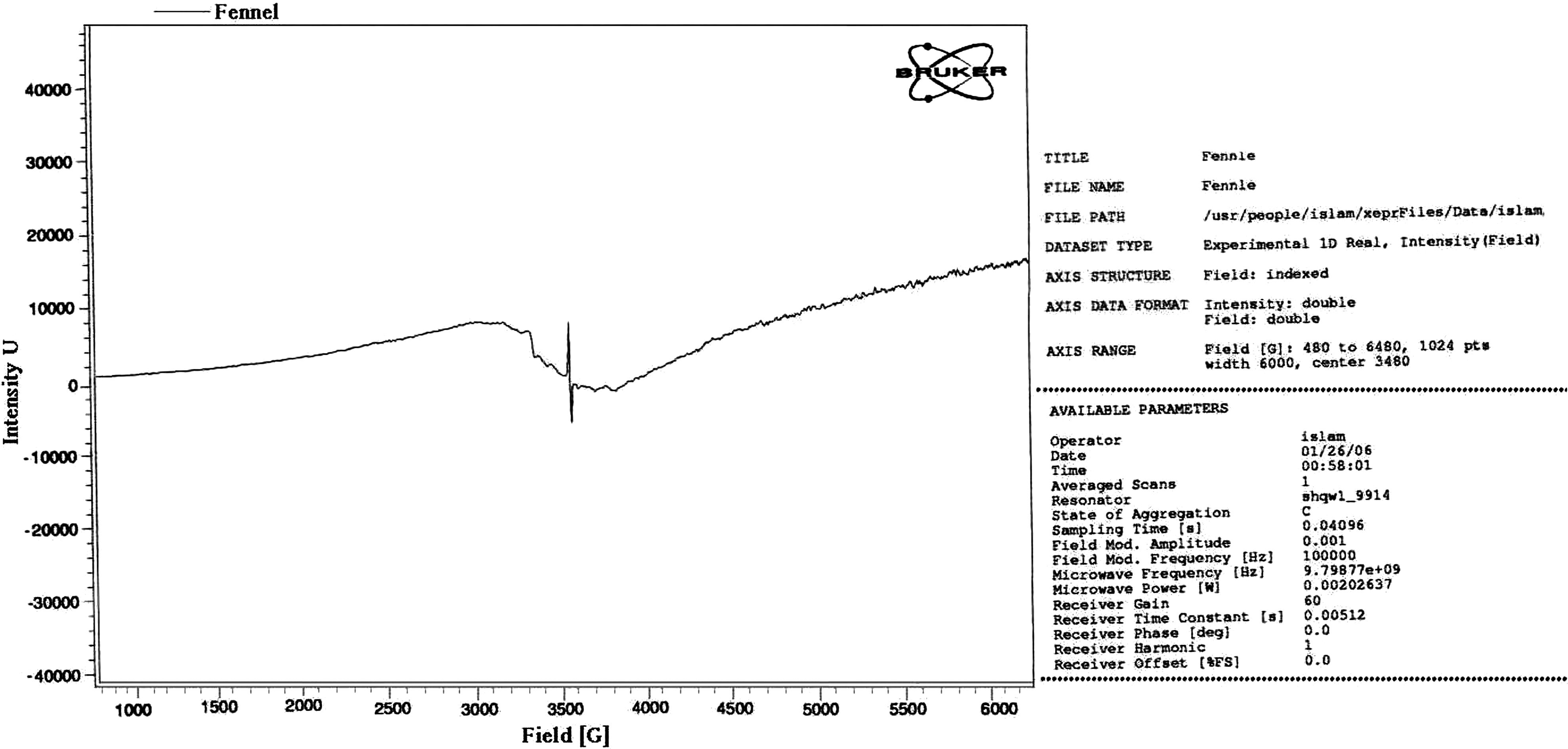
Free radical scavenging activity of fennel seed methanolic extract.
Phenolic compounds identification in FSME
Total phenolic compounds of FSME were separated and identified by HPLC (Table 3). Table 3 shows that five phenolic compounds were separated and four of them were identified. Gallic acid was the most abundant phenolic compound in fennel seeds (18.895 mg/g). Kaempferal, chlorogenic acid, and protocatechuic were found in small amounts (0.123, 0.117, and 0.102 mg/g, respectively). The high level of phenolic compounds seems to be related to the antioxidant activity.
Effect of FSME on oxidative stability of sunflower oil
Table 4 shows the role FSME at levels of 100, 200, 300, 500, and 700 ppm in the shelf-life or oxidative stability of sunflower oil. The results reveal that BHT had the highest antioxidant activity (1.24) compared with FSME levels. Furthermore, the oxidative stability of sunflower oil was increased with increasing levels of FSME until 700 ppm was reached. That concentration exhibited the best oxidative stability. Shelf-life (10.43 to 12.12 months) and antioxidant activity (1.03 to 1.20) of sunflower oil increased gradually as the FSME levels increased.
Antioxidant activity=induction period of sample/induction period of control.
BHT, butylated hydroxy toluene.
In vitro assay
Cytotoxic activity of FSME
Table 5 shows the in vitro screening of the FSME among the 6 tested cell lines. The breast cancer cells (MCF-7) and liver cancer cell line (HEPG-2) were more sensitive than the other cell lines.
MCF7 was breast carcinoma cells; Hepg2 was liver carcinoma cell line; HT-29 was adenocarcinoma of human colon cell line; H460 was lung carcinoma cell line; Hela was cervical carcinoma cell line; and U251 was brain tumor cell line. Each IC50 value represents mean±SEM of 6 replicates.
IC50, Dose that reduces survival to 50%.
Evaluation of antitumor activity in vitro
Activity of FMSE at levels of 25, 50, and 100 μg/mL against EAC was evaluated (Table 6). A level of 25 μg/mL was sufficient to render 80% of the cells nonviable.
FSME, fennel seed methanolic extract.
In vivo assay
Acute toxicity of FSME
As shown in Table 7 and Figure 4, FSME caused no deaths, and no significant difference in body weights were seen up to a dose of 100 mg/kg. However, some side effects occurred (loss of appetite and piloerection) at FSME doses up to 500 mg/kg.
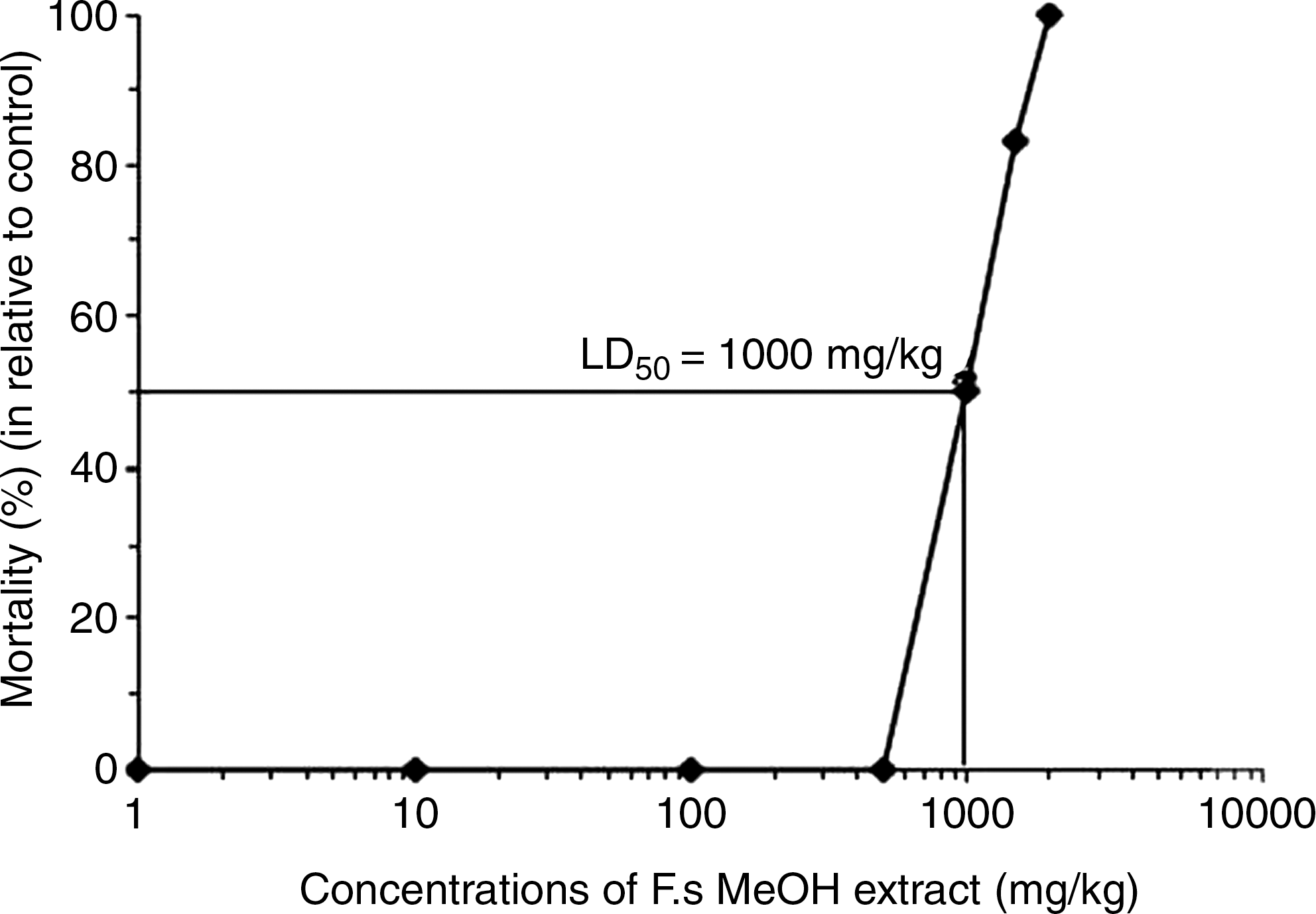
Dose–mortality curve of mice treated with fennel seed methanolic extract.
All treated animals were carefuly examined for signs of toxicity.
Time elapsed between dosing and death.
At a dose level of 1000 mg, higher mortalities were observed. In general, side effects were observed at 24 hours after injection (sedation, movement disorders, unresponsiveness to external stimulation, hind limb weakness, and transient hyperactivity occurred). In mice that survived the treatment, these symptoms disappeared one week after injection.
Acute toxicity in mice receiving FSME and radiation
As shown in Figure 5, regression analysis revealed that 100 mg of FMSE per kg body weight was the optimum dose against 6 Gy of radiation. No deaths occurred with this dose, and the maximum survival rate was 70%. The radiation dose rate of 6 Gy was selected on the basis of maximum survivability. The survival percentage was significantly increased in FSME-treated mice subsequently exposed to irradiation and in FSME-pretreated irradiated mice. When survival data were fit on a regression line equation, the LD50/30 values for control (irradiation only) and experimental (FSME plus irradiation) groups were computed as 6.47 and 11.23 Gy, respectively. On the basis of these LD50/30 values, FSME pretreatment produced a dose reduction factor of 14.3.
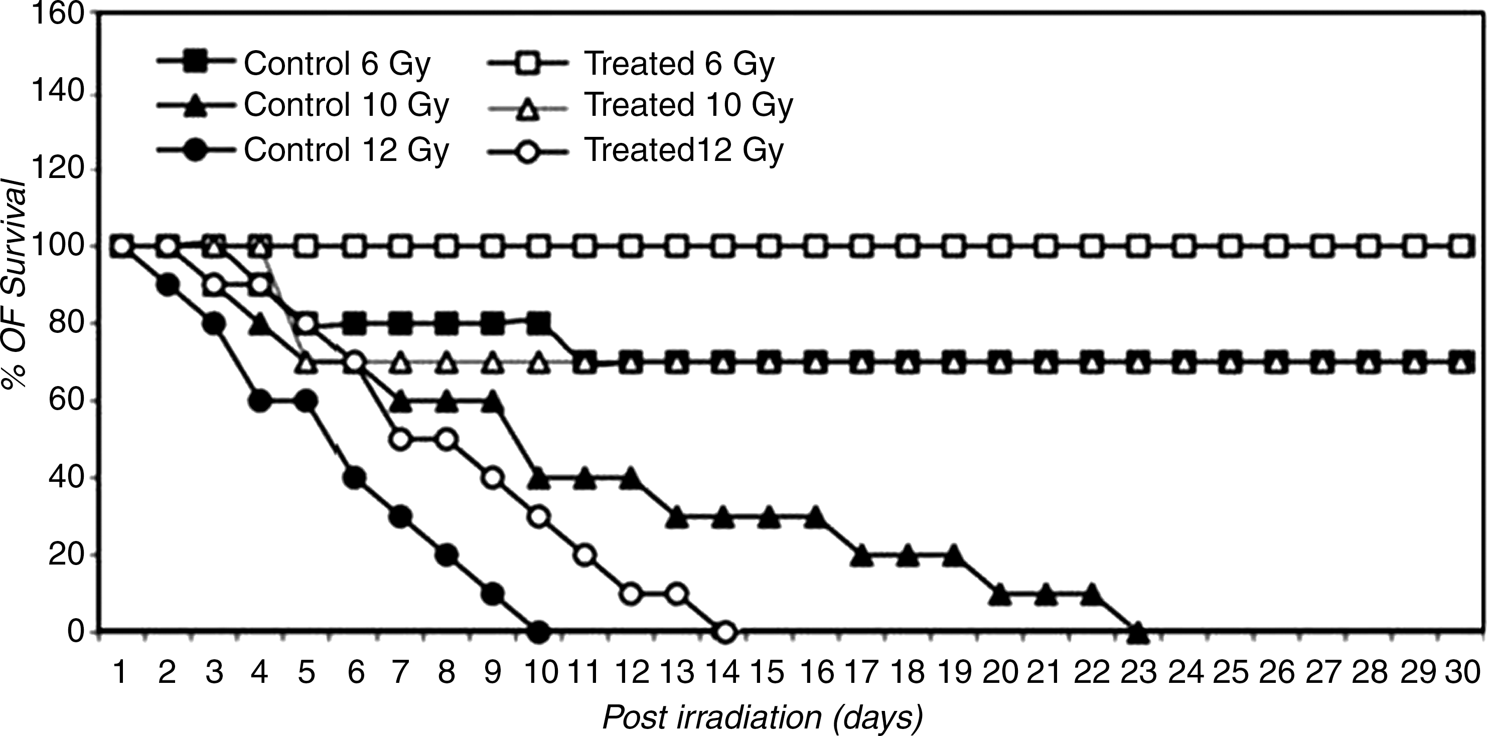
Survival–dose–response curves for determination of 50% lethal dose at 30 days. Control mice were irradiated and not treated with fennel seed methanolic extract; other groups of mice were irradiated and injected intraperitoneally with 100mg/kg fennel seed methanolic extract. All mice were checked daily until day 30.
Inhibition of radiation- and EAC-induced change in total body weight by FSME
As shown in Table 8, normal mice showed a progressive increase in body weight equivalent to the normal growth of mice (20.72–26.23 g) at the end of the experiment. Mice bearing EAC showed a rapid increase in body weight (20.03–30.41 g) which is equivalent to normal growth plus the increase due to the accumulation of ascitic fluid.
Values are expressed as the mean±SD.
Significant at * P<.01, ** P<.001, *** P<.0001.
5-FU, 5-fluoroucil; EAC, Ehrlich ascites carcinoma.
At day 14 the mortality rate was 100%. Irradiated mice showed a progressive decrease in body weight, and on day 30 the mortality rate was 100%. However, in all groups treated with FSME, the survival rate was improved, no deaths had occurred on day 30 (the end of the experiment). As in Table 9 the group administered EAC and FSME had significantly lower mean food consumption than the control group according to Eq. (1). The group administered EAC or radiation alone also had significantly lower food consumption compared with the control and EAC plus FSME groups.
Values are expressed as the mean±SD.
Means having different letter are significantly different, with a P<.05 significant, b P<.001 highly significant.
Table 10 shows the percentage of tumor growth in the group inoculated with EAC and treated with FSME. The decrease in this group, 5.54%, is considered more significant (P<.01) than the changes in the other treated groups. The percentage of inhibition of tumor growth, 94.46%, was significantly greater (P<.01) than the inhibition in the other groups.
Data are expressed as means±SD. Each group consisted of 30 animals.
5-FU, 5-fluoroucil; EAC, Ehrlich ascites carcinoma; FSME, fennel seed methanolic extract.
P<.01 compared with EAC control group.
Table 11 indicates that fractionated gamma irradiation at a total dose of 6 Gy significantly decreased the levels of GSH in liver tissue homogenates and ascitic fluid. The MDA levels and catalase activity in liver homogenate tissues and ascitic fluid provide a rough index for a balance between free radical generation and scavenging. MDA content in liver and tumor tissues was significantly elevated among the mice bearing tumor; this change was accompanied by an imbalance in lipids in the tumor tissue compared with the peroxidation level and antioxidant system of the control group. Moreover, the mice bearing tumor exhibited a highly significant decrease (P<.001) in catalase activity in liver and tumor tissues compared with those of control group.
The data are expressed as means±SD.
Non-significant.
Significant at * P<.01, ** P<.001, *** P<.0001.
These changes were ameliorated and values reached almost normal levels after treatment with FSME plus irradiation.The concentration of serum iron, TIBC, transferrin, and total protein in liver and tumor were determined (Table 12). Serum iron was significantly increased (P<.001) in mice bearing tumor. Serum TIBC and transferrin significantly decreased (P>.05), and total protein in the tumor tissue in mice bearing tumor was significantly increased compared with values in the control group. Treatment with irradiation alone (group II) significantly increased (P<.001) serum iron levels (172.5 μg/dL), as well as total protein in liver and tumor tissue.
The data are expressed as means±SD of values of 6 mice.
Nonsignificant.
Significant at * P<0.05, ** P<0.01, *** P<0.001.
The same group showed significantly decreased serum TIBC and transferrin compared with the control group. Inoculation of EAC in female Swiss albino mice led to deleterious changes in serum iron, TIBC, transferrin, and tumor total protein. Treatment of mice bearing tumor with a combination of FSME and irradiation led to marked improvement in serum iron (116.11 μg/dL), but the level was still higher than the control value (94.11 μg/dL) (Table 12). In the same group, the decrease in serum TIBC and transferrin was highly significant, and tumor total protein was significantly increased.
Discussion
This study showed that the alcohols (estragole) and monoterpene hydrocarbons (L-limonene) were the major groups identified in fennel seed volatile oil fractions. The oxygenated derivatives commonly called terpenoids are considered an important flavor compound. Benzene,1-methyl-4-(1-methylethyl), α- and β-pinene, and terpene hydrocarbons were also detected in small amounts. These results agree with those reported by Biljana et al. 5 and Mitja et al. 8
The high level of total phenolic compounds may contribute to retardation of lipid peroxidation as a natural antioxidant. Therefore, FSME and its content of phenolic compounds had the highest antioxidant activity. These results are similar to those of the study performed by Hamburger, 36 who reported that fatty foods are easily affected by oxidation. Autoxidation of fats and oils not only lowers the nutritional value of food but is also associated with damage to aging membranes, heart disease, stroke, emphysema, and cancer.
Phenolic compounds in FSME are very important plant constituents because of their scavenging ability (which, in turn, is due to their hydroxyl groups). 6 We found 5 phenolic compounds in FSME and identified 4 of them: gallic acid, kaempfera, chlorogenic acid, and protocatechuic acid. The high content of phenolic compounds appears to be related to the antioxidant activity; this finding is accordance with those reported by Oktay et al. 7 and Mitja et al. 8
We studied the radical scavenging activity of FSME. The findings (Figs. 2 and 3) agree with those reported by Oktay et al. 7 Those investigators found that both water and methanolic fennel seed extracts have effective reducing power and free radical–scavenging, superoxide anion radical–scavenging, hydrogen peroxide–scavenging, and metal-chelating activities.
Addition of FSME has a role in the oxidative stability of sunflower oil. BHT had the highest antioxidant activity. In addition, a positive correlation was seen between the oxidative stability of sunflower oil and the increase in FSME levels. These data concur with those of a study performed by Addis and Warner. 37 Therefore, FSME could be used as a safe and easily accessible source of natural antioxidants to improve the oxidative stability of fatty foods during storage.
We found that the intraperitoneal inoculation of EAC into female Swiss albino mice caused deleterious changes in serum iron, TIBC, tranferrin, ferretin, and tumor total protein; a significant increase of MDA in liver and tumor tissue; and a significant decrease of catalase and GSH activity in the tested tissues. Many studies have indicated that tumor growth can cause antioxidant disturbances and acceleration in lipid peroxidation in liver and tumor hosts. 38 In one study, lipid peroxide levels decreased dramatically among patients with malignant tumors. 39 The hepatic injury due to tumor growth appeared to be associated with oxidative stress–mediated mechanisms, as evidenced by increased lipid peroxidation and decreased catalase activity in liver (both indices of oxidative stress). We found a significant decline in catalase activity in mice bearing EAC reported here; this result is similar to those reported by Sun et al. 40 and Marklund et al., 41 who found a depression in catalase activity in 7 lines of neoplastic tumor cells. The decrease of catalase activity as a result of tumor growth may be attributed to its inactivation by superoxide radical by conversion to the ferroxy and ferryl states of the enzyme. 42 Iron is normally conserved and reutilized. It is supplied to the bone marrow by serum protein ferretin transferrin, subsequently reappears in hemoglobin, and is eventually recycled to transferrin after the processing of senescent red cells by macrophages. Iron within circulation is in equilibrium with iron in all body tissues. 43 In addition, the small amount of iron present in all tissues is essential for many vital enzymes, including the cytochromes involved in oxidative energy production; catalase, which acts as a free radical scavenger; and cyclooxygenase, which converts arachidonic acid to prostagaladin-2. 44
Iron balance is maintained by regulation of iron absorption. It has been postulated that the need for iron in the homeopathic system is increased during tumor growth to remain within the normal range. Therefore, iron absorption is accelerated by the intestinal mucosa. 45 Excess body iron may result in hemochromatosis, in which tissue is damaged. The hepatic injury occurring during tumor growth causes the release of many constituents into plasma; among these is iron, released from liver stores. 46
Tietz
47
repoted that the transport of iron from one organ to another is accomplished by plasm transport protein called apotransferrin (β1–globulin). The apotransferrin
Heininger and McNeely also stated that the TIBC is the measurement of the maximum concentration of iron that serum proteins (principaly apotransferrin) can bind. The serum of TIBC varies in disorder iron metabolism. It is often increased in iron deficiency and decreased in malignancies, chronic inflammatory disorder, and hematochromatosis. 48
The levels of TIBC are often decreased in animals treated with FSME, which contains limonene and estragol. These data are similar to those reported by Fairbanks and Klee, 45 who found that carvone and limonene given 3 times (at 5 and 10 mg/animal with chronic inflammatory disorders or malignancies) every 2 days induced the detoxifying enzyme glutathione-S-transferase. Similar results were also reported by Kono and Fridovich, 49 who found that transferrin levels and glutathione-S-transferase were decreased in the target tissues of female mice treated with carvone and limonene. All tumors in female mice contained decreased levels of transferrin and glutathione-S-transferase in the target tissues. 49,50 In hypoxic cells, tumor growth was inhibited, disrupting release of iron and implicating the extracellular space in both the initiation and the promotion stages. 51
Our results revealed that administration of FSME alone to normal mice resulted in significant increase of serum iron, but it had no significant change in the levels of TIBC, transferrin, or total protein in liver and tumor. These data are in agreement with those reported by Jamindar and Dawson, 52 who found that the natural product increase iron content, and release of iron in the extracellular spaces in many tissues of the body during the tumor growth. Under this condition iron can act as a chemopreventive agent that can inhibit or reverse tumorigenesis to prevent malignancy.
Efforts should be made to treat small tumors, which are frequently cured by radiation alone. The standardized protocol for evaluating the chemopreventive response of large solid tumors to radiation therapy with different agents as inhibitors of both initiation and progression stages to overcome hypoxia. 52
Our study revealed that the protective effect of FSME administered to mice bearing tumor, either alone or in combination with fractionated doses of gamma irradiation, is a complex process.
Enhancement of cellular immune response to irradiation has the tendency to normalize MDA content in liver; nevertheless, it is accompanied by inhibition of catalase and GSH activity in tumor tissue. This means that FSME enhances the radiation effect through increasing cytotoxicity and decreasing protection factors. In this respect, it has been elucidated that cytotoxic drug scheduling that does not enhance the radiation injury to normal tissues must be used. 53
In our study, superoxide dismutase and catalase activities of MDA and GSH were determined as measures of oxidative stress; the data showed that fennel seeds significantly increased the plasma superoxide dismutase and catalase activities and high-density lipoprotein cholesterol level. On the other hand, MDA (as a measure of lipid peroxidation level) was significantly decreased. This finding agrees with that of Choi et al. 54 Furthermore, FSME was evaluated for its radical scavenging activity by the DPPH, NBT/hypoxanthine superoxide, and OH/luminol chemiluminescence methods. The antioxidant activity was assessed by using the β-carotene balancing test. As a result, the distilled plant material exhibited better antioxidant and radical scavenging activities than the nondistilled material. 55 Essential oil of fennel has also been observed to have strong radical scavenging activities and protects against lipid peroxidation when compared with a standard compound such as Trolox®, vitamin E, and tert-butyl hydroxyl anisol. 56
The present data also agree with those in a study performed by Singh and Kale. 57 Those investigators reported that measures of peroxidative damage and lactate dehydrogenase activity were significantly reduced at all doses of a test diet of F. vulgare Mill (fennel) seed. These findings indicate the chemopreventive potential of fennel against carcinogens.
This means that combined treatment of FSME with radiation exposure showed an improvement in therapeutic gain, which may be an enhancement of tumor response and minimization of normal tissue damage. Previous studies demonstrated that the essential oil content of dried leaves is mainly linalool (55.57%), camphor, borneol, geraniol, carvone, terpinene, limonene, and other constituents. 57
Many substances have been identified in FSME, including estragol, flavonoid glycosides, flavonoid aglycons, quercetin, eriocitrin, rutin, rosmarinic acid, and caffeoylquinic acid; most of these substances are antioxidants in fennel seed extract. 58
The present data are similar to those in previous studies that demonstrated the antiperoxidative effect of FSME and showed increasing activities of antioxidant enzymes. 59 Moreover, FSME has chemopreventive potential against carcinogenesis. 60 However, in radiotherapeutic practice, FSME still awaits further investigation on the optimal dose to be used in clinical trials and whether it will be of value when used in fractionated radiation therapy on reserve iron-binding capacity.
In conclusion, FSME has a remarkable anticancer potential against breast cancer cell lines (MCF-7) and liver cancer cell line (Hepg-2). It also exhibited strong free radical–scavenging activity. Thus, it may present a strategy to reduce oxidative stress and protect mice cells from damage caused by reactive oxygen species. In addition, it could be used as a safe, effective, and easily accessible source of natural antioxidants to improve the oxidative stability of fatty foods during storage. FSME exhibited antitumor effect by modulating lipid peroxidation and augmenting the antioxidant defense system in EAC-bearing mice with or without exposure to radiation. Successful application of FSME in radiotherapeutic practice still awaits further investigation on the optimal dose to be used in clinical trials and whether it will be of value when used with fractionated radiation therapy.