




Abstract
Methanolic extracts of 10 selected Nigerian medicinal plants—Psidium guajava, Alstonia boonei, Cassia alata, Newbouldia laevis, Spondias mombin, Globimetula cupulatum, Chromolaena odorata, Securidaca longepedunculata, Ocimum gratissimum, and Morinda lucida—widely used in ethnomedicine, were assessed for phytochemical constituents and antioxidant and free radical scavenging activities using seven different antioxidant assay methods. Phytochemical screening gave positive tests for steroids, terpenoids, and cardiac glycosides, alkaloids, saponins, tannins, and flavonoids contained in the extracts. P. guajava contained the highest amount of total phenolics (380.08 ± 4.40 mg/L gallic acid equivalents), and the highest amounts of total flavonoids were found in the leaf extracts of C. alata (275.16 ± 1.62 μg/mL quercetin equivalents [QE]), C. odorata (272.12 ± 2.32 μg/mL QE), and P. guajava (269.72 ± 2.78 μg/mL QE). Percentage 2,2-diphenyl-1-picrylhydrazyl (DPPH) radical scavenging activity was highest in S. mombin (88.58 ± 3.04%) and P. guajava (82.79 ± 2.84%) and compared with values obtained for ascorbic acid and gallic acid. All the extracts, generally, had low nitric oxide radical scavenging activities, and G. cupulatum had the highest hydroxyl radical scavenging activity (63.84 ± 0.97%). The extracts in general demonstrated high lipid peroxidation inhibitory activity, with only M. lucida (38.74 ± 1.99%) and A. boonei (47.16 ± 0.59%) being exceptions. The reductive potential was highest in P. guajava (0.79 ± 0.04) and least in S. longepedunculata (0.26 ± 0.00). DPPH assay correlated well with total phenolic contents (r 2 = 0.76) and reductive potential (r 2 = 0.81) and fairly with lipid peroxidation inhibitory activity (r 2 = 0.51). There was a good correlation between total phenolic contents and reductive potential (r 2 = 0.79) and a fair correlation between total phenolic contents and lipid peroxidation inhibitory activity (r 2 = 0.55). These results suggest that the methanolic extracts of the studied plant parts possess significant antioxidant and radical scavenging activities that may be due to the phytochemical content of the plants and as such make them potential candidates as natural chemoprophylactic agents. In addition, multiple assay methods should be used in comparing antioxidant capacities of samples to have a reliable result.
Introduction
G
Plants contain many antioxidant compounds that act as their major defense against radical-mediated toxicity by preventing or attenuating the damages caused by free radicals. 3 The consumption of plant foods such as fruits and vegetables has been associated with a lower risk of degenerative diseases that come with aging such as cancer, cardiovascular disease, cataracts, and immune dysfunction. 4 This protection can be explained by the capacity of antioxidants in the plant foods 5 to scavenge free radicals, which are responsible for the oxidative damage of lipids, proteins, and nucleic acids. 4 Studies have reported the carcinogenic and toxic properties of some synthetic antioxidants such as butylated hydroxyanisole, butylated hydroxytoluene, and tert-butylated hydroxyquinone at higher levels. 6 Therefore recent research has shifted attention to the potential applications of natural antioxidants from herbs and spices for stabilizing foods against oxidation as well as for therapeutic intervention against free radical-mediated diseases. 6
Phytochemicals, especially phenolics in fruits and vegetables, are suggested to be the major bioactive compounds responsible for their health benefits. Most of these beneficial effects are due to the antioxidant and metal chelating abilities of phenolic compounds. Phenolics have been shown to be highly effective scavengers of most types of oxidizing molecules, including singlet oxygen and other free radicals produced by lipid peroxidation. 7,8
A plethora of methods have come into common use for screening antioxidant activity of various classes of compounds. This is due to the search for novel natural antioxidants in medicinal plants and vegetables that may be relevant in pathologies involving reactive oxygen species, as well as preservation of food substances against oxidation in food industries. These include 2,2-diphenyl-1-picrylhydrazyl (DPPH) reactivity, total phenolic analysis, Trolox equivalent antioxidant capacity, ferric reducing antioxidant power, and oxygen radical absorbance capacity. 9
In Nigeria numerous food plants are used as herbs and health foods and for therapeutic purposes. This is the first study of its kind to report the antioxidant effectiveness of some Nigerian indigenous plants (Psidium guajava, Alstonia boonei, Cassia alata, Newbouldia laevis, Spondias mombin, Globimetula cupulatum, Chromolaena odorata, Securidaca longepedunculata, Ocimum gratissimum, and Morinda lucida) (Table 1). Therefore, the antioxidant and free radical scavenging capacities of selected plants from Nigeria, having medicinal properties, have been evaluated by using seven methods. The level of correlation among the methods was also examined.
Materials And Methods
Sample preparation and extraction
Plant materials were obtained from farmlands in Akure, Southwestern Nigeria, in the latter part of 2005. They were dried under active ventilation at room temperature, packed in paper bags, and stored. The plant materials were later pulverized with a Retsch Muhle (Haan, Germany) blending machine. The powdered samples (200 g) were extracted by maceration in 500 mL of a solution of methanol and water (4:1 vol/vol) for 72 hours. The mixtures were filtered, first with a mesh and then with Whatman (Maidstone, UK) No. 1 filter paper. The filtrate was concentrated using a rotary evaporator (Resona, Gossau, Switzerland) and then lyophilized with a Modulyo (Edwards, Crawley, UK) SB4 freeze-dryer. The lyophilysates were preserved in dessicators at −4°C.
Phytochemical screening
Extracts were screened for the presence of specific phytochemicals like alkaloids, tannins, cardiac glycosides, terpenoids, flavonoids, and steroids as previously described. 10
Test for alkaloids
Plant extract (0.5 g) was added to 5 mL of aqueous HCl (1%) on a steam bath. The solution was filtered, and the filtrate was treated with a few drops of Dragendorff's reagent. Turbidity or precipitate indicated the presence of alkaloids.
Test for saponins
Plant extract was shaken with 5 mL of water in a test tube and warmed. Frothing indicated the presence of saponins.
Test for tannins
About 0.5 g of extract was stirred with 10 mL of distilled water. The mixture was filtered, and the filtrate was treated with ferric chloride. A blue-green–black-green precipitate indicated the presence of tannins.
Test for phlobatannins
The extract was boiled with 1% aqueous HCl. A red precipitate showed the presence of phlobatannins.
Test for anthraquinones
About 0.5 g of extract was shaken with 10 mL of benzene and filtered. Five milliliters of 10% ammonia solution was added to the filtrate. The mixture was shaken. The presence of pink, red, or violet color in the ammoniacal lower phase indicated the presence of free anthraquinones.
Test for steroids
Acetic acid (2 mL) was added to 0.5 g of extract. Two milliliters of H2SO4 was then added. A violet to blue-green color showed the presence of steroids.
Test for terpenoids
The extract was mixed with 2 mL of chloroform. Three milliliters of concentrated H2SO4 was then carefully added to form a thin layer. A reddish brown coloration at the interface indicated a positive result for terpenoids.
Test for flavonoids
Dilute ammonia solution was added to the extract followed by addition of concentrated H2SO4. A yellow coloration that disappeared on standing indicated the presence of flavonoids.
Assay for total phenolic content (TPC)
The TPC of the extracts was assessed as described by McDonald et al. 11 Serial dilutions of 50 mg/L, 100 mg/L, 150 mg/L, 200 mg/L, and 250 mg/L were prepared from a standard gallic acid (Sigma Chemical Co., St. Louis, MO, USA) solution. Gallic acid solution (0.1 mL) or extract solution (0.1 mL, 20 mg/mL) was added to 0.2 mL of Folin-Ciocalteu reagent (Sigma) and diluted 10-fold, and 2 mL of distilled water was added. After a few minutes, 1 mL of 15% Na2CO3 was thoroughly mixed with the solution. The solutions were then incubated at 40°C for 30 minutes, after which absorbance was read at 760 nm using a Jenway (Stone, Staffordshire, UK) UV-Vis spectrophotometer. The total content of phenolic compounds in plant methanolic extracts was expressed in gallic acid equivalents (GAE) (in mg/L).
Assay for total flavonoid content (TFC)
The TFC of extracts was estimated using the aluminium chloride colorimetric method of Chang et al. 12 with a slight modification. Each plant extract (0.5 mL, 1 mg/mL) in methanol was separately mixed with 0.1 mL of 10% AlCl3 · 6H2O, 0.1 mL of NaCN, and 2.8 mL of distilled water. The absorbance of the reaction mixture was measured at 415 nm after 30 minutes. TFC was expressed as quercetin equivalents (QE) (in μg/mL).
Evaluation of DPPH radical scavenging activity
The DPPH radical scavenging activity of the extract was determined according to the method of Mensor et al. 13 DPPH methanol solution (1 mL, 3 mM) was added to 1 mL of 300 μg/mL methanolic solution of extract and allowed to react at room temperature. The absorbance was read after 30 minutes and converted into percentage antioxidant activity.
Evaluation of nitric oxide (NO) radical scavenging activity
NO scavenging activity was determined spectrophotometrically as previously described. 14 In brief, the reaction mixture (3 mL) containing sodium nitroprusside (10 mM) in phosphate-buffered saline and the extract (1 mg/ml) was incubated at 25°C for 150 minutes. Then 0.5 mL of the reaction mixture was removed, and 0.5 mL of Griess reagent was added. The absorbance of the chromophore formed was evaluated at 546 nm. The results were expressed in percentage radical scavenging activity.
Deoxyribose (DOR) assay
Hydroxyl radical scavenging activity was evaluated according to the protocol described by Neergheen et al. 15 The method is based on studying the competition between DOR and the extracts for hydroxyl radical generated by the Fe3+/ascorbate/EDTA/H2O2 system. The reacting mixture contained, in a final volume of 1 mL, 200 μL of KH2PO4-KOH, 200 μL of 15 mM DOR, 200 μL of 500 μM FeCl3, 100 μL of 1 mM EDTA, sample (100 μL, 1.5 mg/mL), H2O2 (100 μL, 10 mM), and 100 μL of 1 mM ascorbic acid. The reaction mixture was incubated at 37°C for 1 hour, after which 1 mL of 1% (wt/vol) thiobarbituric acid (TBA) was added to the mixture followed by addition of 1 mL of 2.8% (wt/vol) trichloroacetic acid. The solution was heated in a water bath at 80°C for 20 minutes to develop a pink color characteristic of malondialdehyde–(TBA)2 adduct. This compound was then extracted into 2 mL of butan-1-ol. The absorbance measured at 532 nm was converted into percentage inhibition of DOR degradation.
Evaluation of lipid peroxidation inhibitory activity (LPIA)
A modified TBA-reactive substances assay was used to measure the lipid peroxide formed using egg yolk homogenate as lipid-rich medium. 16 Egg homogenate (0.5 mL, 10% [vol/vol]) was added to 0.1 mL of extract (1 mg/mL), and the volume was made up to 1 mL with distilled water. Then 0.05 mL of 0.07 mM FeSO4 was added, and the mixture was incubated for 30 minutes. Thereafter 1.5 mL of acetic acid (pH 3.5, 20%) was added, followed by addition of 1.5 mL of 0.8% (wt/vol) TBA in sodium dodecyl sulfate (1.1%). The resulting mixture was vortex-mixed and heated at 95°C for 60 minutes. After cooling, 5 mL of butan-1-ol was added, and the mixture was centrifuged at 2,300 g for 10 minutes. The absorbance of the organic upper layer was measured at 532 nm and converted to percentage inhibition of lipid peroxidation.
Evaluation of reductive potential (RP)
The method of Oyaizu 17 was used. Extract (1 mL, 150 μg/mL) was mixed with 2.5 mL each of phosphate buffer and potassium ferricyanide. The mixture was incubated at 50°C for 20 minutes. Trichloroacetic acid (2.5 mL, 10% [wt/vol]) was then added, and the mixture centrifuged at 1,000 g for 10 minutes. Thereafter, 2.5 mL of the upper layer of the solution was mixed with 2.5 mL of distilled water and 0.5 mL of 1% (wt/vol) FeCl3. The absorbance was read at 700 nm. Higher absorbance values indicate higher RP of the extracts.
Statistical analysis
All statistical analyses were performed using the GraphPad InStat version 3 software (GraphPad InStat Software Inc., San Diego, CA, USA). Results are expressed as mean ± SEM values (n = 3). One-way analysis of variance was used for data analysis. Significant differences between groups were detected in the analysis of variance using Duncan's multiple range test at P < .05. Statistical differences between mean values of individual tests were detected using independent-sample Student's t test.
Results
Phenolics are one of the largest and the most widely studied groups of phytochemicals. They are widely reported to possess remarkable antioxidant and medicinal properties. Flavonoids account for approximately two-thirds of the phenolics in our diet 18 and are the major focus of researches investigating phenolics. Most of the antioxidant and medicinal properties credited to phenolics have been attributed to the flavonoids. TPC measures the total amount of phenolics, which include flavonoids. TFC is a specific test to quantify the amount of flavonoids. The TPC and TFC of extracts are gross indices of promising medicinal and nutritional benefits. DPPH and hydroxyl radical scavenging activities are different radical scavenging assays. The DPPH radical scavenging assay evaluates the ability of the extracts to quench the DPPH radical, whereas the hydroxyl radical scavenging assay evaluates the ability of extracts to inhibit DOR degradation by the hydroxyl radicals generated via Fenton's reaction.
Phytochemical screening gave positive results for steroids, terpenoids, and cardiac glycosides in all extracts. Alkaloids, tannins, and flavonoids were also detected in many of the extracts (Table 2). The results show that the studied plants are rich in diverse phytochemicals, which are probably responsible for their medicinal properties (Table 1).
Phl, phlobatannins; Anth, athraquinones; CG1, cardiac glycoside with steroidal ring; CG2, cardiac glycoside with deoxy sugar. (–) = absent; (+) = present.
P. guajava extract showed consistently high values in all assays except NO (21.68 ± 1.51%), where it had the least value among all the studied plants. It had the highest values for TPC (380.08 ± 4.40 mg/L GAE), LPIA (70.82 ±0.90%), and RP (0.79 ± 0.04). In the DPPH and TFC assays, its values were not significantly different from those of the extracts of S. mombin (88.58 ± 3.04%) and C. alata (275.16 ± 1.62 μg/mL QE), which recorded the highest values, respectively (Table 3).
Data are mean ± SEM values (n = 3).
Data with the same superscript letters in a column are not significantly different (P > .05).
S. mombin and G. cupulata were second and third, respectively, behind P. guajava in order of ranking.
C. alata and O. gratissimum also have high values of antioxidant indices in many of the assays. The NO (44.88 ±0.55%) value for A. boonei is remarkably higher than the values for the other plant extract. C. odorata showed a remarkably high value for TFC (272.12 ± 2.32 μg/mL QE) and hydroxyl radical scavenging activity (56.53 ± 0.86%). The NO value for S. longepedunculata was high (43.90 ± 0.04%). Only M. lucida appears to have consistently low values in the assays.
The correlation coefficients confirm that there is a high level of agreement between pairs of some of the assays (Figs. 1 –4). DPPH assay had an extremely significant correlation with total phenolic content (r 2 = 0.76, P = .001) and RP (r 2 = 0.81, P < .05) (Fig. 1) and a significant correlation with LPIA (r 2 = 0.41, P < .05). There was also an excellent significant correlation between TPC and RP (r 2 = 0.79, P = .0006) and a significant correlation between TPC and LPIA (r 2 = 0.55, P = .01) (Fig. 2). A significant correlation was also observed between TPC and TFC content (r 2 = 0.43, P < .05) (data not shown). A fair correlation was observed between LPIA and DPPH (r 2 = 0.50, P < .05) and LPIA and RP (r 2 = 0.40, P < .05) (Fig. 3), whereas a low correlation was observed between hydroxyl radical scavenging activity and LPIA (r 2 = 0.31) and hydroxyl radical scavenging activity and TPC (r 2 = 0.33; P > .05) (Fig. 4). The values of P. guajava, S. mombin, G. cupulata, C. alata, and O. gratissimum for DPPH free radical scavenging capacity, TPC, LPIA, and RP reflect this observation. The trend of the results in the four assays for the five plants is apparently the same. However, the levels of agreement between some other pairs of assay methods are insignificant (Tables 3 and 4).

Relationship between (
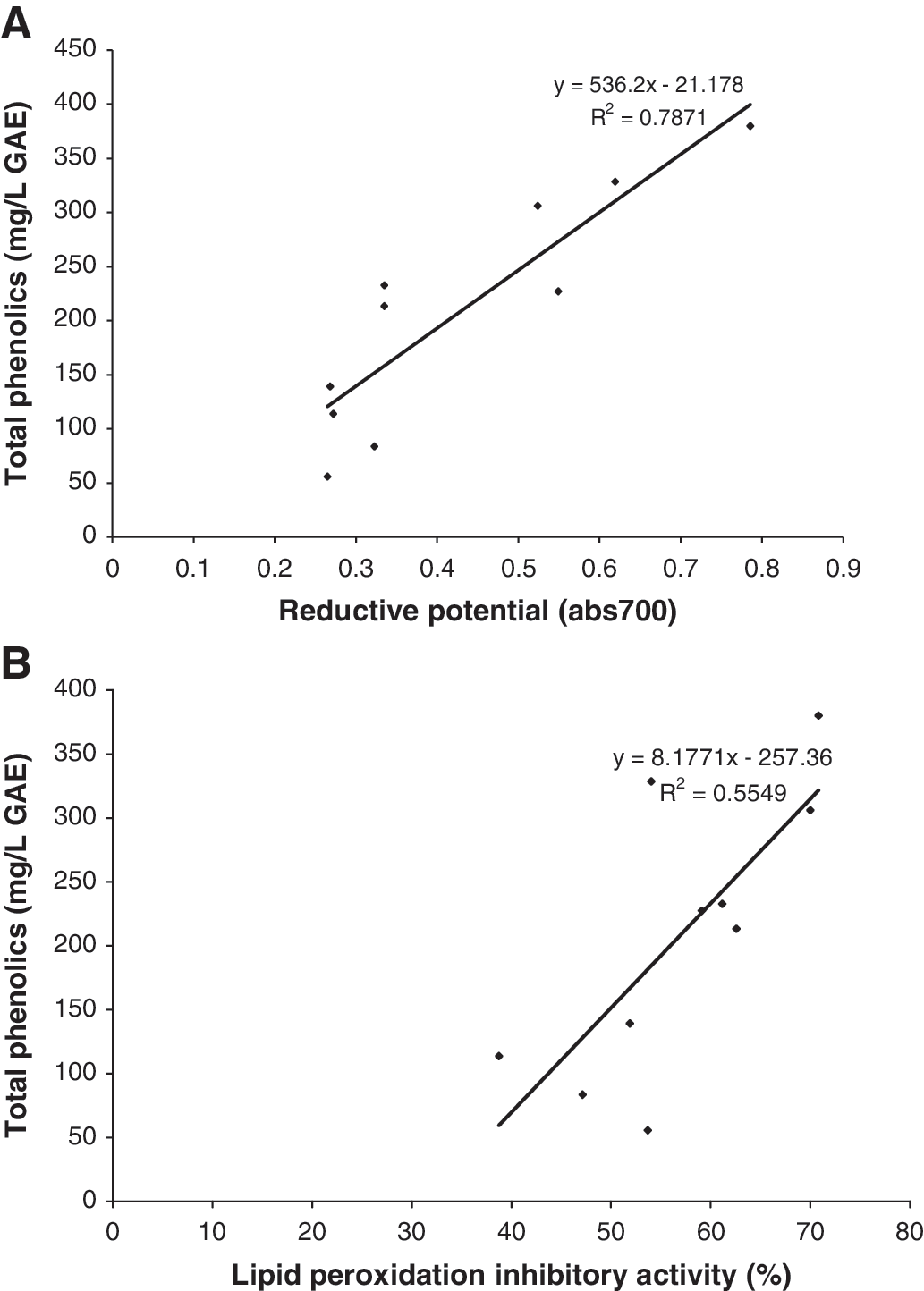
Relationship between (
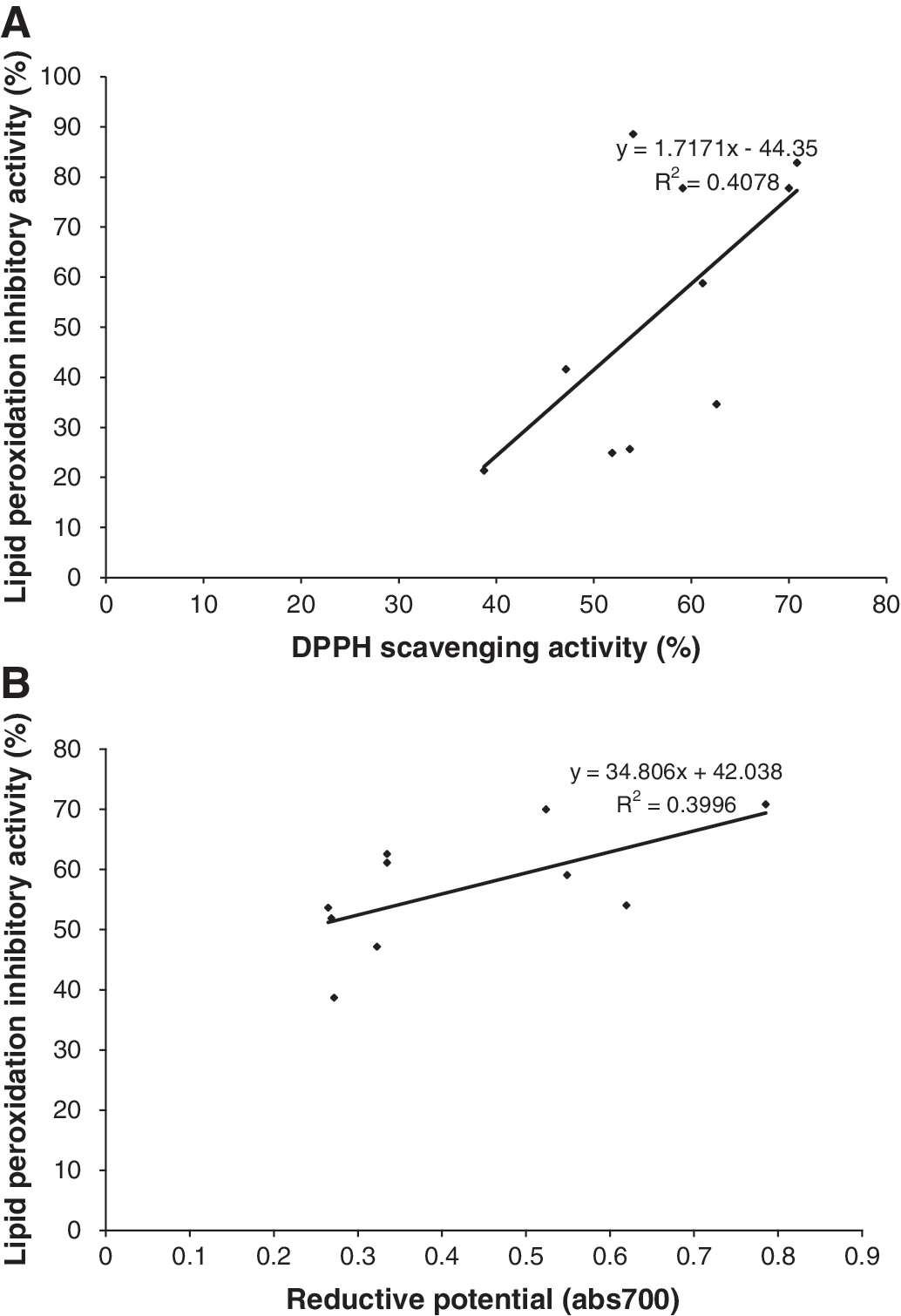
Relationship between (
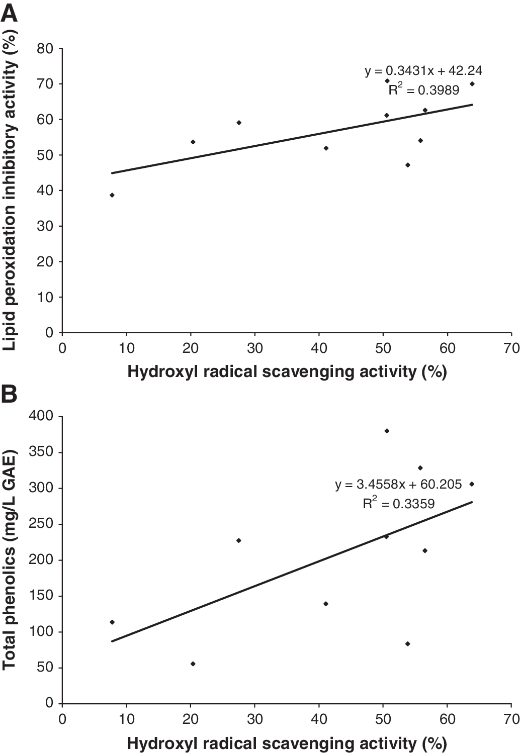
Relationship between (
Discussion
It is well recognized in plant chemistry that the mode of preparation and administration of herbal remedies are often crucial variables in determining efficacy for pharmacological evaluations. 13 In the traditional use of these plants, decoctions or infusions of the relevant parts are usually made with either water or alcohol as the solvent. The nature of solvent may influence the medicinal or other effects exhibited by plants because solvents extract antioxidant components to different degrees.
Antioxidant activity in higher plants has often been associated with phenolic compounds. 19 In addition to their roles in plants, phenolic compounds in our diet may provide health benefits associated with reduced risk of chronic diseases. 18 Flavonoids are the largest group of phenolics. They have been identified in fruits, vegetables, and other plant parts and linked to reducing the risk of major degenerative diseases. More than 4,000 distinct flavonoids have been identified. 18 The antioxidant activity of plant extracts has been reported to correlate with their phenolic content. 20,21 Data from the present work indicate that this correlation is dependent on the nature of the antioxidant assay used. The results of this work clearly illustrate that different methodologies can provide completely different responses with respect to the antioxidant capacity of a pure compound or a mixture of compounds.
Significant correlations were observed between some of the assay methods. DPPH free radical scavenging activity had an excellent correlation with TPC and RP (Fig. 1). These three methods have a similar underlying mechanism of reaction. The DPPH assay evaluates antioxidant activity by testing the ability of compounds to act as free radical scavengers or hydrogen donors. 22 The antioxidant activity of phenolics is mainly due to their redox properties, which allow them to act as reducing agents, hydrogen donors, and singlet oxygen quenchers. They also have a metal chelating potential. 23 The RP assay also has to do with the redox properties of substances being investigated. Each assay or group of assays with a similar underlying mechanism may be specific for a particular group of antioxidant substances, and where this group occurs in a substantial amount, such tests will yield high values. Exceptions can occur where these groups are bound or masked, leading to their nondetection by the specified assay(s). There are different types of antioxidants in plants. There are metals like selenium, vitamins such as ascorbic acid, and phytochemicals such as carotenoids, phenolics, organosulfur compounds, and nitrogen-containing compounds. The nature and position of functional groups in some antioxidant compounds, e.g., the hydroxyl groups of flavonoids, influence their reactivity and consequently their activity.
Odabasoglu et al. 24 reported that there was no correlation between antioxidant activity and TPC of extracts of some lichen species, a contradiction to previous reports. 20,21 The present investigation also clearly contradicts this submission of Odabasoglu et al. 24 There were strong correlations between TPC on the one hand and DPPH, RP, and LPIA assays on the other (r 2 = 0.76, 0.81, and 0.55, respectively). Odabasoglu et al., 24 however, reported a strong correlation between reducing power and total antioxidant activity. The present investigation also revealed significant correlations between reducing power on one hand and DPPH and TPC on the other. The authors noted that individual phenolics may have distinct antioxidant activities, and there may be antagonistic or synergistic interactions between phenolics and other compounds like carbohydrates and proteins.
Miliauskas et al. 25 reported a good correlation between antiradical activity (DPPH) and TPC. Our finding in the present work is in harmony with theirs. The results of the present work also confirm their findings that there was low correlation between TFC and DPPH assay and between TPC and TFC. The values for the correlation coefficients between TFC and DPPH assay and between TPC and TFC in our own study (r 2 = 0.21 and 0.43, respectively) were similar to those obtained by Miliauskas et al. 25 (0.32 and 0.43, respectively). The results of the present work showed only a low correlation between TFC and TPC (Table 4) and also between TFC and radical scavenging assays. For example, the correlation coefficient between the DPPH assay and TFC was 0.21, and that of hydroxyl radical scavenging capacity and TFC was 0.03 (Table 4). These results are also in agreement with the findings of Miliauskas et al. 25
The present investigation goes further to show that DOR and NO showed no strong correlation with any of the other assays that were carried out.
Although previous investigators used very few assays or few plants for the purpose of investigating correlations, the present study used seven assays and 10 plants to ensure more accurate results.
Investigators need to be more specific when reporting antioxidant activities of phytochemicals. Terms like “total antioxidant capacity” or “total antioxidant activity” are too general and could be misleading. Tests used for assessment should be clearly indicated to leave no room for ambiguity.
It has been observed that only flavonoids of a certain structure and, in particular, the hydroxyl position in the molecule determine antioxidant properties. These properties, in general, depend on the ability to donate hydrogen or electron to a free radical. Miliauskas et al. 25 found, in the same study, some correlation between TPC and flavonols. In support of the above observations, Choi et al. 26 reported that the interaction of a potential antioxidant with DPPH depends on its structural conformation and that this structural requirement is correlated with the presence of hydroxyl groups on the flavonoids. Cos et al. 27 reported that allopurinol showed remarkable activity in inhibiting xanthine oxidase and scavenging superoxide radical, whereas taxifoline showed relatively weak activity. The difference in activities was attributed to variation in the location of the hydroxyl groups and double bonds.
Choi et al. 28 found that the scavenging activity of flavonoids on peroxynitrite was governed by the position of the hydroxyl group. o-Hydroxyl structures increased the scavenging activity on peroxynitrite. Structural comparison of the flavonols in their study and their scavenging activities clearly shows that the C-3 hydroxyl group plays a pivotal role in the observed scavenging activity. These authors inferred that the higher scavenging potency of galangin compared with galangin 3-O-methyl ether may suggest that C-3 methoxylation reduced the scavenging effect of flavonols.
Phytochemicals are complex in nature. Therefore, the antioxidant activities of plants extracts cannot be evaluated by only a single method. The antioxidant defense system of the body is composed of different antioxidant components. The antioxidant capacities of these antioxidant components depend upon which free radicals or oxidants are produced in the body. 26 The various methods used in evaluating the antioxidant activity of samples can give varying results depending on the specificity of the free radical being used as a reactant. 22 The results of the present work highlight the diversity and complexity of phytochemicals present in plant extracts and the specificity of different assay protocols for specific antioxidant species based on the mechanism of reaction.
The DPPH assay appears to be a reliable method of measuring total antioxidant capacity of substances or systems. Its values correlated well with about three other methods used in this study (Fig. 1). As noted by Prakash, 22 it is rapid, simple, and inexpensive, and its value applies to the overall antioxidant capacity of the sample and is not specific to any particular antioxidant component.
Using the frequency of high antioxidant capacity values as the basis, results obtained in this work reveal that P. guajava, S. mombin, and G. cupulata are the plants that demonstrated consistent high activities in the various assays, followed by C. alata and O. gratissimum. It must be noted that some of the remaining plants have higher activities than the ones in the list above in some of the assays. For example, the NO radical scavenging activity of A. boonei (44.88 ±0.55%) was higher than that of P. guajava (21.68 ± 1.51%) and O. gratissimum (30.576 ± 1.61%) (P < .05).
Overall, the results obtained in this study indicate that Nigerian indigenous medicinal plants could be a source of natural chemoprophylactic antioxidants against reactive oxygen species and as such could be relevant in the treatment of cardiovascular disease, cancer, arthritis, and other pathologies in which free radical mechanisms have been implicated. In view of the potential beneficial properties of the studied plants, our results warrant further investigations on the identification of novel chemoprophylactic compounds in these plants.
Footnotes
Author Disclosure Statement
No competing financial interests exist.