




Abstract
The aim of the present study was to investigate the protective role of Ginkgo biloba leaf extract against uranium (U)-induced toxicity in Swiss albino mice. The mice were randomly divided into six groups, each consisting of six animals: Group I (control) received tap water alone, Group II received U at a dose of 5 mg/kg of body weight, Group III received G. biloba at a dose of 50 mg/kg of body weight, Group IV received G. biloba at a dose of 150 mg/kg of body weight, Group V received G. biloba (50 mg/kg of body weight) and U (5 mg/kg of body weight), and Group VI received G. biloba (150 mg/kg of body weight) and U (5 mg/kg of body weight) by oral gavage for 5 days. Serum aspartate aminotransferase (AST), alanine aminotransferase (ALT), blood urea nitrogen (BUN), and creatinine levels were determined to assess liver and kidney function, respectively. Also, liver and kidney samples were taken for the determination of tissue malondialdehyde (MDA) and reduced glutathione (GSH) levels, and histopathological changes in liver and kidneys were investigated. The results indicated that there was a significant increase (P < .05) in selected serum parameters. Serum AST, ALT, BUN, and creatinine levels significantly increased in mice treated with U alone when compared to the other groups. Moreover, U-induced oxidative damage caused a significant decrease in GSH levels and a significant increase in MDA levels of liver and kidney tissues. Treatment with G. biloba produced amelioration in biochemical indices of hepatotoxicity and nephrotoxicity according to Group II. Each dose of G. biloba provided significant protection against U-induced toxicity, and its strongest effect was observed at a dose of 150 mg/kg of body weight. In vivo results showed that G. biloba extract is a potent protector against U-induced toxicity, and its protective role is dose-dependent.
Introduction
U
In the present study, uranyl acetate was given to mice during a period of 5 days by oral gavage. This duration was preferred because Ozmen and Yurekli 10 reported that uranyl acetate treatment for 5 days causes various effects on food and water consumption, body weight, blood urea nitrogen (BUN) and creatinine concentrations, and activities of alkaline phosphatase, aspartate aminotransferase (AST), alanine aminotransferase (ALT), glutathione S-transferase, and catalase in Swiss albino mice.
The use of certain materials may help to decrease of the toxicity created by chemical or radioactive agents such as U. 11,12 Recently, biopolymers of various biological materials such as lycopene, grape seed, royal jelly, and Ginkgo biloba have been used for this aim. 13,14 G. biloba is considered the oldest tree species to survive on earth, with a history dating back over 200 million years. Some Ginkgo trees have been known to live well over an average of 1,000 or more years. G. biloba leaf extract is the most widely sold phytomedicine in Europe, where it is used to treat the symptoms of early-stage Alzheimer's disease, vascular dementia, peripheral claudication, and tinnitus of vascular origin. 15
Although there are many published clinical studies on Ginkgo in the literature, unfortunately, the protective role of G. biloba on U-induced toxicity in human and animals is still poorly understood. The aim of the present study was to evaluate the protective role of G. biloba on U-induced hepatotoxicity and nephrotoxicity in Swiss albino mice.
Materials and Methods
Animals
The experiments were carried out on 36 male Mus musculus var. Albino (12–14 weeks old, weighing 25–30 g). The healthy mice were obtained from the Animal Research Center of the Refik Saydam Hifzissiha Institute (Ankara, Turkey). The mice were housed in stainless steel cages (approximately 26 cm long × 15 cm wide × 50 cm deep) and kept under controlled conditions at 22 ± 3°C and 55 ± 5% relative humidity, and they were adapted to the experimental condition of 12:12-hour light–dark cycles. The mice were acclimatized for 1 week prior to the planned experiments and fed with a standard pellet diet (Samsun Food Industry, Samsun, Turkey) ad libitum. In this study, the methods and techniques applied to mice were carried out in accordance with the ethical standards of Kirikkale University Faculty of Veterinary Medicine and with the guidelines set by the World Health Organization (Geneva, Switzerland).
Product and chemicals
G. biloba leaf extract was obtained from Health Genesis Corp. (Bay Harbor Island, FL, USA). Uranyl acetate dihydrate (Sigma) was purchased from Interlab A.S., Istanbul, Turkey. Commercial test kits for AST, ALT, BUN, and creatinine were purchased from Teco Diagnostics (Anaheim, CA, USA).
Experimental protocol
Albino mice were randomly divided into six groups, each consisting of six animal:. Group I served as the tap water–treated control, Group II animals received U at a dose of 5 mg/kg of body weight, Group III animals received G. biloba at a dose of 50 mg/kg of body weight, Group IV animals received G. biloba at a dose of 150 mg/kg of body weight, Group V animals received G. biloba (50 mg/kg of body weight) + U (5 mg/kg of body weight), and Group VI animals received GB (150 mg/kg of body weight) + U (5 mg/kg of body weight) by oral gavage for 5 consecutive days. The dose of U was selected as 5 mg/kg of body weight. This dose was chosen because it is known to induce a high frequency of pathological damage, which essential to determine the protective role of G. biloba. 16,17 G. biloba doses determined favorable for daily consumption in an amount recommended by practitioners of nutritional medicine for support optimal health (50 and 150 mg/kg of body weight) were used as effective doses to evaluate the protective role of G. biloba. 18
Serum analysis
For serum isolation, whole blood samples were collected by cardiac puncture with the animal under mild ether anesthesia. Blood samples were transferred directly into plain Vacutainer tubes (BD Vacutainer Systems, San Jose, CA, USA), centrifuged at 1,200 g for 10 minutes at 4°C, and stored at −20°C until analysis. Blood BUN (catalog number B549-150, Teco Diagnostics) and creatinine (catalog number C513-480, Teco Diagnostics) concentrations and AST (AST/GOT liquid reagent, catalog number A559-150, Teco Diagnostics) and ALT (ALT/GPT liquid reagent, catalog number A524-150, Teco Diagnostics) activities were measured by commercially available kits using an autoanalyzer (Model 99 M Chemistry Analyzer, Medispec, Germantown, MD, USA).
GSH and MDA analysis
At the end of the 5-day treatment period, animals were sacrificed after an overnight fast by exsanguinations under ether anesthesia. The liver and kidney tissues of each animal were removed, cleaned, dried, and processed for biochemical measurements. The tissues were then homogenized in ice cold 0.15 M KCI by a homogenizer (Ultra-Turrax® type T25-B, IKA Labortechnik, Staufen, Germany) at 16,000 rpm for 3 minutes. The homogenates were centrifuged at 5,000 g for 15 minutes at 4°C. The supernatants were stored at −40°C until they were analyzed. Tissue GSH and MDA contents were colorimetrically measured by the methods of Beutler et al. 19 and Yoshoiko et al., 20 respectively, using an ultraviolet/visible spectrophotometer (UVmini-1240, Shimadzu, Kyoto, Japan).
Histopathological examinations
For light microscopic examination, fresh tissue samples including liver and kidneys were fixed in 10% neutral buffered formalin solution for routine processing, embedded in paraffin wax, sectioned at 5 μm, and stained with hematoxylin and eosin. Histopathological changes were assessed semiquantitatively under a light microscope with an ocular with grids and 4 ×, 10 ×, and 40 × objective, respectively. A total of 10 high-power fields were randomly chosen in each section. Changes in the experimental histopathological parameters for liver and kidney tissues were graded as follows: −, no changes; +, ++, and +++, mild, moderate, and severe changes, respectively.
Statistical analysis
The statistical analysis was carried out using SPSS for Windows version 10.0 statistical software (SPSS Inc., Chicago, IL, USA). Statistically significant differences between the groups were compared using one-way analysis of variance and Duncan's test. The data are displayed as mean ± SE values, and values of P < .05 are considered “statistically significant.”
Results
Table 1 shows the changes in serum AST, ALT, BUN, and creatinine levels in each group after treatment with the different doses of G. biloba (50 and 150 mg/kg of body weight) and U (5 mg/kg of body weight) for 5 days. No significant differences in AST, ALT, BUN, and creatinine levels were observed among the control and the groups treated with G. biloba alone (P > .05). In other words, G. biloba when administered alone did not alter the level of serum parameters compared to the control values. Serum AST, ALT, BUN, and creatinine levels showed a statistically significant (P < .05) increase after U exposure. Enzyme activities after the administration of two different doses of G. biloba again decreased in Groups V and VI compared to Group II. However, enzyme levels were still higher than the control value (P < .05). Also, G. biloba had an effect on renal markers (BUN and creatinine). Serum BUN and creatinine levels significantly decreased in G. biloba–administered mice compared to those of Group II (P < .05). In Group VI, the mean level of BUN was about 1.6-fold lower and the mean level of creatinine was about 1.8-fold lower than in Group II. Moreover, GSH levels were significantly decreased, whereas MDA levels in liver and kidney tissues of the U-treated group were significantly increased compared to those of the control group (Table 2, P < .05). However, oral administration of the 150 mg/kg of body weight dose of G. biloba for 5 days reversed GSH and MDA levels back to the control levels in the tissues compared to U-damaged mice (P > .05), and this effect was lesser for the 50 mg/kg of body weight dose of G. biloba (P < .05).
Group I, control; Group II, U (5 mg/kg of body weight); Group III, G. biloba (50 mg/kg of body weight); Group IV, G. biloba (150 mg/kg of body weight); Group V, G. biloba (50 mg/kg of body weight) + U (5 mg/kg of body weight); and Group VI, G. biloba (150 mg/kg of body weight) + U (5 mg/kg of body weight). Data are mean ± SE values (n = 6).
Statistical significance between means was determined using one-way analysis of variance followed by Duncan's test as a post-analysis of variance test (P < .05). Means with the same letter for different treatment groups for the same parameter are not significantly different.
Group I, control; Group II, U (5 mg/kg of body weight); Group III, G. biloba (50 mg/kg of body weight); Group IV, G. biloba (150 mg/kg of body weight); Group V, G. biloba (50 mg/kg of body weight) + U (5 mg/kg of body weight); and Group VI, G. biloba (150 mg/kg of body weight) + U (5 mg/kg of body weight). Data are mean ± SE values (n = 6).
Statistical significance between means was determined using one-way analysis of variance followed by Duncan's test as a post-analysis of variance test (P < .05). Means with the same letter for different treatment groups for the same parameter are not significantly different.
Histopathology of liver and kidney tissue from the different treatment groups is shown in Figures 1 –12. There was no evidence of a pathological change in Groups I, III, and IV except mild hyperemia in some liver sections of Groups III and IV (Figs. 1, 2, and 5 –8). Histopathological changes were observed in liver and kidney tissues of mice in Group II. There was severe degeneration, pyknosis, and necrosis of hepatocytes in liver (Fig. 3). Kuppfer cell proliferation was also observed in some sections of liver from to this group. In kidney tissues, there was a prominent tubular dilatation, and lumens were filled by albuminoid content (Fig. 4). Histopathological findings in Groups V and VI were moderate, especially Group VI, compared to those in Group II. Hydropic degeneration, pyknosis (Fig. 9), and less hyaline materials within the tubular lumens of kidney (Fig. 10) were observed in Group V. There was a prominent decrease in pyknosis of hepatocytes (Fig. 11), and degeneration and necrosis of renal tubular epithelial cells with limited hyaline materials within the tubular lumens were seen in Group VI (Fig. 12). The severity of histopathological lesions in liver and kidney tissues is semiquantitatively shown in Tables 3 and 4.
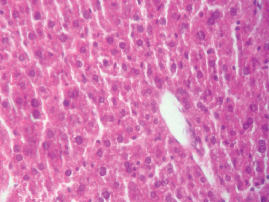
Liver from Group I (control group). Hepatocytes are predominantly nondegenerative. Hematoxylin and eosin stain. Original magnification, × 400. Color images available online at

Kidney from Group I (control group).There was no marked epithelial degeneration and abnormal albuminoid content within the lumens. Hematoxylin and eosin stain. Color images available online at

Liver from Group II (treated with 5 mg/kg of body weight dose of U). Degeneration of the hepatocytes is severe. Pyknotic nuclei (black arrows) and necrotic hepatocytes (white arrows) are indicated. Hematoxylin and eosin stain. Color images available online at

Kidney from Group II (treated with 5 mg/kg of body weight dose of U). Tubular lumens are filled by albuminoid content. Hematoxylin and eosin stain. Color images available online at
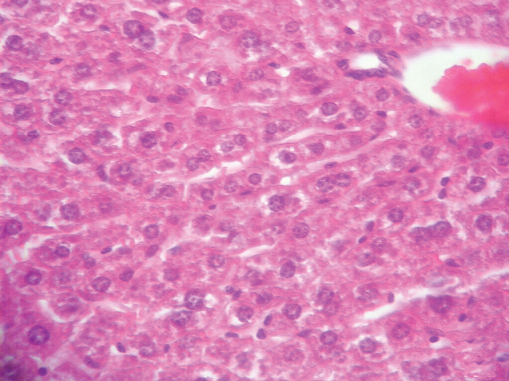
Liver from Group III (treated with G. biloba at a dose of 50 mg/kg of body weight). The appearance is similar to that of control group liver. Hematoxylin and eosin stain. Original magnification, × 400. Color images available online at

Kidney from Group III (treated with G. biloba at a dose of 50 mg/kg of body weight). The appearance is similar to that of control group kidney. There was no marked epithelial degeneration and abnormal albuminoid content within the lumens. Hematoxylin and eosin stain. Color images available online at

Liver from Group IV (treated with G. biloba at a dose of 150 mg/kg of body weight). The appearance is similar to that of control group liver. Hematoxylin and eosin stain. Original magnification, × 400. Color images available online at

Kidney from Group IV (treated with G. biloba at a dose of 150 mg/kg of body weight). The appearance is similar to that of control group kidney. There was no marked epithelial degeneration and abnormal albuminoid content within the lumens. Hematoxylin and eosin stain. Color images available online at

Liver from Group V (treated with G. biloba at a dose of 50 mg/kg of body weight and 5 mg/kg of body weight dose of U). Marked swelling of the hepatocytes (small arrows) and pyknosis (large arrows) are visible. Hematoxylin and eosin. Original magnification, × 400. Color images available online at
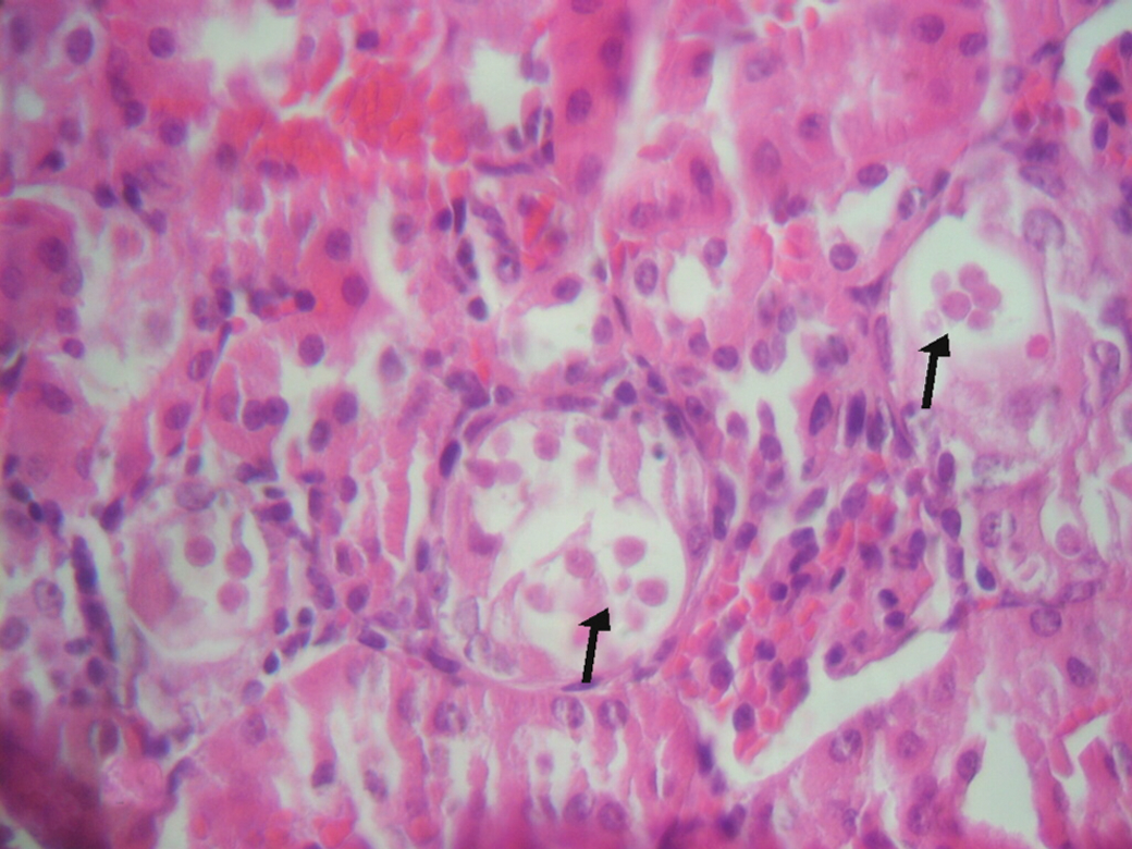
Kidney from Group V (treated with G. biloba at a dose of 50 mg/kg of body weight and 5 mg/kg of body weight dose of U). There are some hyaline materials within the lumens. Hematoxylin and eosin stain. Original magnification, × 400. Color images available online at
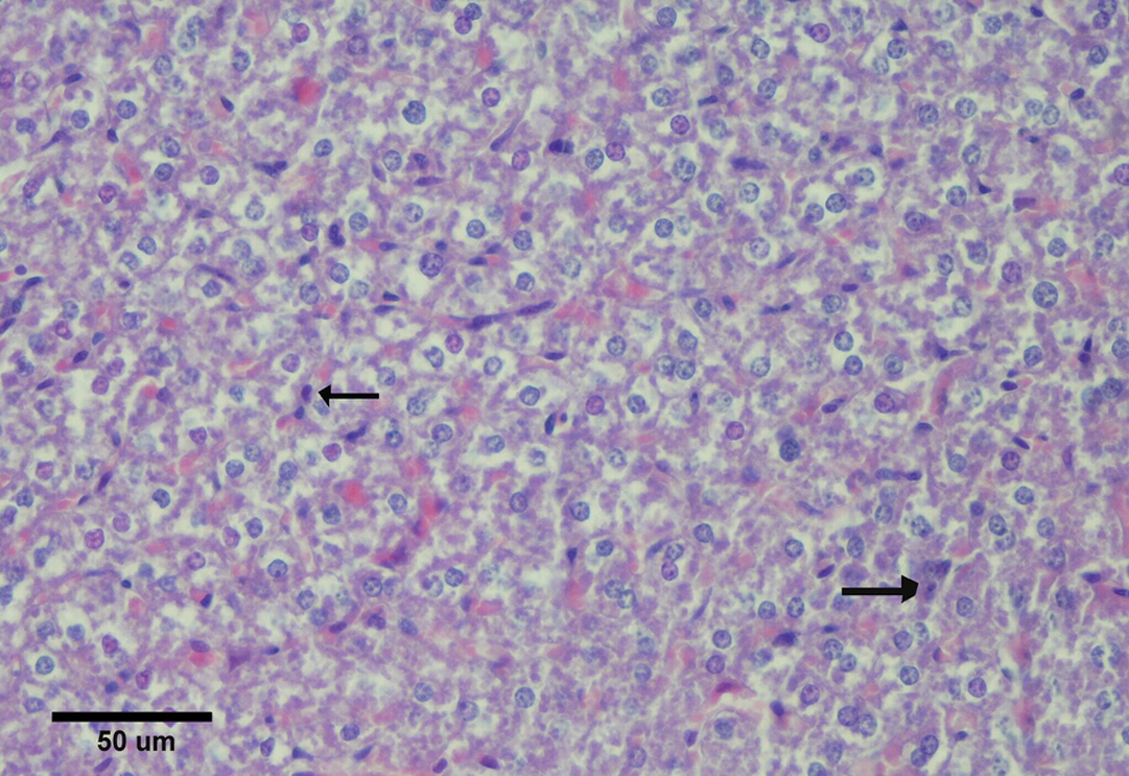
Liver from Group VI (treated with G. biloba at a dose of 150 mg/kg of body weight and 5 mg/kg of body weight dose of U). A section shows many degenerative hepatocytes. The arrows indicate less pyknotic nuclei. Hematoxylin and eosin stain. Color images available online at
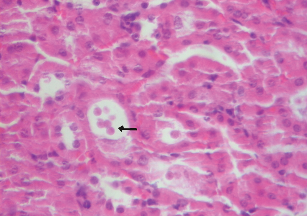
Kidney from Group VI (treated with G. biloba at a dose of 150 mg/kg of body weight and 5 mg/kg of body weight dose of U). There are small amounts of hyaline materials (arrow). Hematoxylin and eosin stain. Original magnification, × 400. Color images available online at
Group I, control; Group II, U (5 mg/kg of body weight); Group III, G. biloba (50 mg/kg of body weight); Group IV, G. biloba (150 mg/kg of body weight); Group V, G. biloba (50 mg/kg of body weight) + U (5 mg/kg of body weight); and Group VI, G. biloba (150 mg/kg of body weight) + U (5 mg/kg of body weight). Changes in the experimental histopathological parameters for liver tissues were graded as follows: −, no changes; +, ++, and +++, mild, moderate, and severe changes, respectively.
Group I, control; Group II, U (5 mg/kg of body weight); Group III, G. biloba (50 mg/kg of body weight); Group IV, G. biloba (150 mg/kg of body weight); Group V, G. biloba (50 mg/kg of body weight) + U (5 mg/kg of body weight); and Group VI, G. biloba (150 mg/kg of body weight) + U (5 mg/kg of body weight). Changes in the experimental histopathological parameters for kidney tissues were graded as follows: −, no changes; +, ++, and +++, mild, moderate, and severe changes, respectively.
Discussion
U is a potentially hazardous metal, which is not physiologically or biochemically essential for mammals. Exposure to U in large doses can cause biochemical and functional changes in the critical organs. 21 For example, Bussy et al. 22 investigated the effects of uranyl nitrate on cholinergic acetylcholinesterase activity and on dopaminergic and serotoninergic metabolisms in several areas of male Sprague-Dawley rat brains. As a result, they showed that uranyl nitrate can cause chronic and progressive perturbations of physiological levels of neurotransmitter systems. Linares et al. 23 investigated the effects of stress on the potential reproductive toxicity of long-term exposure to uranyl acetate dihydrate in adult male rats. As a result, they observed that there was a significant decrease in the pregnancy rate. Moreover, spermatid number/testis was significantly decreased by U administration. In a similar study, the pro-oxidant activity of U was assessed in kidney and testes of male rats. The results suggested that graded doses of U elicit depletion of the antioxidant defense system of the rat and induce oxidative stress in testes and kidneys. 24 In another study, the effects of uranyl nitrate on viability, cell cycle kinetics, micronuclei, chromosome aberrations, and sister chromatid exchanges in Chinese hamster ovary cells were investigated. The results showed that uranyl nitrate decreased the viability of Chinese hamster ovary cells in a dose-related fashion. Also, uranyl nitrate at concentrations ranging from 0.01 to 0.3 mM decreased cell cycle kinetics and increased frequencies of micronuclei and sister chromatid exchanges. Chromosome aberrations were also significantly augmented by uranyl nitrate. 25 In addition, a few published studies have described the distribution of elemental U after chronic exposure, and one study has demonstrated an accumulation in the brain. 26 Nevertheless, bone and kidneys are the primary reservoirs for U. In particular, liver and kidneys are the main target organs for U toxicity. 27,28
Because of high chemical toxicity, radiological effects of U are difficult to study in laboratory animals. 29 U also poses little radioactive danger because it gives off very small amounts of radiation. 30 Both animal experiments and human exposures to high levels of U show that U can causes hair loss, changes in blood counts, genetic effects, reproductive effects, and some cancers, including lung, kidney, and leukemia, when people and animals receive high doses in a short period of time. 31 In the present study, we determined, as U-induced radiological effects, the morphological alterations in shape and the number of blood cells, including erythrocytes and leukocytes. Our findings were generally related to the chemical toxicity of U.
Previous studies have shown that contamination with toxic doses of U induces harmful changes to liver and kidneys. 32 In the present study, the mice treated with U showed a significant liver damage, as indicated by the increased levels of hepatic marker enzymes. The mean serum AST and ALT levels were found to be significantly higher in the U-treated group than those of the control group. This finding confirms the results of pathological findings and probably indicates a disruption of liver function. An increase in the levels of these hepatic enzymes also serves as an indicator of altered cell membrane permeability. 33 U hepatotoxicity is probably effected in two ways: on the one hand, by the occurrence of the inflammatory state, and on the other hand, by direct toxic action of U on liver cells. The increase in the levels of AST and ALT in serum may be due to the leakage of these enzymes from liver cytosol into the bloodstream. 34,35 In the present study, increases in AST and ALT levels may result from liver damage as supported by the pathological findings, characterized by fibrosis in portal area and increased numbers of Kuppfer cells in liver consistent with the findings of similar studies. 36,37 The increases in serum AST and ALT levels are in agreement with previous findings of Ozmen and Yurekli. 10 They investigated subacute effects of uranyl acetate in laboratory mice during a period of 5 days. As a result, they showed that uranyl acetate was accumulated in liver, kidney, and brain tissues. Also, the levels of AST and ALT significantly increased in the exposure group compared to the control animals. Similar results were also showed by Dufour et al. 38 and Domingo et al. 39 However, Souidi et al. 3 observed a decrease in plasma levels of ALT and AST in rats chronically exposed to depleted U in their drinking water for 9 months. This contradictory result is probably due to the quantity of U used and the duration of exposure.
BUN and creatinine are nitrogenous waste products that are eliminated by kidneys, when excretion is suppressed in renal insufficiency. 40 The rise in BUN and creatinine concentrations in serum is used as an indicator of U-induced nephrotoxicity. Table 1 shows the increase in serum BUN and creatinine levels. Also, renal tubular damage and glomerular filtration impairment were observed in kidney exposed to U. These damages may account for the increase in serum BUN and creatinine levels of the animals receiving U. The effect of U on BUN and creatinine levels has also been described by other authors. Ozmen and Yurekli, 10 Tolson et al., 41 and Mizuno et al. 42 reported a significant increase in plasma BUN and creatinine levels after U exposure.
Administration of two different doses of G. biloba together with U for a total of 5 days significantly decreased the levels of AST, ALT, BUN, and creatinine in Groups V and VI compared to group II. Administration of G. biloba also caused less pathological change in liver and kidney tissues. In other words, G. biloba caused significant decreases in renal tubular proteinuria, renal injury, proximal tubular necrosis, and renal dysfunction in kidneys and necrosis, tubular endothelial lesions, cellular degeneration, and loss of the distinct characteristic configuration in liver. Two different doses (50 and 150 mg/kg of body weight) of G. biloba showed a partial improvement in the biochemical parameters and a clear protective effect against pathological damages. It has also appeared that the 150 mg/kg of body weight dose of G. biloba had a greater effect on serum parameters than the 50 mg/kg of body weight dose.
In cells, GSH serves to remove reactive oxygen species such as H2O2 produced because of either cellular respiration or metabolism of toxic substances and to protect cells from oxidative injuries. 43 A change in the GSH ratio is considered as an indicator of the oxidative state of the cell. 44 In the present study, there was a significant reduction in GSH levels of liver and kidney tissues, implicating the presence of oxidative tissue damage. Furthermore, these tissue injuries caused functional impairment as evidenced with renal and hepatic function tests in which elevated serum levels demonstrated the severity of the U-induced systemic inflammatory response. Probably, G. biloba leaf extract, as an antioxidant agent, ameliorated oxidative injury in the tissues and functional deteriorations. In our study, GSH levels of liver and kidney tissues were significantly decreased by U toxicity, and because of its antioxidant activity, G. biloba treatment reduced the U-induced oxidative injury and restored GSH levels significantly.
MDA is a good indicator of the degree of lipid peroxidation. 45 MDA is a product generated during the oxidative breakdown of certain macromolecules such as lipids and is found either in free form or bound to certain tissue structures. Endoperoxides are generated as a result of changes that occur in the molecular structure of fatty acids during their breakdown, and MDA is generated during the breakdown of endoperoxides. MDA is considered to be the most significant indicator of membrane lipid peroxidation, arising from the interaction of reactive oxygen species with cell membranes. Because of the high susceptibility of lipid membranes to peroxidation, the free radicals easily peroxidated the lipid membranes, and MDA was generated as a final product of peroxidation. MDA by itself also causes peroxidation and accelerates peroxidation by the means of synergy with free radicals. 46 In the present study, we found a significant increase in MDA levels during U toxicity, which is in agreement with the previous studies. The increase in MDA levels in liver and kidneys suggests enhanced lipid peroxidation leading to tissue damage and failure of antioxidant defense mechanisms. Our results show that G. biloba treatment significantly inhibits MDA production, implying a reduction in lipid peroxidation and cellular injury that protect the tissues against U-induced oxidative damage.
Consequently, G. biloba had a protective effect against U-induced hepatotoxicity and nephrotoxicity, and this effect was dose-dependent. The protective role of G. biloba on U-induced toxicity may be explainable with the antioxidant properties of G. biloba. Although it is not a general rule, antioxidants and G. biloba share similar mechanisms of protection against the toxicity. Antioxidants act as free radical scavengers, and they trap the free radicals and give up their own electrons. Thus, antioxidants with stated functions protect molecules such as proteins, lipids, enzymes, chromosomes, and DNA against free radical damage. 47,48 Researchers have shown that G. biloba possesses antioxidant properties. G. biloba is also known to be efficient in helping to treat or prevent diseases associated with free radicals. Pharmacologically, there are two groups of substances that are significant compounds found in G. biloba: the flavonoids, such as myricetin and quercitin, which give Ginkgo its antioxidant action, and the terpenes, which help to inhibit the formation of blood clots. These compounds have the capability of decreasing the cell damage that results from the presence of free radicals. Also, these compounds scavenge and destroy free radicals and reactive forms of oxygen, such as O2 −, · OH, and lipid peroxide radicals. 15,49 Human laboratory tests and animal studies have shown that Ginkgo contains antiradical or antioxidant properties. Goksel et al. 50 investigated the possible beneficial effect of G. biloba against toxicity of the analgesic acetaminophen in mice. As a result, they showed that acetaminophen caused oxidative damage in hepatic tissues, but G. biloba extract supplementation, by its antioxidant effects, protected the tissues. In a similar study, the anti-apoptotic effect of G. biloba in gossypol-treated human lymphocytes was investigated. As a result, it was demonstrated that G. biloba extract significantly reduced gossypol-induced apoptosis in human lymphocytes. 51 In another study, the protective effects of G. biloba were investigated on thioacetamide-induced fulminant hepatic failure in rats. The results indicated that G. biloba ameliorated hepatic damage in thioacetamide-induced fulminant hepatic failure. 52
In conclusion, the result of the present study clearly demonstrated that subacute U toxicity induces oxidative damage in liver and kidneys. However, supplementation with G. biloba extract can protects against U toxicity, by reducing the effects of free radicals and preventing lipid peroxidative degradation of biomembranes. Therefore, the antioxidant role of G. biloba may serve as a “toxicity-limiting agent” to reduce environmental effects of chemical and radioactive agents or may provide a new approach for understanding the mechanism of chemical toxicity.
Footnotes
Acknowledgments
This study was supported by grants from Giresun University Scientific Research Projects Department.
Author Disclosure Statement
The authors have declared no conflict of interest.