




Abstract
Antimicrobial and antioxidant activities of mycelia obtained from 10 wild edible mushrooms—Armillaria mellea, Meripilus giganteus, Morchella costata, Morchella elata, Morchella esculenta var. vulgaris, Morchella hortensis, Morchella rotunda, Paxillus involutus, Pleurotus eryngii, and Pleurotus ostreatus—were investigated. For determination of antimicrobial activities of these mushrooms, ethanol extracts were examined with 11 test microorganisms by the agar well diffusion method. P. ostreatus and M. giganteus were the most active species against both bacteria and yeast. Antioxidant properties of ethanol extracts were studied by the 1,1-diphenyl-2-picrylhydrazyl free radical scavenging method. Among the mushroom extracts, M. elata showed the most potent radical scavenging activity. This research has shown that these 10 wild macrofungi have potential as natural antioxidants and antibiotics.
Introduction
M
Infectious diseases remain one of the major threats to human health. Although numerous antibiotics have been used against pathogens, antimicrobial resistance is an increasing public health problem. The antibiotics in mushrooms are less well documented in the discovery of new antimicrobial agents with different structural types. Mushrooms need antibacterial and antifungal compounds to survive in their natural environments. Therefore, antimicrobial compounds could be isolated from many mushroom species and could be of beneficial for humans. 3 Most of the medicinal extracts from mushrooms are different forms of polysaccharides, and all of them are strengtheners of the immune system with few or no side effects. 4
The degenerative diseases associated with aging include cancer, cardiovascular disease, immune system decline, brain dysfunction, and cataracts. They are also associated with free radicals because oxidative damage to DNA, proteins, and other macromolecules accumulates with age and has been postulated to be a major type of endogenous damage leading to aging. 5 The consumption of plant foods, such as fruits, vegetables, red wines, and juices, provides protection against various diseases, including cancer and cardio- and cerebrovascular diseases. This protection can be explained by the capacity of antioxidants in the plant foods to scavenge free radicals, which are responsible for the oxidative damage of lipids, proteins, and nucleic acids. 6 Like plants, mushrooms accumulate a variety of secondary metabolites, including phenolic compounds, polyketides, terpenes, and steroids. Some mushrooms have currently been found to possess antioxidant activity that is well correlated with their total phenolic content. Recently, mushrooms are considered to be a good source of antioxidants such as variegatic acid and diboviquinone, which have been found in mushrooms. 7
This experimental study is part of a program focusing on screening of mycelia obtained from wild mushrooms collected from different parts of Turkey. Wild mushrooms are seasonal, and a particular mushroom may disappear from the initial place of collection for a number of years, appearing in another place beyond reach. Although there are many studies on cultivated and wild mushrooms in the world, there is little information available about antimicrobial and antioxidant activities of wild edible mushrooms of Turkey, and this is the first study on the pharmacological statue of many of the mushrooms used in this study. Our objective was to evaluate the antimicrobial and antioxidant properties of mycelia obtained from 10 wild mushrooms collected from different parts of Turkey.
Materials and Methods
Mushrooms and growth of mycelia
Mycelia obtained from 10 wild edible mushroom species (Armillaria mellea, Meripilus giganteus, Morchella costata, Morchella elata, Morchella esculenta var. vulgaris, Morchella hortensis, Morchella rotunda, Paxillus involutus, Pleurotus eryngii, and Pleurotus ostreatus) were grown at 25°C in submerged liquid cultures. The liquid medium (pH 6.6) contained glucose (20 g/L), peptone (10 g/L), and yeast extract (2 g/L). Erlenmeyer flasks that included liquid medium were inoculated with agar plugs (potato dextrose agar, 6 mm in diameter) covered by the mycelium. 8 After 30 days of incubation in the dark, the liquid medium was filtered, and the biomass was separated from the liquid. Voucher specimens of mushroom were deposited in the Fungarium of Microbiology, Department of Biology, Celal Bayar University, Manisa, Turkey.
Test microorganisms and growth conditions
Test microorganisms included the bacteria Bacillus cereus CM 99, Bacillus subtilis ATCC 6633, Enterobacter aerogenes ATCC 13048, Enterobacter cloacae ATCC 13047D, Enterococcus faecalis ATCC 29212, Escherichia coli ATCC 39628, Proteus vulgaris ATCC 8427, Salmonella typhimurium CCM 5445, Sarcina lutea ATCC 9341NA, and Staphylococcus aureus ATCC 6538P and the yeast Candida albicans ATCC 10231. Cultures of these bacteria were grown in Mueller-Hinton broth (Oxoid, Basingstoke, UK) at 37°C for 24 hours, and the yeast studied was incubated in glucose yeast extract broth at 30°C for 48 hours. 9 Test microorganisms were obtained from the culture collection of the Basic and Industrial Microbiology Department, Faculty of Science, Ege University, Izmir, Turkey.
Antimicrobial activity assay
The dried and powdered mycelia were reduced to coarse powder. Two grams of each species was extracted with 20 mL of ethanol at room temperature with stirring for 3 days (125 cycles/minute). The ethanol was evaporated to dryness after the extraction process. Sample solutions were prepared by dissolving the extracts in ethanol (1 mL). In vitro antimicrobial studies were carried out by the agar well diffusion method against test microorganisms. Bacterial strains grown on nutrient agar at 37°C for 24 hours were suspended in a saline solution (0.85% NaCl) and adjusted to the turbidity of the 0.5 MacFarland standard (106 colony-forming units/mL). In brief, a 50-μL inoculum (containing approximately 105 bacteria/mL and 104 yeast/mL) was added to 25 mL of melted Mueller-Hinton agar and potato dextrose agar medium cooled at 45°C. This was then poured into 90-mm diameter Petri dishes and maintained for 1 hour at room temperature. Small wells (6 mm in diameter) were cut in the agar plate using a cork borer; 60 μL of extract concentration with a negative control (ethanol, 60 μL) was loaded in the wells. The dishes were preincubated at 4°C for 2 hours to allow uniform diffusion into the agar. After preincubation, for bacteria the plates were incubated at 37°C for 24 hours; and for yeast, 30°C for 48 hours was used. 9 The antimicrobial activity was evaluated by measuring the inhibition zone diameter observed. In addition, commercial antibiotics (penicillin G [10 IU], nalidixic acid [30 μg], novobiocin [30 μg], and nystatin [10 μg]) were used as positive controls to determine the sensitivity of the strains. 10 All experiments were performed in triplicate.
Antioxidant activity assay
The hydrogen atom or electron donation abilities of the extracts were measured from the bleaching of the purple-colored methanol solution of 1,1-diphenyl-2-picrylhydrazyl (DPPH). This spectrophotometric assay uses the stable radical DPPH as a reagent.
11
One thousand microliters of a 1 mg/mL concentration of the extracts in ethanol was added to 4 mL of 0.004% methanol solution of DPPH. After a 30-minute incubation period at room temperature, the absorbance was read against a blank at 517 nm. Inhibition of free radical by DPPH in percentage (I%) was calculated as follows:
where A blankis the absorbance of the control reaction and A sample is the absorbance of the test compound. α-Tocopherol (TOC) was used for comparison. Tests were carried out in triplicate.
Statistical analysis
The mean values were statistically analyzed with the Minitab Release 13.20 program (Minitab, Inc., State College, PA, USA) by the general one-way (unstacked) analysis of variance to find out the most effective extracts and the most sensitive test microorganisms. Percentage similarity of microorganisms in relation to their susceptibility to the mushroom extracts was analyzed by the multivariate cluster analysis according to the data obtained from the well diffusion assay.
Results and Discussion
Antimicrobial activity of mycelia
The antimicrobial activity of mycelium culture extracts of wild mushroom species was studied by the agar well diffusion method. All extracts were tested against 10 species of bacteria and one yeast. Antimicrobial activity was observed in all mushroom species included in the study. The data relating to the antimicrobial activities of samples are summarized in Table 1. Overall, 10 ethanol extracts from different mycelia were examined; antibacterial and antiyeast activities were detected in seven of these samples.
Data are mean values (n = 3). Values of 0 and 6 indicate no inhibitory activity.
Inhibition zone diameter (in mm), not including well diameter (6 mm) and if equal to negative control inhibitions or under recorded as 0, but including disk diameter (6 mm) for standard antibiotics.
Bacteria were tested in Mueller-Hinton agar medium; yeast was tested in potato dextrose agar.
, partial inhibition; ND, not determined; R, resistant; NC, negative control. EC, E. coli; SA, S. aureus; SL, S. lutea; ST, S. typhimurium; CA, C. albicans; EF, E. faecalis; PV, P. vulgaris; BS, B. subtilis; BC, B. cereus; EA, E. aerogenes; ECL, E. cloacae.
According to results of the antimicrobial screening assay, some of the mushrooms studied are potentially a rich source of antimicrobial agents, but many of the mycelia have weak activities. The most active species were P. ostreatus and M. giganteus, which showed broad-spectrum antimicrobial activity against Gram-positive and Gram-negative bacteria, whereas the least active species were M. elata and M. hortensis. P. involutus and M. costata demonstrated antiyeast activity against C. albicans. Extract of M. rotunda only showed antiyeast activity against C. albicans and not the other organisms tested.
The maximum antibacterial effect in tested macrofungi was shown by ethanol extracts of P. ostreatus against S. lutea as 30 mm. A large inhibition zone diameter against C. albicans was obtained by P. involutus (15 mm) (Table 1).
Sensitivity of test strains was, in decreasing order, C. albicans > S. lutea > E. coli > S. aureus > S. typhimurium > E. cloacae > B. subtilis > B. cereus > E. aerogenes > P. vulgaris > E. faecalis (Fig. 1). Figure 2 summarizes the similarity of microorganisms in relation to their susceptibility to the mushroom extracts.

Mean values of microorganisms in relation to their susceptibility to the mushroom extracts. The bar indicates SE. *Means are indicated by solid circles. EC, E. coli; SA, S. aureus; SL, S. lutea; ST, S. typhimurium; CA, C. albicans; EF, E. faecalis; PV, P. vulgaris; BS, B. subtilis; BC, B. cereus; EA, E. aerogenes; ECL, E. cloacae.
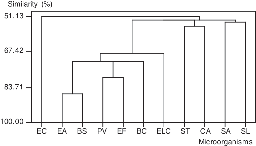
Similarity (%) of microorganisms in relation to their susceptibility to the mushroom extracts. EC, E. coli; SA, S. aureus; SL, S. lutea; ST, S. typhimurium; CA, C. albicans; EF, E. faecalis; PV, P. vulgaris; BS, B. subtilis; BC, B. cereus; EA, E. aerogenes; ECL, E. cloacae.
Some of the results reported in this study are consistent with those from earlier studies. 3,12,13 For instance, it was found that extracts from mycelial cultures of Lepista nuda and several Ganoderma species have antibacterial activity. 12 Rosa et al. 13 found high antimicrobial activity from several Basidiomycetes species (Agrocybe perfecta, Hexagonia hydnoides, Irpex lacteus, Nothopanus hygrophanus, Pycnoporus sanguineus, and Tyromyces duracinus) against bacteria and yeasts. Also, Yamaç and Bilgili 3 reported that Clavariadelphus truncatus has wider antibacterial properties.
Intraspecific genetic differences have already been observed. 12 The production of distinct secondary metabolites by cospecific isolates in fungi has been reported in the literature. 14,15 Thus, it is important to keep and screen for antimicrobial activity of different samples/isolates of the same species of the Basidiomycetes in the collections. It is known that several macrofungi such as Ganoderma, 16 Lentinus, 8 Pleurotus, 13 and Stereum 17 produce bioactive metabolites in culture. However, it is also apparent from this study that the biological activity of most species has not previously been studied.
Antioxidant activity of mycelia
The ethanolic extracts of mycelia were subjected to screening for their possible antioxidant activity. The DPPH free radical scavenging method was used for the analysis. The model of scavenging the stable DPPH radical is a widely used method to evaluate antioxidant activities in a relatively short time compared with other methods. DPPH, a stable free radical, has a characteristic absorption at 517 nm. As antioxidants donate protons to these radicals, the absorption decreases. The decrease in absorption is taken as a measure of the extent of radical scavenging. Free radical scavenging values of mycelial extracts as percentages are shown in Table 2.
It can be seen that the mycelium extracts prepared by ethanol exhibited varying degrees of scavenging capacities. M. elata showed the strongest radical scavenging effect (59.22%) in the case of 1 mg/mL. This activity was followed by M. giganteus (43.12%) and M. costata (27.32%), respectively (Table 2). The lowest scavenging activity was exhibited by A. mellea (2.85%). However, the scavenging effect was 91.6% at 0.5 mg/mL for TOC. At 1 mg, M. elata ethanol extract has an equivalent inhibition value of 6.50 μg of TOC. The TOC equivalent inhibition values of all mushroom extracts are shown in Table 2. The scavenging effect of TOC is higher than that of all extracts of mushroom species. In previous studies, the same situations were reported. 18,19 Huang 20 found that the methanolic extracts from mycelia of Antrodia camphorata and Agaricus blazei scavenged DPPH radicals by 97.1% and 98.8% at 5 mg/mL, respectively. At 10 mg/mL, the methanolic extracts from mycelia of Agrocybe cylindracea and Ganoderma tsugae scavenged DPPH radicals by 91.4% and 95.6%, respectively. 21 According to Mau et al., 18 scavenging effects of Termitomyces albuminosus, Grifola frondosa, and M. esculenta mycelia at 10 mg/mL were 78.8%, 79.4%, and 94.1%, respectively. Lee et al. 22 reported that the ethanolic extract of Hypsizigus marmoreus mycelium showed scavenging ability (75.5%) at 5 mg/mL.
These results revealed that ethanolic extracts of mushrooms were free radical scavengers, acting possibly as primary antioxidant. Ethanolic extracts from wild mushrooms might react with free radicals, which are the major initiator of the autooxidation chain of fat, thereby terminating the chain reaction. 23,24
According to the results of this study, it is clearly indicated that the ethanolic extracts of some mushroom species have significant antioxidant and antimicrobial activity in vitro. Moreover, macrofungi can be used as a good source of natural antibiotics and antioxidants and as a possible food supplement. The spectrum of detected biological activities of mushrooms is very broad. Necessary for a use as a drug, food supplement, or other purpose is the continuous production of mycelium in high amounts and in a standardized quality. According to Chang, 25 mycelial products are the “wave of the future” because they ensure standardized quality and year-round production.
As far as our literature survey could ascertain, there is no information about the mycelia of mushroom species except for Pleurotus spp. presented here. Further works could be done on the isolation and purification of the biological active components from the crude extracts of mycelia of wild mushroom species for showing their mode of action.
Footnotes
Acknowledgment
The authors wish to express their profound gratitude to the Scientific and Technological Research Council of Turkey (accessing code number TBAG-107T668) for financial support.
Author Disclosure Statement
The authors report no conflicts of interest. The authors alone are responsible for the content and writing of this article.